
Iterative optical based histology

Inactive Publication Date: 2005-02-17
RGT UNIV OF CALIFORNIA SAN DIEGO
View PDF12 Cites 23 Cited by
  • Summary
  • Abstract
  • Description
  • Claims
  • Application Information

AI Technical Summary

Benefits of technology

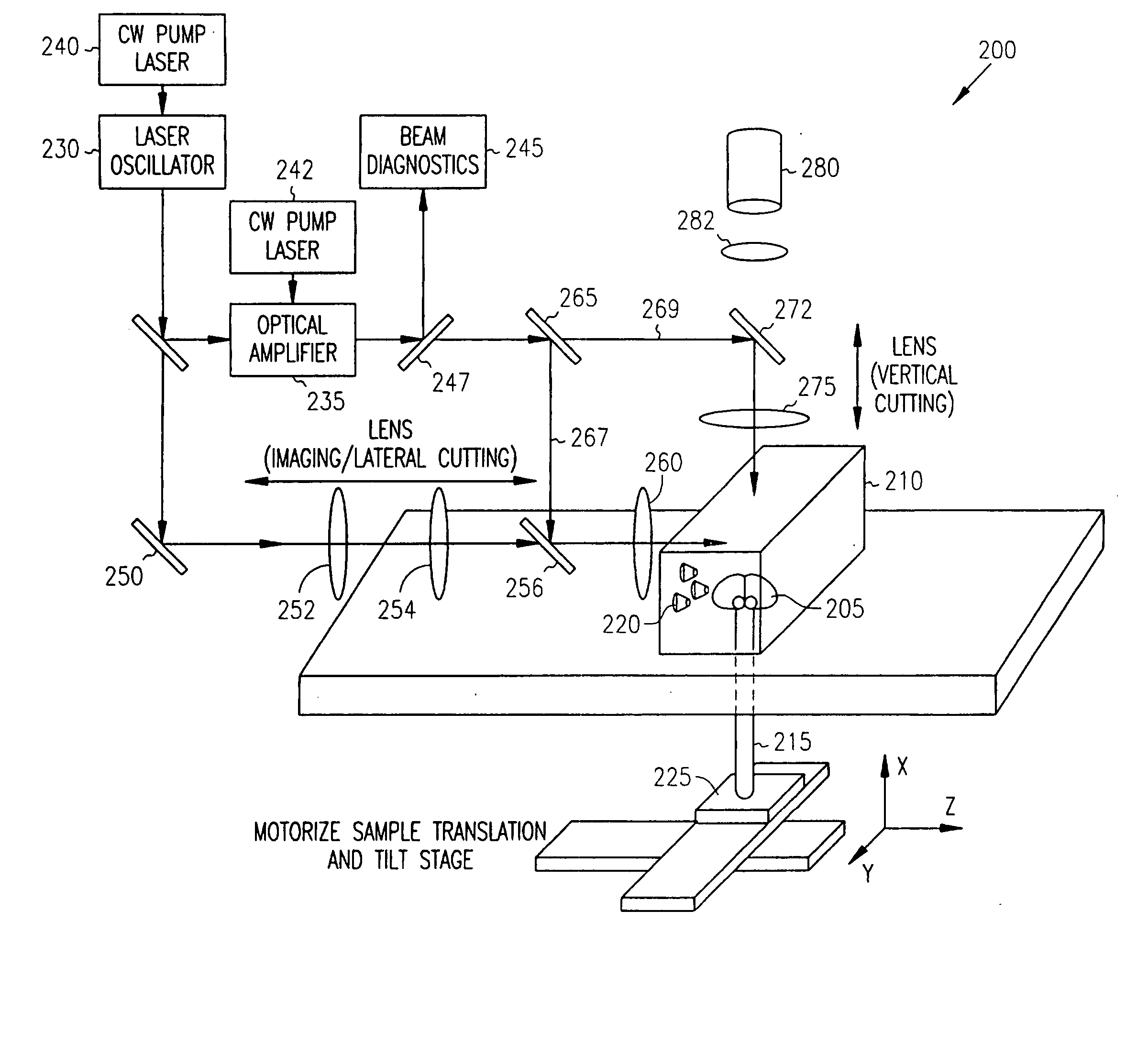
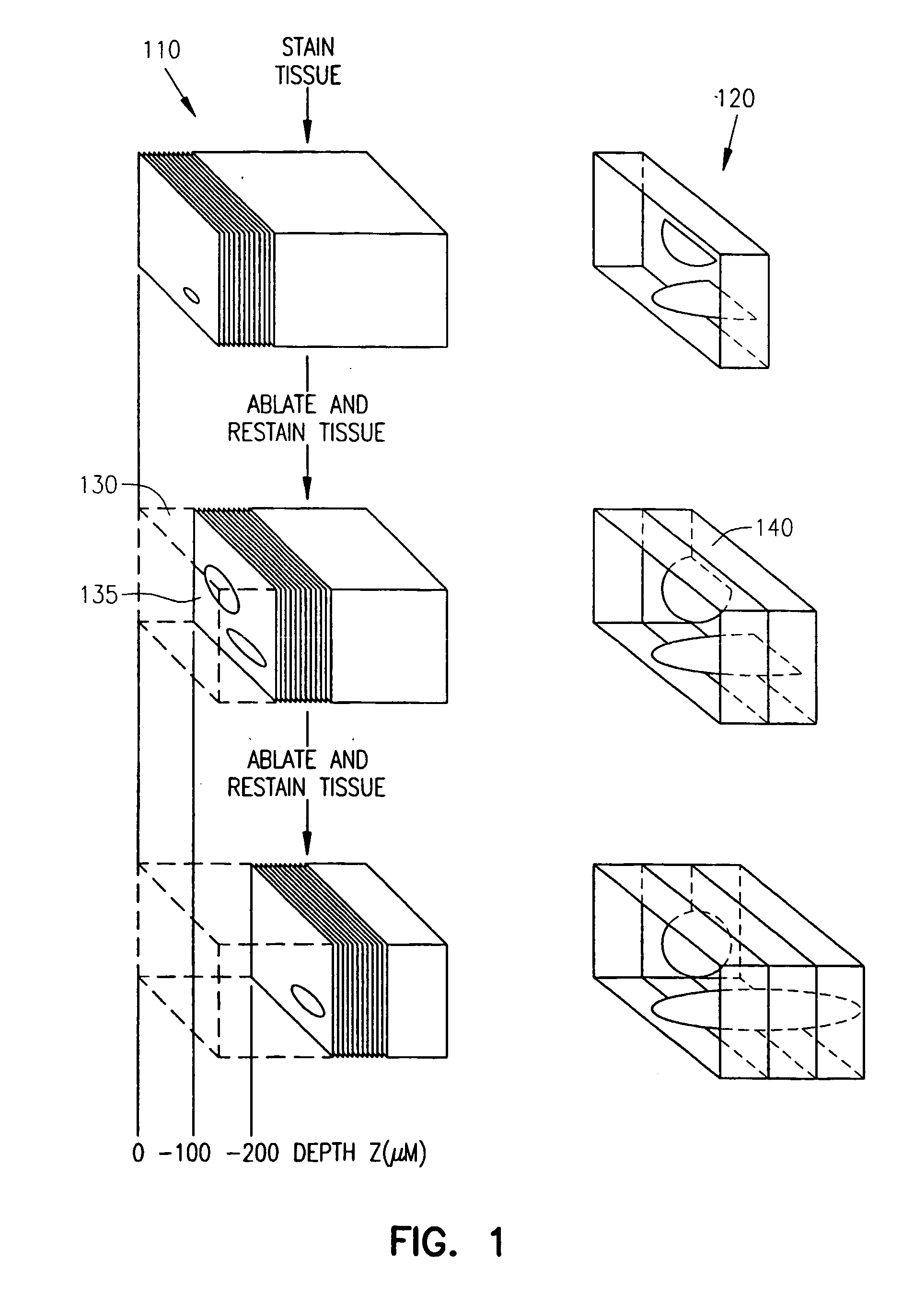
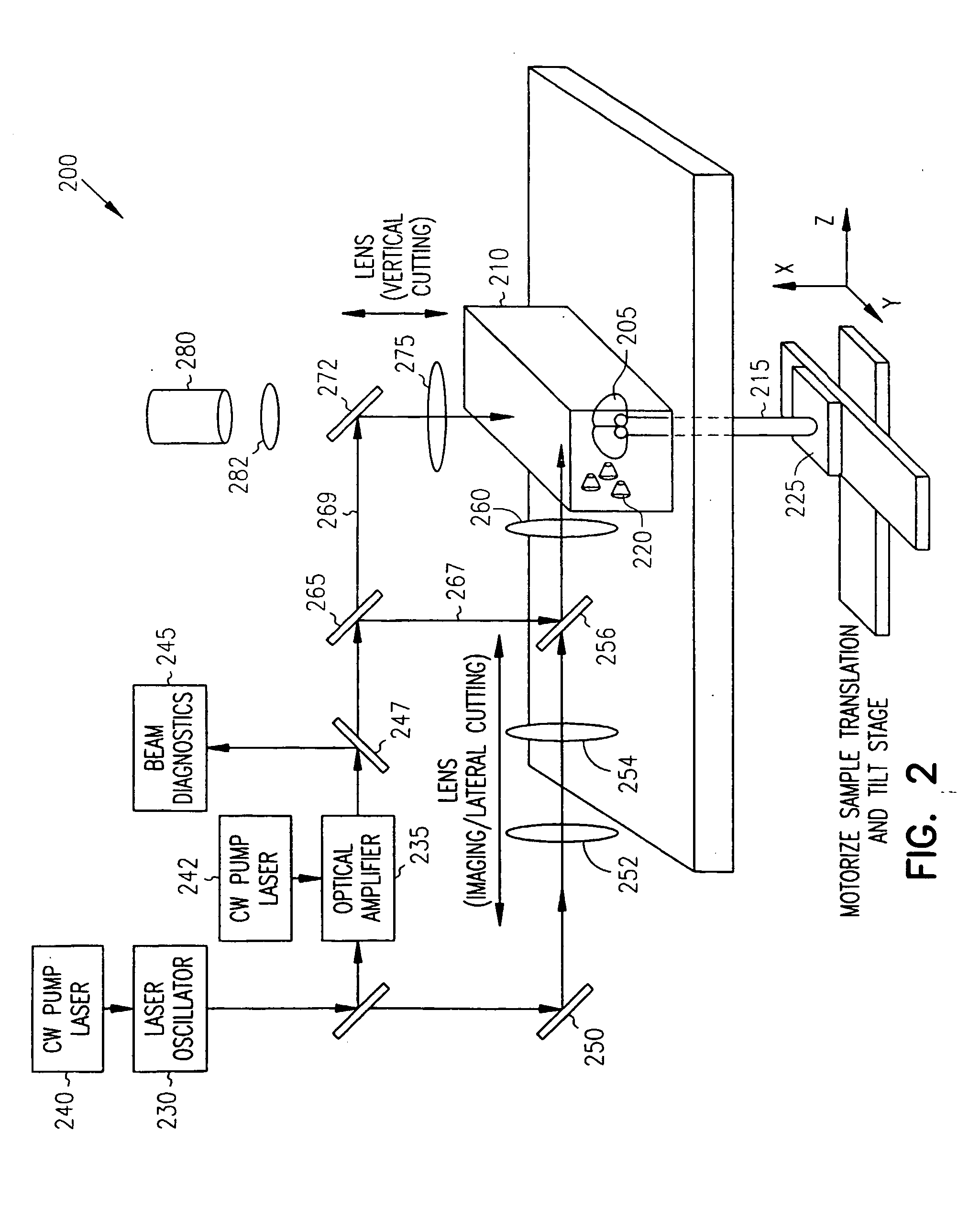
[0007] Femtosecond laser pulses are used to iteratively cut and image fixed as well as exsanguinated fresh tissue. Such images help to automate three-dimensional histological analysis of biological tissue. Cuts are accomplished with 0.3 to 100 microjoule pulses to ablate tissue with one-micrometer precision. Permeability, immunoreactivity, and optical clarity of the remaining tissue is retained after pulsed laser cutting. Samples from transgenic mice that express fluorescent proteins retained their fluorescence to within micrometers of the cut surface.
[0008] Imaging of exogenous dyes that bind to specific proteins or nucleic acids, or imaging of endogenous fluorescent labels is

Problems solved by technology

This results in shrinkage and distortion of the tissue and thus the loss of information contained in the slice.
The relatively large thickness of each slice, typically 25 micrometers or more and the relatively low resolution of the imaging apparatus led to a loss in information.
This leads to heating and collateral tissue damage.

Method used


Image

  • Iterative optical based histology
  • Iterative optical based histology
  • Iterative optical based histology

Examples


Embodiment Construction

[0017] In the following description, reference is made to the accompanying drawings that form a part hereof, and in which is shown by way of illustration specific embodiments in which the invention may be practiced. These embodiments are described in sufficient detail to enable those skilled in the art to practice the invention, and it is to be understood that other embodiments may be utilized and that structural, logical and electrical changes may be made without departing from the scope of the present invention. The following description is, therefore, not to be taken in a limited sense, and the scope of the present invention is defined by the appended claims.

[0018] Functions or algorithms described herein are implemented in software or a combination of software and human implemented procedures in one embodiment. The software comprises computer executable instructions stored on computer readable media such as memory or other type of storage devices. The term “computer readable me...


PUM


Abstract

Femtosecond laser pulses are used to iteratively cut and image fixed as well as exsanguinated fresh tissue. Such images help to automate three-dimensional histological analysis of biological tissue. Cuts are accomplished with approximately 0.3 to 100 microJoule pulses to ablate tissue with one-micrometer precision. Permeability, immunoreactivity, and optical clarity of the remaining tissue is retained after pulsed laser cutting. Samples from transgenic mice that express fluorescent proteins retained their fluorescence to within micrometers of the cut surface.

Description

RELATED APPLICATIONS [0001] This application is a continuation under 35 U.S.C. 111 (a) of International Application No. PCT / US03 / 00777 filed Jan. 10, 2003 and published in English as WO 03 / 060477 A2 on Jul. 24, 2003, which claims priority from U.S. Provisional Application Ser. No. 60 / 347,625 filed Jan. 10, 2002, which applications and publication are incorporated herein by reference.GOVERNMENT FUNDING [0002] The invention described herein was made with U.S. Government support under Grant Number NS41096 awarded by the National Institute of Neurological Disease and Stroke and under Grant Number DBI-9987257 awarded by the National Science Foundation. The United States Government has certain rights in the invention.FIELD OF THE INVENTION [0003] The present invention relates to histology, and in particular to a laser-based optical histology. BACKGROUND OF THE INVENTION [0004] To obtain three dimensional representations as by current methods of histology of biological tissue, such as brai...

Claims


Application Information

Patent Timeline
IPC IPC(8): G01N1/04G01N1/06G01N33/483
CPCG01N1/06G01N2001/2886G01N2001/045
Inventor KLEINFELD, DAVIDSQUIER, JEFFREYTSAI, PHILBERT
Owner RGT UNIV OF CALIFORNIA SAN DIEGO
Learn More