
System and method for determining compactness in images

a compactness and image technology, applied in the field of image processing, can solve the problems of complex task of three-dimensional object detection, impractical technique, and further compounding of 3-d recognition, and achieve the effect of rapid detection of such nodules and computationally less intensive procedures

Inactive Publication Date: 2005-03-17
SIEMENS MEDICAL SOLUTIONS USA INC
View PDF7 Cites 4 Cited by
  • Summary
  • Abstract
  • Description
  • Claims
  • Application Information

AI Technical Summary

Benefits of technology

[0009] An intersection technique is applied to determine the presence of the candidates in the three dimensional image. The mask is applied to a section of the foreground region. The number of intersections between the mask and the section of the foreground region are counted. The presence of a candidate can be inferred if there is zero or only one intersection. This indicates that the nodule is either fully covered by the mask or else is attached to a wall or vessel. However, if the intersections are more than one then the image section is not a candidate.
[0010] In an embodiment, the three dimensional image is a image captured from a human or other living organism. Here, the candidate is a medical structure to be searched such as a lung nodule, tumor, etc. The mask is typically a cube shaped mask that is shaped and sized to cover a single nodule (or other structure / object). Rapid detection of such nodules in possible because only the voxels of the foreground image region are tested and not the voxels of the whole image. Further, the determination of intersections is a computationally less intensive procedure as compared to testing the surface properties of all voxels in the image.

Problems solved by technology

Detection of three dimensional (3-D) objects in volumetric images is a complex task.
However, the problem of 3-D recognition is further compounded when the speed of recognition is a paramount concern.
However, this would be an impracticable technique, given the sheer amount of voxels present even in a relatively medium size image.
For example, detecting lung nodules automatically in a CT (Computed Tomography Scan) lung scans is a difficult process.
However, calculation of such features is computationally time consuming.
Typically, the time constraints of the medical diagnosis process render the thorough testing of each voxel of a 3-D medical image to be an inefficient technique.
However, if the intersections are more than one then the image section is not a candidate.

Method used

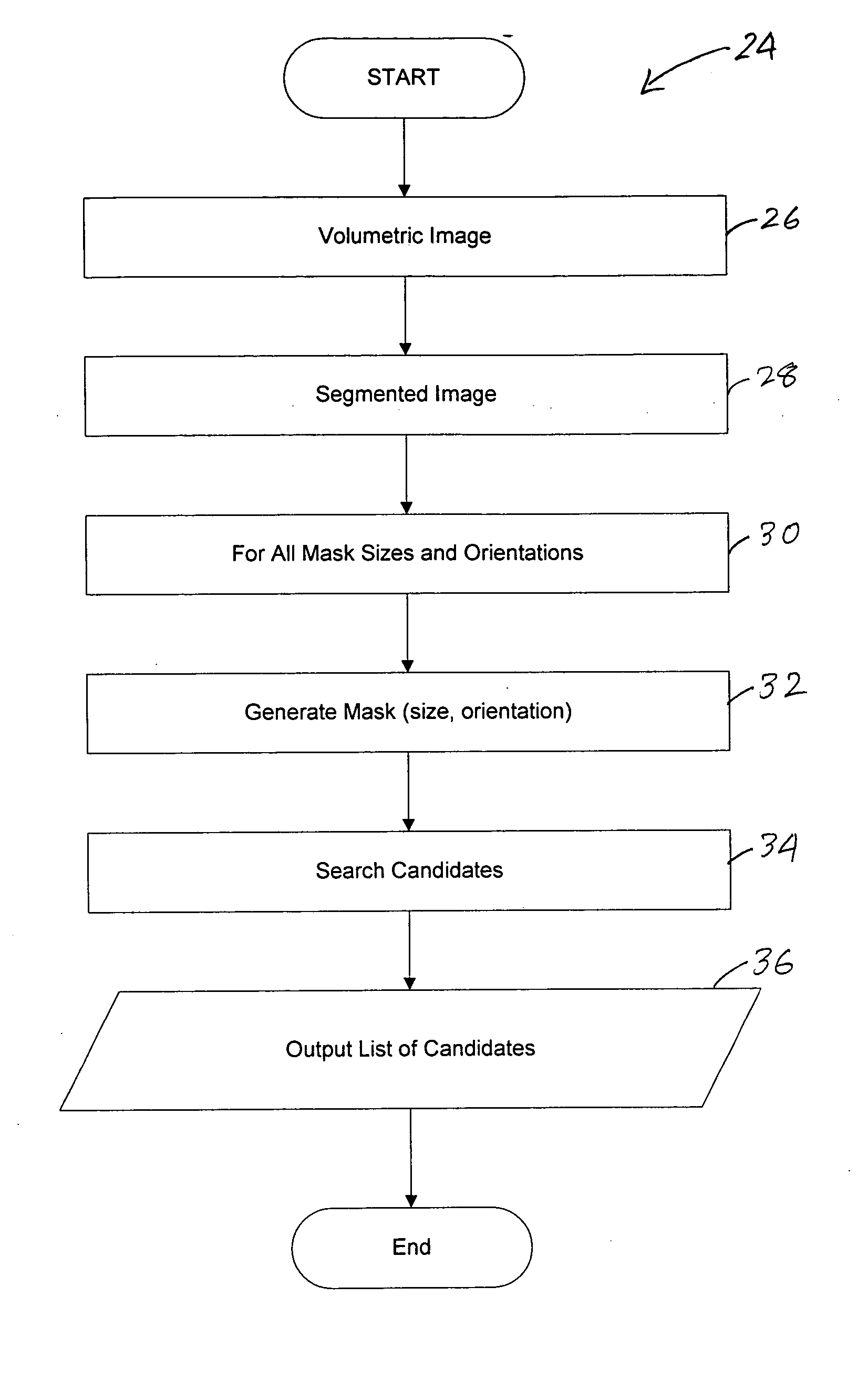
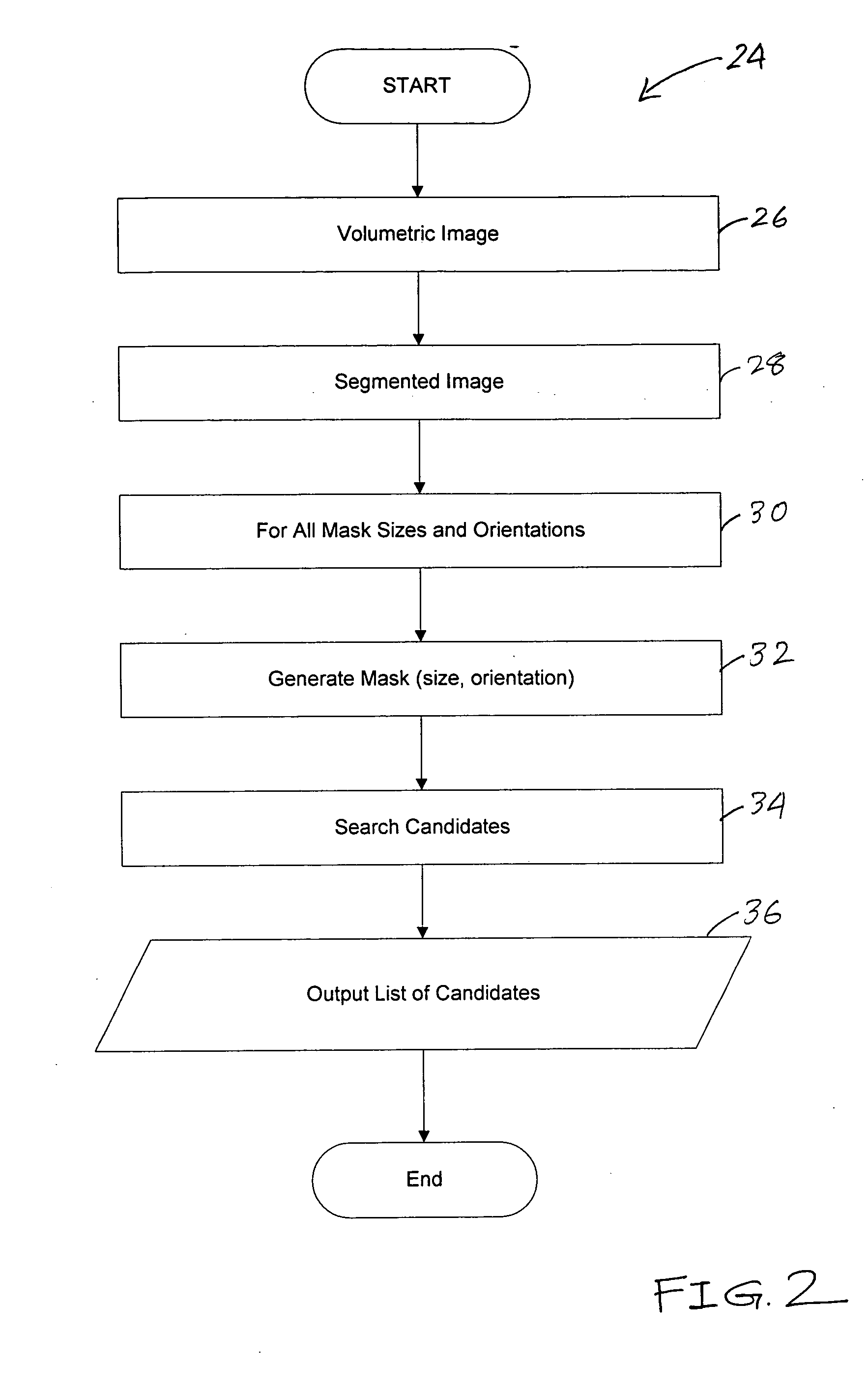

Image

  • System and method for determining compactness in images
  • System and method for determining compactness in images
  • System and method for determining compactness in images

Examples


Embodiment Construction

[0022] The preferred embodiments of the present invention will be described with reference to the appended drawings.

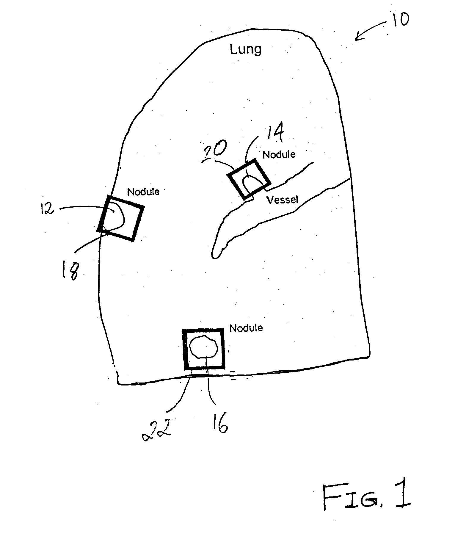
[0023]FIG. 1 is an illustrative diagram showing lung nodules with exemplary masks. At least one embodiment of the present invention can be utilized to detect solid pulmonary nodules. However, the those skilled in the art will realize that the pulmonary nodules are only used as a typical example to illustrate, and any structure with a known geometry in a volumetric image can be detected. For example, tumors, cysts, etc., can be detected. Further, although the appended drawings are in two dimensions for clarity in illustration, the embodiments of the present invention operate on three-dimensional images.

[0024] A typical image captured by a medical imaging device is a volumetric image. The cross-section 10 of an exemplary volumetric image is a part of a lung scan. Three illustrative nodules 12, 14 and 16 are shown along with corresponding masks 18-22. The input image fr...


PUM


Abstract

An image processing system for recognizing image features in three dimensional images, which can be medical images, uses a mask generator for generating masks that are used by a candidate searcher to search for candidate images in the three dimensional image. The candidate searcher applies the mask to a section of a foreground region of the image to determine the presence of a structure / object by counting the number of intersections between the mask and the section of the foreground region.

Description

CROSS-REFERENCE TO RELATED APPLICATION [0001] This application claims the benefit of U.S. Provisional Application No. 60 / 495,304, filed on Aug. 14, 2003, a copy of which is herein incorporated by reference.TECHNICAL FIELD [0002] This invention relates to the field of image processing and particularly to the detection of three dimensional objects in binary images or label images. DISCUSSION OF THE RELATED ART [0003] The field of image processing has progressed beyond the basic functions of capturing images and applying fundamental image transformations to the captured images. In general, processing of two dimensional images is relatively less complex as compared to the processing of three dimensional (3-D) images. The 3-D image recognition is a complex process given that surface, shape and size detection in 3-D images require a processing of a large number of parameters before a 3-D shape can be recognized. [0004] Detection of three dimensional (3-D) objects in volumetric images is a...

Claims


Application Information

Patent Timeline
Patent Type & Authority Applications(United States)
IPC IPC(8): G06T5/00G06T7/00G06V10/36
CPCG06K9/56G06K2209/05G06T7/0012G06T2207/30064G06T2207/10081G06T2207/20144G06T7/004G06T7/70G06T7/194G06V10/36G06V2201/03
Inventor WOLF, MATTHIAS
Owner SIEMENS MEDICAL SOLUTIONS USA INC
Learn More