Methods of diagnosis using pulse volume measurement

Active Publication Date: 2005-03-31
SMITHMARKS INC
View PDF8 Cites 20 Cited by
  • Summary
  • Abstract
  • Description
  • Claims
  • Application Information

AI Technical Summary

Benefits of technology

It will be readily apparent from the above that a need exists in the art to extend the use of plethysmography to diagnose more conditions. To that end, the present invention is directed to varied techniques using plethysmography to detect conditions that have previously not been detected or have been detected using more complicated techniques. Such conditions include blood loss, septic shock, cardiogenic shock, neonatal s

Problems solved by technology

That is clearly not a techniq

Method used


Image

  • Methods of diagnosis using pulse volume measurement
  • Methods of diagnosis using pulse volume measurement
  • Methods of diagnosis using pulse volume measurement

Examples


Embodiment Construction

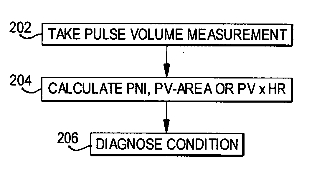
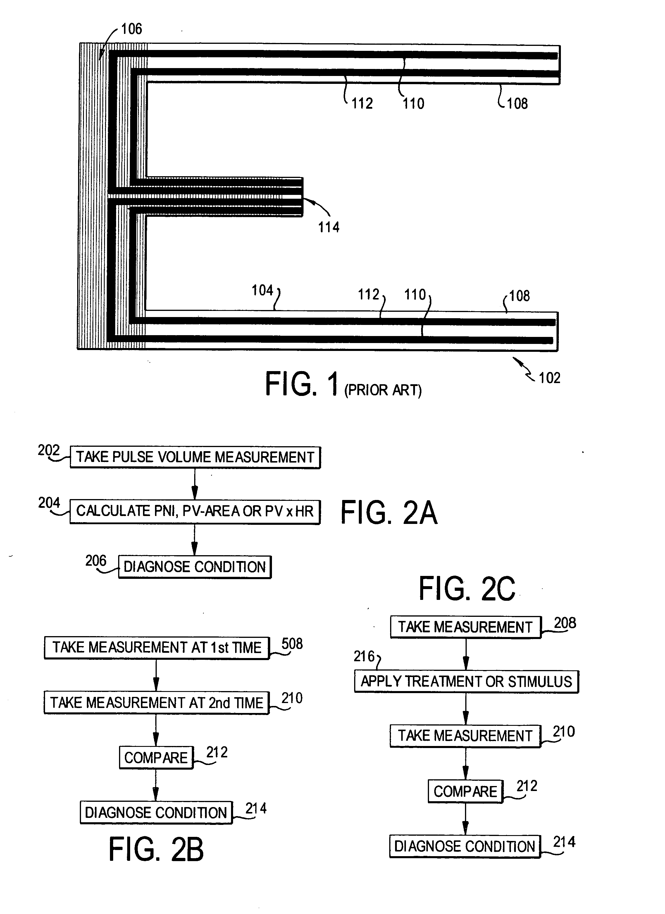
Preferred embodiments are set forth in detail herein with reference to the drawings. First, three general procedures will be outlined. Then, their application to various uses will be described in detail.

The most general procedure is shown in the flow chart of FIG. 2A. A pulse volume measurement is taken once or more than once in step 202, using a suitable device, such as, for example, any of the devices disclosed in the above-cited Marks patent and Smith et al patent application. Then, if required, the PNI (or PV-area OR PV×HR) is calculated in step 204. The condition is diagnosed in step 206.

Some conditions, as will be explained below, must be diagnosed in accordance with a change in time of the pulse volume or the PNI (or PV-area or PV×HR). In that case, as shown in FIG. 2B, the measurements are taken at different times in steps 208 and 210 and compared in step 212; then the diagnosis is made in step 214.

Still other conditions, as well as evaluation of treatment and testing...


PUM


Abstract

The utility of pulse volume measurement is expanded to detection of many conditions which have previously not been detected or have been detected using more complicated techniques. Such conditions include blood loss, septic shock, cardiogenic shock, neonatal sepsis, patent ductus arteriosus, limb ischemia, intra-aortic balloon pump performance, peripheral vascular disease, congestive heart failure, the effectiveness of vasoactive medications, syncope, dehydration, pre-eclampsia, deep vein thrombosis, thermal injuries, vascular instability due to renal dialysis, compromising of circulation to the hand caused by radial artery harvesting, changes in cardiac output, and hypertension. According to the present invention, such diagnoses can be performed by taking one measurement, by taking measurements over time to detect a change or by taking measurements before and after application of a treatment or stimulus.

Description

FIELD OF THE INVENTION The present invention is directed to methods of diagnosis using pulse volume measurement and more particularly to diagnostic methods which expand the utility of pulse volume measurement. DESCRIPTION OF RELATED ART The measurement of peripheral blood flow is important in medicine, since there are many specific diseases in which peripheral blood flow is impaired, e.g., diabetes and atherosclerosis. Also, the peripheral blood flow is altered as the total cardiac output is increased or decreased. Cardiac output is particularly important in patients who are under anesthesia, are in the post-operative state, or are critically ill or unstable. As blood flow from the heart falls, the peripheral blood flow is dramatically reduced to preserve flow to the brain and vital organs. Blood flow to an extremity can be measured painstakingly and invasively by dissecting out the main blood vessels to the limb (e.g., brachial artery in an arm) and encircling it with an electro...

Claims


Application Information

Patent Timeline
IPC IPC(8): A61B5/053
CPCA61B5/412A61B5/0535
Inventor MARKS, LLOYDSMITH, MICHAEL
Owner SMITHMARKS INC
Why Eureka
Try Eureka