Ultrasonic diagnostic equipment and imaging processing apparatus

a diagnostic equipment and ultrasonic technology, applied in ultrasonic/sonic/infrasonic image/data processing, applications, tomography, etc., can solve the problems of shortening the duration of the contrasting effect by ultrasound projection, weakening the contrasting effect instantly, and non-uniform enhancement, etc., to achieve easy to see, compare more accurately, and accurately and easily set rois

Active Publication Date: 2005-05-12
TOSHIBA MEDICAL SYST CORP
View PDF30 Cites 46 Cited by
  • Summary
  • Abstract
  • Description
  • Claims
  • Application Information

AI Technical Summary

Benefits of technology

[0042] As described above, in accordance with the ultrasonic diagnostic equipment and the image processing apparatus according to the invention, the images before and after stressing can be compared more accurately.
[0043] Moreover, in accordance with the ultrasonic diagnostic equipment and the image processing apparatus according to the invention, the ROIs can be set accurately and easily.
[0044] Furthermore, the invention has the advantage of obtaining the ultrasonic diagnostic equipment and the image processing apparatus in which the operated results are easily visible.

Problems solved by technology

In relation to the property of the bubbles of the contrast medium, however, such a situation occurs that the duration of the contrasting effect is shortened by ultrasound projection.
In consideration of these facts, the bubbles will disappear in succession before intensity enhancement based on the contrast medium is confirmed on the image of such a diagnosis, with the result that the contrasting effect will weaken instantly.
In myocardial contrast echocardiography, however, enhancement is nonuniform due to nonuniformity in the acoustic field of a sector probe, and nonuniformity in an acoustic field as is induced by an in-vivo structure.
The nonuniformity forms an obstacle to the diagnosis of myocardial ischemia or a myocardial infarction part.
However, the above countermeasure is directed toward the case of the B-mode image, and it does not suppose the case of tissue harmonic imaging (THI) having come into the limelight in recent years, or harmonic imaging such as contrast echo imaging, so that drawbacks as stated below are involved.
On the other hand, the “nonuniformity of the acoustic field attributed to the in-vivo structure” is induced in such a manner, for example, that a rib exists in close proximity to the probe, so the transmission and reception of ultrasounds are hampered by the rib.
Therefore, it becomes difficult to detect a morbid part, such as myocardial ischemia or myocardial infarction part, which is clinically to be found.
That is, even with the stress echocardiography, it is very difficult to judge an ischemic region from a single image.
Even with this method, however, the visual inspection of a diagnostician has its limit, and if a certain part is morbid is disputable and suspicious.
When the arithmetic operation is directly executed without making positional corrections, such a situation takes place where a motion artifact appears or where quite no operated result is obtained.
In this regard, in a case where both the set ROIs are inaccurate, that is, where they do not satisfactorily correspond to each other, an accurate operated result cannot be expected.

Method used


Image

  • Ultrasonic diagnostic equipment and imaging processing apparatus
  • Ultrasonic diagnostic equipment and imaging processing apparatus
  • Ultrasonic diagnostic equipment and imaging processing apparatus

Examples


first embodiment

[0067] Now, this invention will be described with reference to the drawings as to an ultrasonic diagnostic equipment in which the data of a blood flow movement are obtained on the basis of the degree of enhancement of a contrast medium flowing into a myocardium, so as to identify an abnormal part. Incidentally, the ultrasonic diagnostic equipment according to the invention is also applicable to any region of interest, an abdominal or other organs, for example, the liver in a case where the ultrasonic contrast medium is given to a patient and where a blood flow state is observed on the basis of the degree of enhancement.

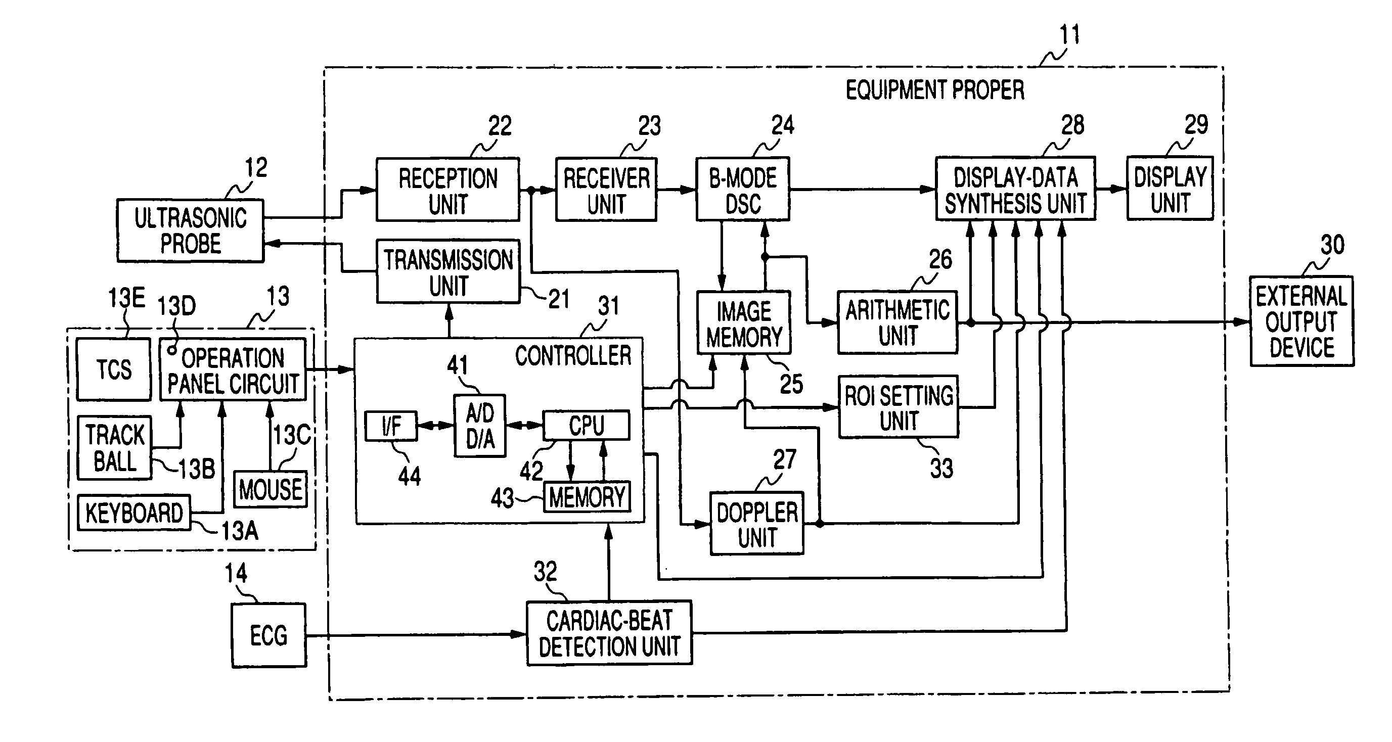
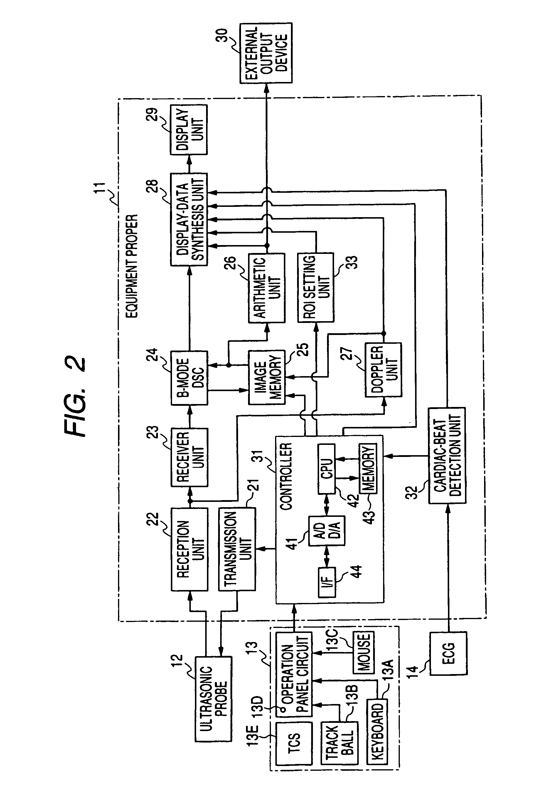
[0068]FIG. 2 schematically shows the general construction of the ultrasonic diagnostic equipment according to the first embodiment. The ultrasonic diagnostic equipment shown in FIG. 2 includes the equipment proper 11, and an ultrasonic probe 12, an operation panel 13 and an ECG (electrocardiograph) 14 which are connected to the equipment proper 11.

[0069] The operatio...

second embodiment

[0125] Subsequently, the ultrasonic diagnostic equipment according to the invention will be described with reference to the accompanying drawings by similarly taking as an example the case where the blood flow velocity of a myocardial tissue is diagnosed.

[0126] Also the ultrasonic diagnostic equipment in this embodiment is identical or equivalent in construction to the ultrasonic diagnostic equipment according to the first embodiment as shown in FIG. 2, and its constituents shall be omitted from description by assigning the same reference numerals and signs thereto.

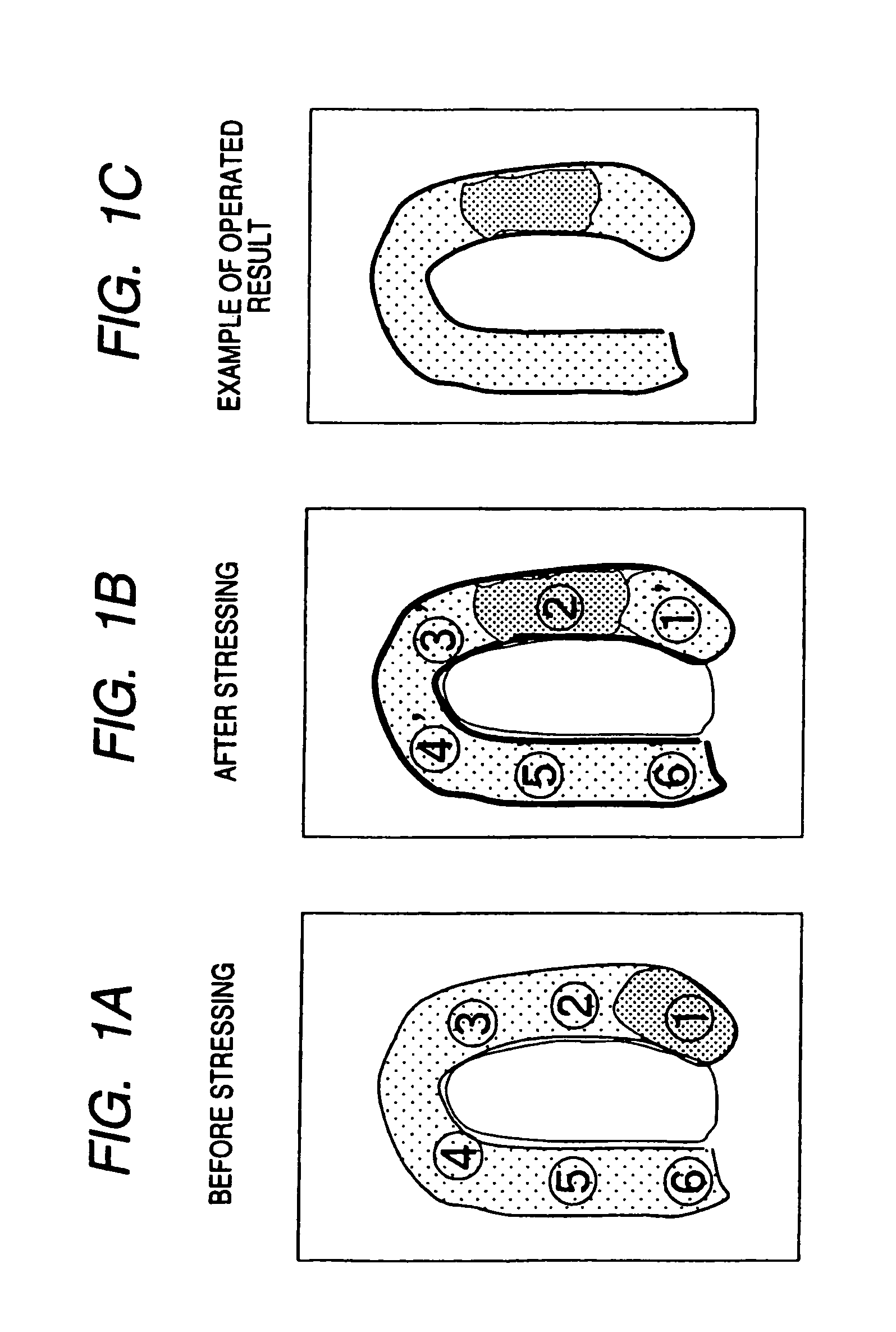
[0127] In this embodiment, unlike in the first embodiment, images after stressing are adopted as two images which are arithmetically operated.

[0128] In the case of the ultrasonic diagnostic equipment according to the first embodiment, a frame rate (the time interval of transmission) is controlled to be constant during observation. Of course, in a case where a depth or a scan line density has been altered, the frame rate...


PUM


Abstract

Each of an ultrasonic diagnostic apparatus and an image processing apparatus according to the present invention is characterized by comprising image acquisition means for acquiring image data by scanning a patient into whom an ultrasonic contrast medium has been injected, with ultrasonic beams, same-part correspondence means for bringing same parts into correspondence with each other, between a plurality of images concerning the same parts of the identical patient and acquired by the image acquisition means, arithmetic operation means for arithmetically operating image information items on changes of intensities between the plurality of images by using the intensities of those corresponding pixels of the plurality of images which have been brought into correspondence by the pixel correspondence means, and display means for displaying results operated by the arithmetic operation means. According to the construction, it is possible to provide the ultrasonic diagnostic equipment and the image processing apparatus which can compare the images before and after injection of a vasodilator drug, more accurately in contrast echo imaging.

Description

BACKGROUND OF THE INVENTION [0001] 1. Field of the Invention [0002] The present invention relates to an ultrasonic diagnostic equipment and an image processing apparatus, and more particularly to an ultrasonic diagnostic equipment and an image processing apparatus which offer information reflective of the blood flow velocity of a myocardial tissue by employing contrast echoes. [0003] 2. Description of the Prior Art [0004] In the field of ultrasonic diagnostic equipments, the spotlight of attention has recently been focused upon contrast echocardiography wherein, in examining the heart, an abdominal organ or the like, an ultrasonic contrast medium is injected from a vein so as to estimate a blood flow movement. The technique of injecting the contrast medium from the vein is less invasive than the technique of injecting the same from an artery, and diagnoses based on the estimation method are coming into wide use. The ultrasonic contrast medium is principally constituted by microbubbl...

Claims


Application Information

Patent Timeline
Patent Type & Authority Applications(United States)
IPC IPC(8): A61B8/08A61B8/00A61B8/06
CPCA61B8/06A61B8/543A61B8/481A61B8/13Y02A90/10
Inventor KAWAGISHI, TETSUYAKAMIYAMA, NAOHISA
Owner TOSHIBA MEDICAL SYST CORP
Why Eureka
Try Eureka