
Method for evaluating magnetic resonance spectroscopy data using a baseline model

a baseline model and magnetic resonance spectroscopy technology, applied in the field of magnetic resonance spectroscopy data evaluation, can solve the problems of reducing the signal-to-noise ratio of the modeled magnetic resonance signal portion, affecting and limiting the parameterization of voigt lines and model spectra to special applications, so as to improve the smoothness of the baseline and optimize the baseline modeling

Active Publication Date: 2005-06-23
SIEMENS HEALTHCARE GMBH
View PDF5 Cites 23 Cited by
  • Summary
  • Abstract
  • Description
  • Claims
  • Application Information

AI Technical Summary

Benefits of technology

[0017] An object of the present invention to provide a method to evaluate the data of a magnetic resonance signal in the time domain that enables a low-noise, exact modeling of the metabolite signals of interest.
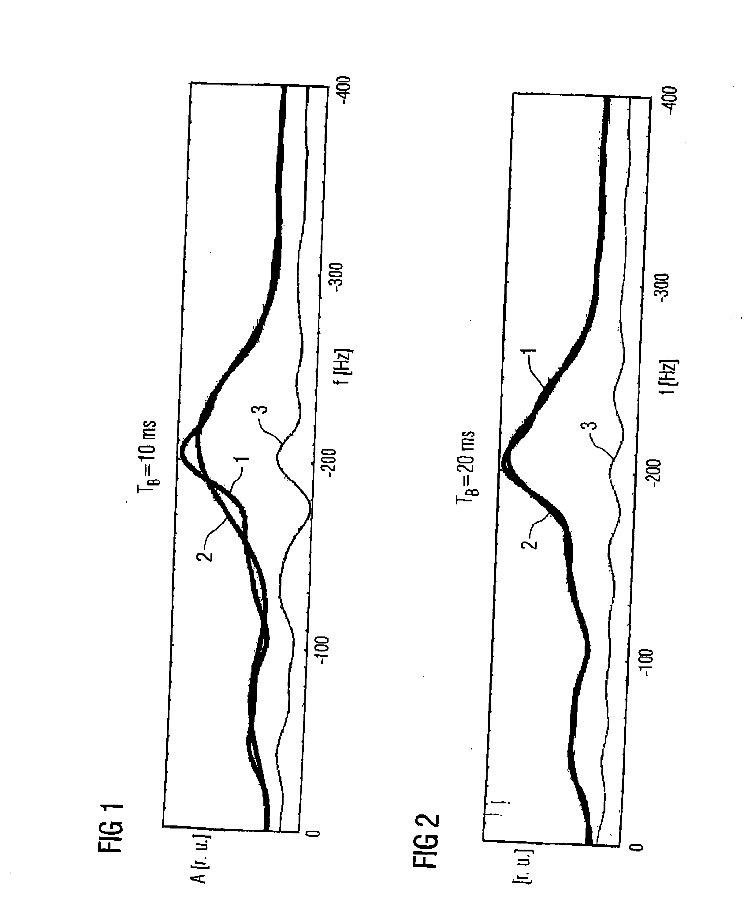
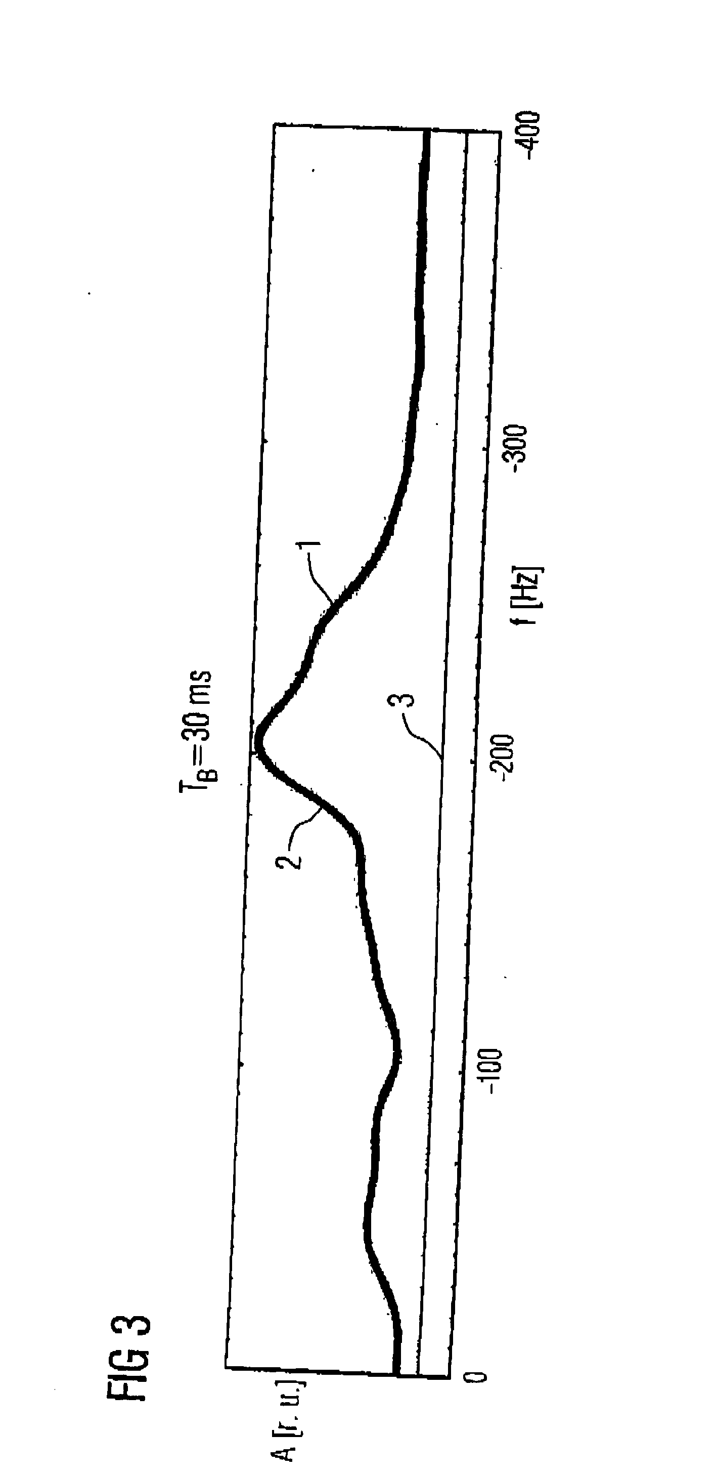
[0020] At the beginning of a magnetic resonance signal acquired in vivo, as it is shown in the time domain, the portion of the components that are associable with the baseline outweigh the portion of the components which are associable with the metabolite signals. Furthermore, the portion of the baseline at the MR signal decays significantly faster than the portion of the metabolite spectrum. The portion of the baseline at the MR signal after 30 ms of a total duration of 200 ms can thus already have decayed to 13% of its original value; but the portion of the metabolite signal can still be present at 75%. Use of the first MR data of a magnetic resonance signal therefore already allows the modeling of a baseline with good precision. Furthermore, since these first MR data are not lost for modeling the metabolite signals of interest, the inventive evaluation method also achieves a low-noise and exact modeling of these metabolite resonances. The modeling of the baseline also ensues exclusively on the basis of the data of the MR signal in the time domain, such that no additional Fourier transformations back and forth are necessary, and very short calculation times are achieved. The method exhibits no limitation to special applications and thus can be used universally.
[0023] An overshadowing of the metabolite resonances not corresponding to reality can be prevented by restringing at least one parameter to optimize the modeling of the baseline, the restringing preferably being accomplished by the introduction of parameter limits or by limiting the portion of the baseline on the overall signal.
[0025] To improve the smoothness of the baseline in the frequency domain representation, the modeled baseline can be regularized.

Problems solved by technology

The parameterization by Voigt lines and model spectra is hereby limited to special applications, since each requires foreknowledge about the examined tissue.
This method, however, leads to a worsening of the signal-to-noise ratio, meaning the signal-to-noise ratio of the modeled magnetic resonance signal portion drops and the adapted parameters exhibit a greater uncertainty.
In the case of narrowly adjacent or strongly overlapping metabolite signals, this method actually breaks down entirely when an insufficient number points are omitted, since contributions of adjacent metabolites can mix, and the individual metabolite signals are overshadowed.

Method used


Image

  • Method for evaluating magnetic resonance spectroscopy data using a baseline model
  • Method for evaluating magnetic resonance spectroscopy data using a baseline model
  • Method for evaluating magnetic resonance spectroscopy data using a baseline model

Examples


Embodiment Construction

[0031] A magnetic resonance spectroscopic examination of a tissue provides a damped, nuclear magnetic resonance signal (MR signal), what is known as the free induction decay (FID) signal, oscillating periodically with the Lamour frequency, The Fourier-transformed FID signal is generally designated as a resonance curve, but the term absorption spectrum also has become common in MR spectroscopy with for this curve. Below, the representation of the nuclear magnetic resonance signals in the time domain is designated as an MR signal, and the representation of the nuclear magnetic resonance signals in the frequency domain is designated as a resonance curve.

[0032] For a digital representation of an MR signal, it is sampled with at least double the Lamour frequency, such that it is available as an MR data set for extensive further processing. In the following the terms MR signal and MR data set are used synonymously, since digital processing, meaning evaluation of a nuclear magnetic resona...


PUM


Abstract

In a method for evaluating data of a magnetic resonance signal, a baseline is modeled and at least one metabolite signal is modeled. The modeling of the baseline ensues based on magnetic resonance data in the time domain, and using only data that begins with the magnetic resonance signal and having a length that is shorter than the duration of the overall magnetic resonance signal.

Description

BACKGROUND OF THE INVENTION [0001] 1. Field of the Invention [0002] The present invention concerns a method to evaluate magnetic resonance spectroscopy data with a determination of the baseline superimposed on the resonance signals. [0003] 2. Description of the Prior Art [0004] Magnetic resonance spectroscopy (MRS) has been used for more than four decades in physical, chemical and biochemical basic research, for example as an analysis technique or for structural clarification of complex molecules. Like magnetic resonance tomography, magnetic resonance spectroscopy (MR spectroscopy) is based on the principle of nuclear magnetic resonance. Imaging is not the goal of MR spectroscopy, but rather the generation of magnetic resonance spectra (MR spectra) for analysis of substances such as for a structure clarification. The resonant frequencies of isotopes that possess a magnetic moment (such as, for example, 1H, 13C, 19F or 31P) are dependent on the chemical environment of these isotopes....

Claims


Application Information

Patent Timeline
IPC IPC(8): G01R33/46
CPCG01R33/4625
Inventor WEILAND, ELISABETH
Owner SIEMENS HEALTHCARE GMBH
Learn More