Method for the analysis of methylation patterns within nucleic acids by means of mass spectrometry

Inactive Publication Date: 2005-07-28
EPIGENOMICS AG
View PDF5 Cites 6 Cited by
  • Summary
  • Abstract
  • Description
  • Claims
  • Application Information

AI Technical Summary

Benefits of technology

[0046] (4) contacting the double stranded nucleic acid with enzymes and / or agents under conditions conducive to the methylation of the synthesised strand such that the CpG dinucleotides within the synthesised strand are methylated according to the methylation status of the corresponding CpG dinucleotide on the template strand thereby preserving the genomic methylation pattern,
[0111] In a particularly preferred embodiment of the method the mass spectrum obtained is compared to the mass spectrum of fragments obtained from known samples of either methylated or unmethylated versions of the target nucleic acid. These known spectra are referred to as “reference” spectra. A simple comparison of the sample spectrum vs. reference spectra enables the determination of the methylation status of the sample.

Problems solved by technology

The analysis of biological compounds such as peptides and nucleic acids by mass spectrometry has been hampered by the difficulty in achieving ionisation of large molecules.
However, although MALDI-TOF spectrometry is well suited to the analysis of peptides and proteins, the analysis of nucleic acids has proved somewhat more difficult (Gut I G, Beck S. DNA and Matrix Assisted Laser Desorption Ionisation Mass Spectrometry.
The problems include a lack of resolution of high molecular weight DNA fragments, DNA instability, and interference from sample preparation reagents.
For nucleic acids having a multiply negatively charged backbone, the ionization process via the matrix is considerably less efficient.
However, although several responsive matrixes are suitable for DNA analysis, the difference in sensitivity has not been reduced.
However, the emphasis on fields such as proteomics, genomics and bioinformatics has meant that analysis of epigenetic variations has not received as much scientific attention.
However, 5-methylcytosine positions cannot be identified by sequencing since 5-methylcytosine has the same base pairing behaviour as cytosine.
Moreover, the epigenetic information carried by 5-methylcytosine is completely lost during PCR amplification.
However none of the currently used methods for the analysis of methylation patterns directly utilise mass spectrometry, as the analysis of minute samples of nucleic acids is not practical without prior amplification of said sample.
No currently known methods of nucleic acid amplification conserve said methylation patterns therefore currently the application of mass spectrometry to the analysis of methylation patterns is not possible on readily available biological samples.

Method used


Image

  • Method for the analysis of methylation patterns within nucleic acids by means of mass spectrometry
  • Method for the analysis of methylation patterns within nucleic acids by means of mass spectrometry
  • Method for the analysis of methylation patterns within nucleic acids by means of mass spectrometry

Examples


example 1

Methylation Retaining PCR Amplification

[0133] Genomic DNA commercially available from Promega is used in the analysis. A CpG rich fragment of the regulatory region of the GSTPi gene is used in the analysis. The DNA is firstly artificially methylated at all cytosine 5 positions within the CpGs (upmethylation). The upmethylated DNA is then amplified using one round of PCR. The resultant amplificate is then divided into two samples, Sample A (the control sample) is amplified using conventional PCR. Sample B is amplified according to the disclosed method. The two samples are then compared in order to ascertain the presence of methylated CpG positions within Sample B. The comparison is carried out by means of a bisulphite treatment and analysis of the treated nucleic acids.

Upmethylation

Reagents:

[0134] DNA [0135] SssI Methylase (concentration 2 units / μl). [0136] SAM (S-adenosylmethionine) [0137] 4.5 μl Mss1-Buffer (NEB Buffer B+ (10 mMole Tris-HCl 300 mMole NaCl, 10 mMole Tris-HCl,...

example 2

Mass Spectrometric Analysis of a Methylated Oligonucleotide

[0158] A test sample (not from a patient) of an oligonucleotide of known sequence and unknown methylation status is provided. It is required that the methylation status of the sample be ascertained, this may be fully methylated, fully unmethylated or a mixture of the two. The sequence of the oligonucleotide is AACACGGGCATTGATCTGACGT (SEQ ID NO: 3).

Reference Spectrum

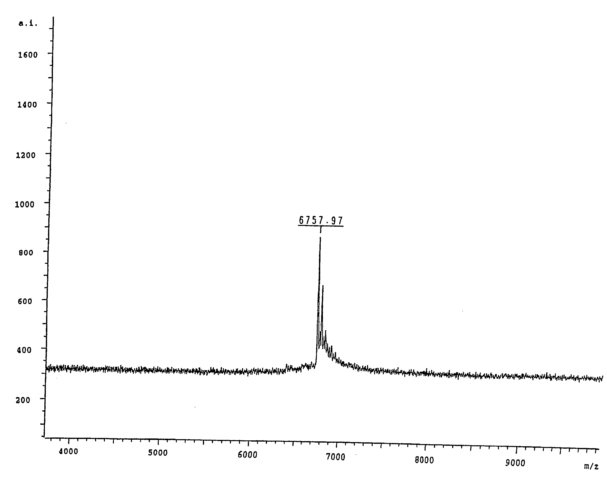
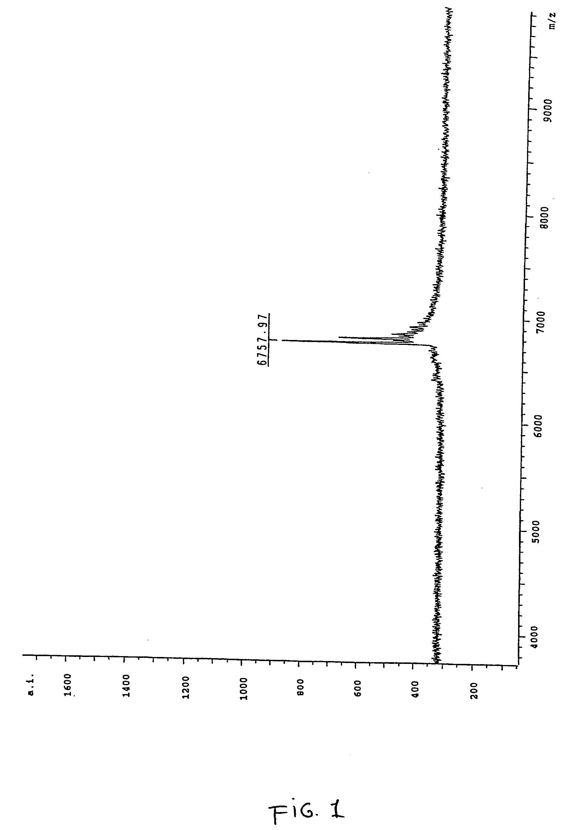
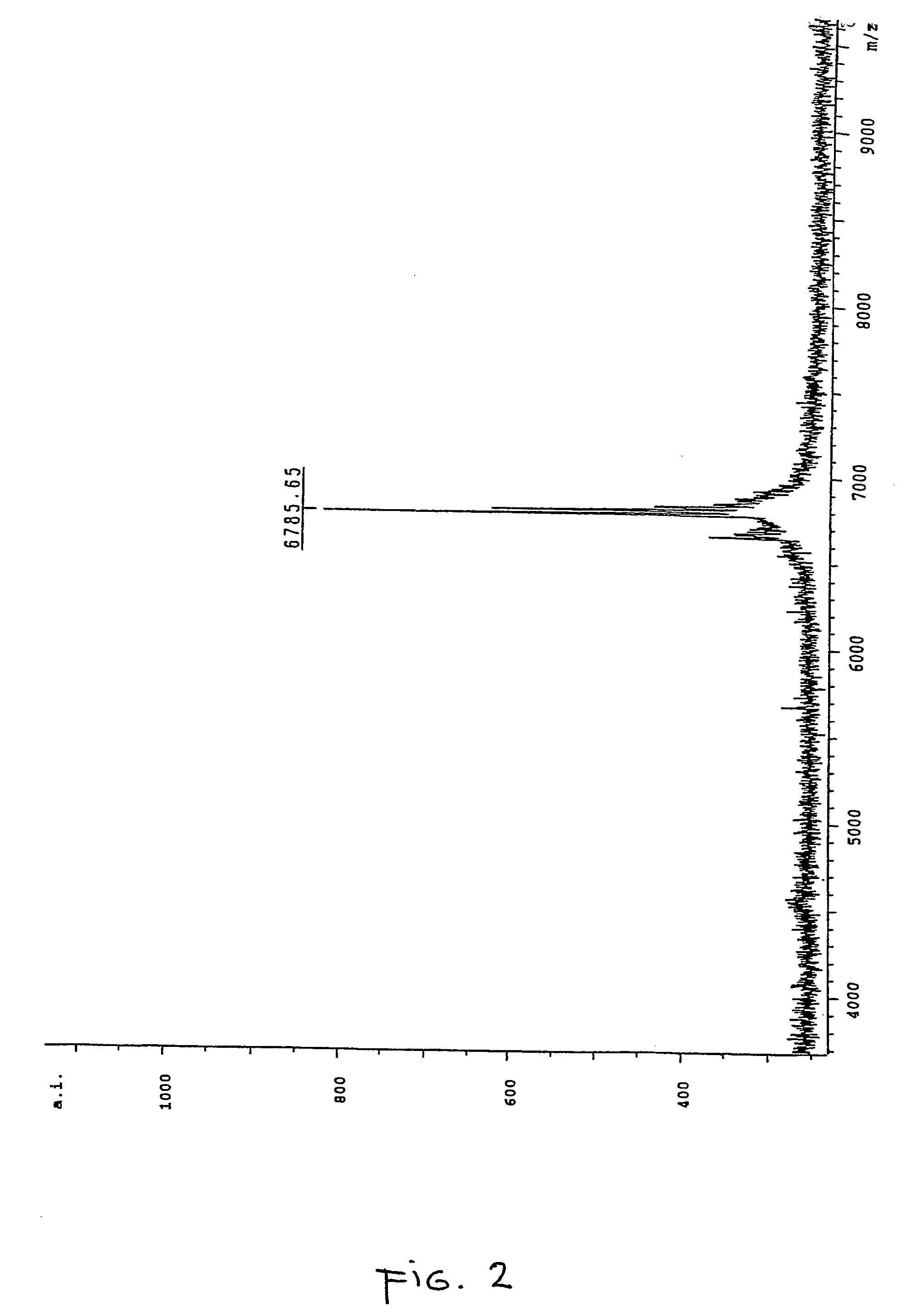
[0159] In order to correctly identify the methylation status of the sample it was necessary to ascertain the spectra of two control samples. Accordingly an oligonucleotide according to SEQ ID NO:3 was ordered from a commercial supplier, one sample being methylated at each cytosine within the oligonucleotide and the other being fully unmethylated. A sample of the unmethylated oligonucleotide was analysed by MALDI-TOF mass spectrometry and the resultant spectrum can be seen in FIG. 1, the mass of the oligonucleotide was measured as 6757.97 daltons. The fully me...


PUM

PropertyMeasurementUnit
Lengthaaaaaaaaaa
Massaaaaaaaaaa
Molecular weightaaaaaaaaaa

Abstract

The present invention describes a method for the analysis of methylation patterns comprising the following steps: a) isolation of genomic nucleic acids from a biological sample, b) amplification of one or more target nucleic acids of said genomic nucleic acids in a manner whereby the methylation patterns of said genomic nucleic acids are maintained in the amplificate nucleic acid, c) performing mass spectrometry on said amplified nucleic acid or fragments thereof to obtain mass spectra; d) evaluating the obtained mass spectra and e) determining the methylation pattern and / or methylation status of the sample. The disclosed invention provides novel methods for the analysis of cytosine methylation patterns within genomic DNA samples. Said method comprises a methylation retaining enzymatic amplification of a test nucleic acid sample, followed by mass spectrometric analysis of the amplificate nucleic acids.

Description

PRIOR ART [0001] Developments in the field of molecular biology over the last decade have in particular been focused upon the analysis of the human genome. The perceived benefits of the understanding of the functioning of the genome have led to the development of a variety of techniques suitable for the analysis and manipulation of nucleic acid sequences. Of particular interest are techniques that decrease the cost and increase the speed of genetic analysis. One such technique is the application of mass spectrometry to the analysis of nucleic acid sequences. [0002] Mass spectrometry is an analytical technique with multiple applications in the field of chemistry and biology. Its uses include the accurate determination of molecular weights, identification of chemical structures by means of their fragmentation properties, determination of the composition of mixtures and qualitative elemental analysis. In a mass spectrometer a sample is first ionised, different species of ions are then ...

Claims


Application Information

Patent Timeline
IPC IPC(8): C12Q1/68
CPCC12Q1/6858C12Q2565/627C12Q2537/164C12Q2521/125
Inventor BERLIN, KURT
Owner EPIGENOMICS AG
Why Eureka
Try Eureka