
Treatment instrument for EMR, and EMR device

Inactive Publication Date: 2005-07-28
KAIGEN +2
View PDF15 Cites 36 Cited by
  • Summary
  • Abstract
  • Description
  • Claims
  • Application Information

AI Technical Summary

Benefits of technology

[0005] The present invention is devised to solve the above problem, and has for its object to provide a treatment instrument for EMR for excising lesion portions while reducing complications and burdens to patients in an endoscopic mucosal resection, and an EMR device with the treatment instrument for EMR.
[0017] According to the treatment instrument for EMR of the first aspect and the EMR device of the second aspect of the present invention as above, the separating and swelling member is included, so that the portion to be dissociated including the lesion portion, and the non-affected portion without including the lesion portion located under the portion to be dissociated are separated and dissociated from each other, and moreover the lesion portion is swollen by the separating and swelling member. Therefore, the treatment instrument for EMR and the EMR device can prevent a situation that the swelling is disappeared simultaneously with the start of cutting the mucosal layer as in the conventional art, and the need of swelling the affected portion by a plurality of the number of times is eliminated. The possibility of generation of the complication such as the perforation and bleeding, and leaving the lesion portion without removed are nearly avoided. Thus the treatment instrument for EMR and the EMR device enable easier and safer endoscopic mucosal resection in comparison with the conventional art, can greatly shorten an operation time in comparison with the conventional art, and can reduce the burden of patients.
[0018] The separating and swelling member exerts the separating and dissociating action, whereby the need of separating and cutting a wide range by, e.g., a diathermy knife or the like so as to preliminarily form a part where the separating and swelling member is to be inserted in the submucosal layer is eliminated. The danger of perforation, bleeding, and leaving the lesion portion without removed, etc. associated with the cutting operation can be avoided.
[0019] In the case of separating and dissociating the portion to be dissociated and the non-affected portion from each other by the separating and swelling member as above, it is difficult in some cases to insert the large separating and swelling member into the submucosal layer located below the lesion portion at a time. For solving the problem, the separation and dissociation between the portion to be dissociated and the non-affected portion is carried out beforehand for a part of the submucosal layer with the use of the insertion member which facilitates the insertion of the separating and swelling member into the submucosal layer, and the injection part for the separating and swelling member is formed. The insertion and the swelling operation of the separating and swelling member are thus facilitated, so that the treatment becomes easy and moreover, the danger of perforation, bleeding, leaving the lesion portion without removed, and the like can be avoided.

Problems solved by technology

As a result, the submucosal layer 2 and the muscular layer 3 below the submucosal layer 2 cannot be spaced fully, which may lead to the danger of perforation at the time of cutting and also the danger of leaving the lesion portion 4 without being removed.
While the loss of the pharmaceutical solution makes it necessary to swell the lesion portion 4 by a plurality of the number of times during the treatment, it is difficult to swell the submucosal layer 2 by injecting the pharmaceutical solution into the same layer within the submucosal layer 2 as that of the first swelling operation for the second time and afterward, and consequently the submucosal layer is often swollen from a much deeper layer.
Visual recognition of the lesion portion 4 is obstructed in many cases if the leading end of the endoscope is disposed at the side face of the swollen part, and the difficulties in visual recognition result in the complication of the perforation and bleeding, and leaving the lesion portion 4 without being removed in some cases.

Method used


Image

  • Treatment instrument for EMR, and EMR device
  • Treatment instrument for EMR, and EMR device
  • Treatment instrument for EMR, and EMR device

Examples


Embodiment Construction

[0052] A treatment instrument for EMR and an EMR device as a preferred embodiment of the present invention will be described below with reference to the attached drawings throughout which like parts are designated by like reference numerals. In the specification, an endoscope represents a general instrument for observing, diagnosing and treating lesions in a body cavity directly by naked eyes or images.

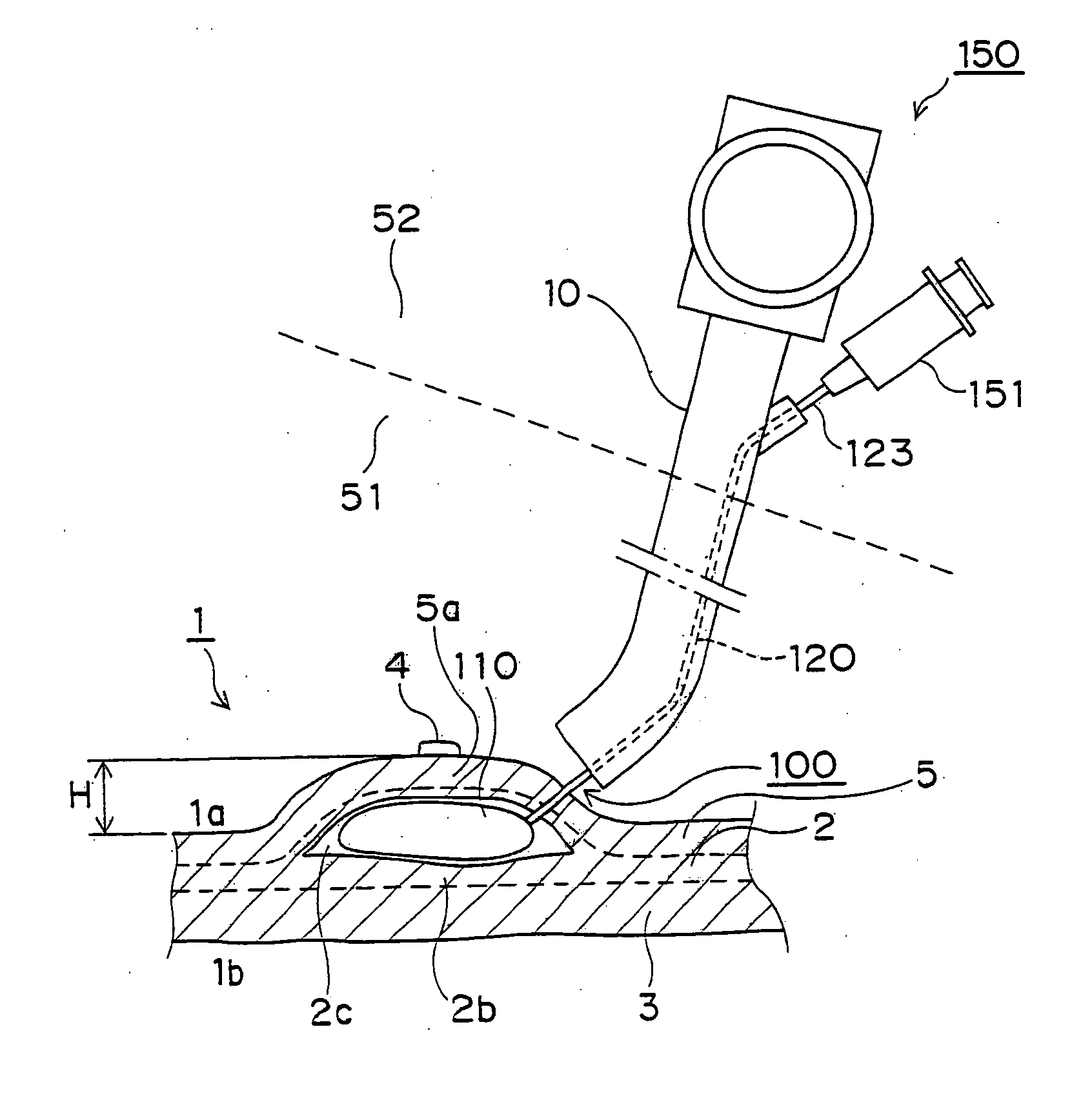
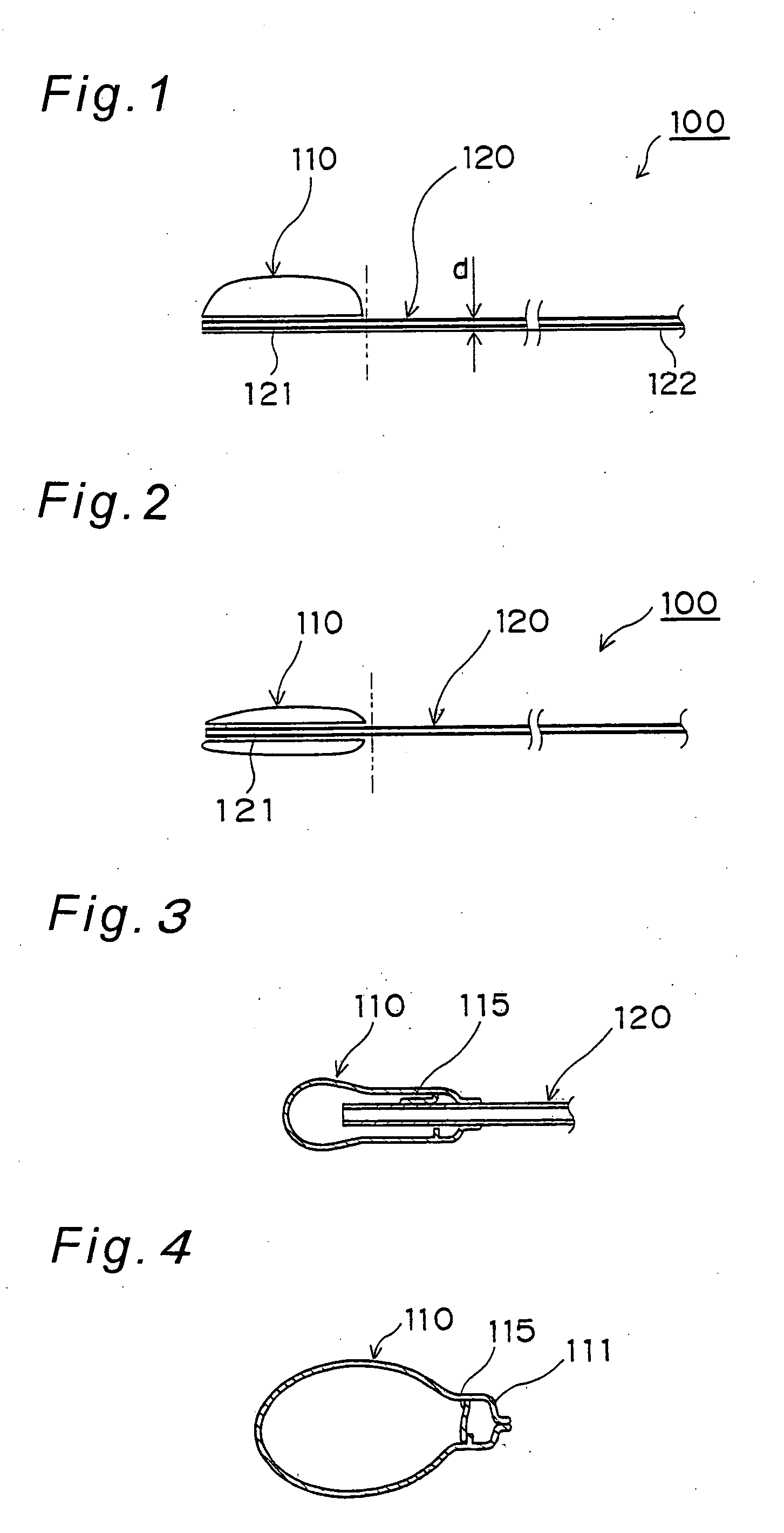
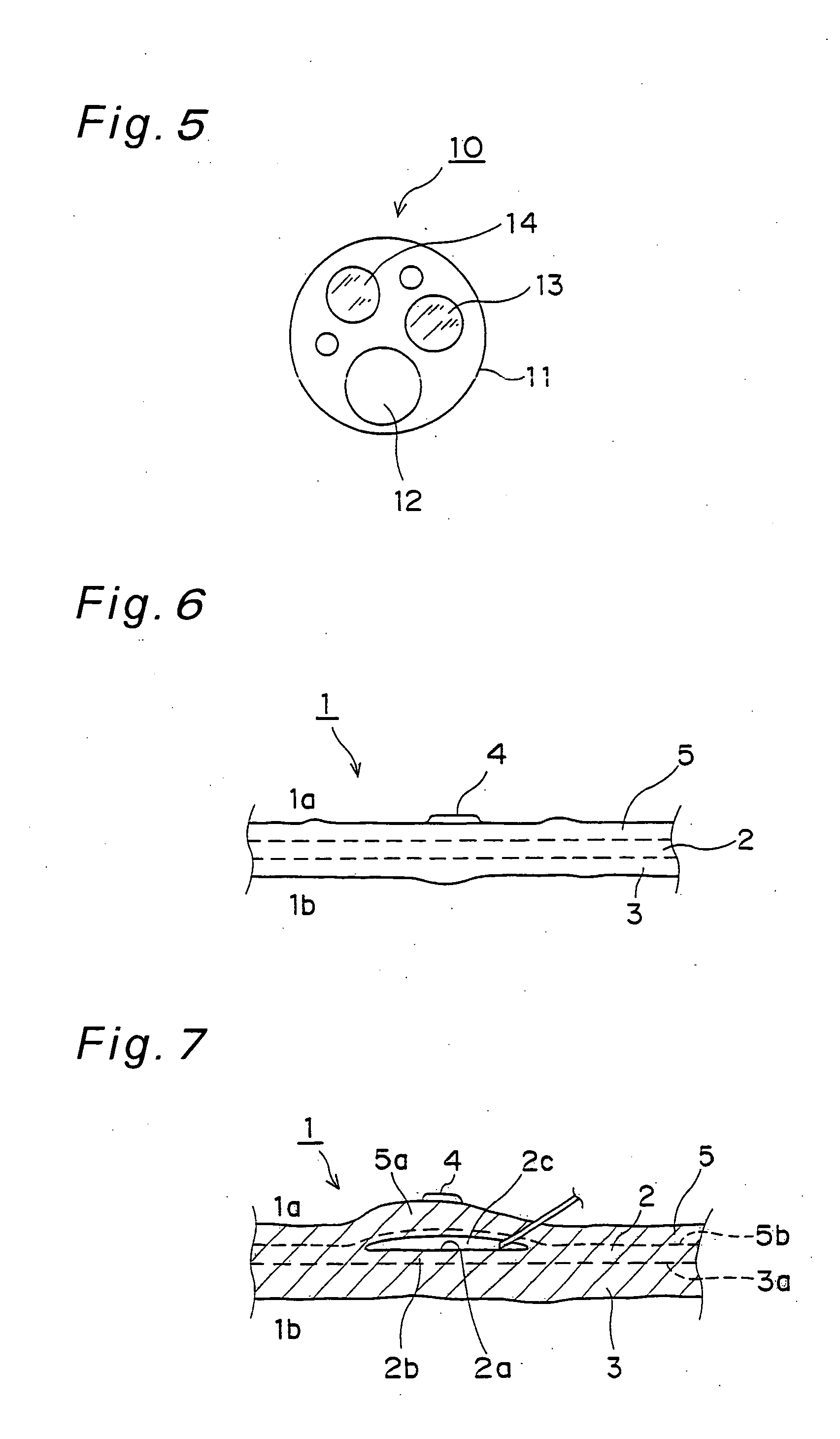
[0053]FIG. 1 shows a treatment instrument for EMR (Endoscopic Mucosal Resection) 100 of the present embodiment. The treatment instrument for EMR 100 comprises a separating and swelling member 110 and a first introduction member 120 in this embodiment. The separating and swelling member 110 is named also a balloon for EMR, which is inserted into a body cavity such as the stomach, large intestine, small intestine or esophagus through a clamp channel 12 of FIG. 5 included in an endoscope 10 such as shown in FIG. 16, and then inserted in a submucosal layer 2 below a lesion portion 4 pres...


PUM


Abstract

A treatment instrument for EMR and an EMR device are provided for excising a lesion portion while reducing complications and burdens to patients in an endoscopic mucosal resection. A separating and swelling member is included, so that a portion to be dissociated including the lesion portion, and a non-affected portion are separated and dissociated from each other by the separating and swelling member, and the lesion portion is swollen. The treatment instrument for EMR and the EMR device enable the endoscopic mucosal resection to be carried out more easily as compared with the conventional art, thereby greatly shortening an operation time, improving safety of the operation and moreover, reducing the burden of patients in comparison with the conventional art.

Description

BACKGROUND OF THE DISCLOSURE [0001] The present invention relates to a treatment instrument for EMR used for EMR (Endoscopic Mucosal Resection), and an EMR device equipped with the treatment instrument for EMR. [0002] These days, to malignant and benign lesion portions within mucous membranes in a body cavity such as the esophagus, stomach, small intestine, large intestine or the like an endoscopic mucosal resection is carried out by inserting a diathermy knife or the like as a kind of an electric knife through a tube 11 into the body cavity with the use of an endoscope 10 such as shown in FIG. 16, and excising the lesion portions by the diathermy knife or the like without executing ventrutomy. According to the endoscopic mucosal resection as above, conventionally, a pharmaceutical solution such as physiological saline, 5% glycol solution, glycerol epinephrine solution, 0.5% hyaluronic acid or the like is injected to a submucosal layer 2 present under a mucosal layer 5 where a lesio...

Claims


Application Information

Patent Timeline
IPC IPC(8): A61B1/018A61B17/32A61B17/34A61B18/14A61B19/00A61M29/00
CPCA61B1/018A61B17/32A61B17/320016A61B17/3478A61M29/02A61B19/24A61B2017/00269A61B2017/320048A61B18/14A61B90/02A47J19/02A47J19/06A47J43/075
Inventor TAKIMOTO, YUKINOBU
Owner KAIGEN
Features
  • Generate Ideas
  • Intellectual Property
  • Life Sciences
  • Materials
  • Tech Scout
  • Unparalleled Data Quality
  • Higher Quality Content
  • 60% Fewer Hallucinations
Learn More