Method and device for medical image reconstruction

a medical image and reconstruction technology, applied in the field of medical image reconstruction, can solve the problems of inability to apply a generic method in the x-ray field, provide only a basic structure of the vascular path, and achieve the effect of ease of execution

Inactive Publication Date: 2005-10-06
SIEMENS AG
View PDF3 Cites 60 Cited by
  • Summary
  • Abstract
  • Description
  • Claims
  • Application Information

AI Technical Summary

Benefits of technology

[0018] The present method and the associated image reconstruction device permit local 3D reconstruction of a locally bounded object area on a cardiology system. A particular advantage of the method is the ease of execution, since ECG data is not requi

Problems solved by technology

However, these technologies provide only a basic structure of the vascular path.
This method cannot thus be applied to a generic method in the X-ray f

Method used


Image

  • Method and device for medical image reconstruction
  • Method and device for medical image reconstruction
  • Method and device for medical image reconstruction

Examples


Embodiment Construction

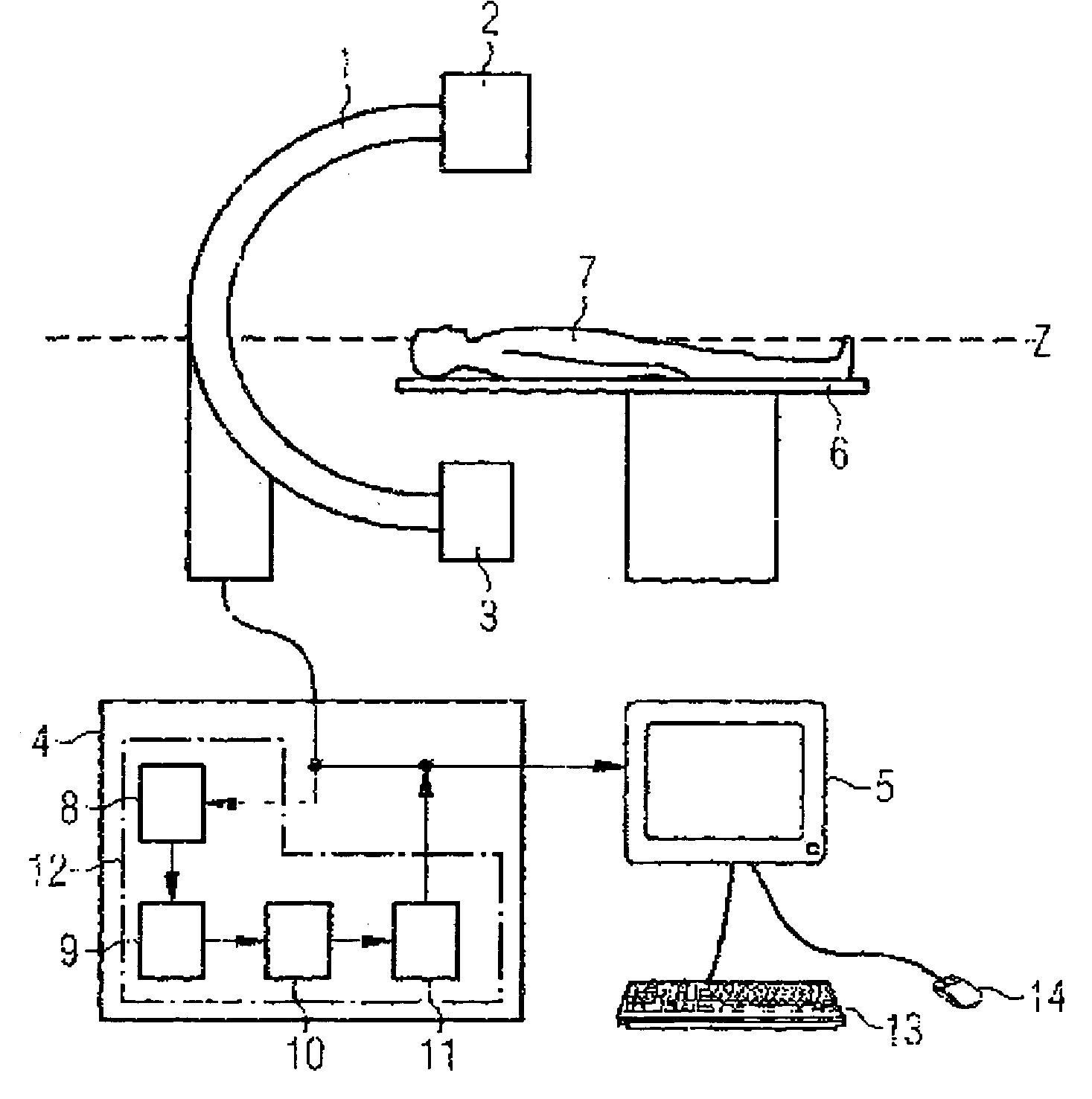
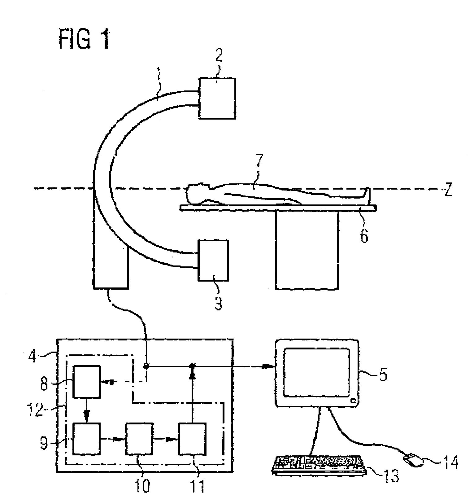
[0022]FIG. 1 shows in highly diagrammatic form a C-arm device for registering the 2D X-ray images. The C-arm device has a C-arm 1 that can rotate around the z-axis and to which are fixed an X-ray tube 2 and a detector 3 opposite the X-ray tube. The image data registered by the detector 3 at different rotational positions of the C-arm 1 is transmitted to the image processing unit 4, which is connected to a monitor 5 for displaying images of the registered or reconstructed images. This image processing unit 4 comprises, besides normal, not explicitly shown processing units, an image reconstruction device 12 with a detector module 8, an object tracking module 9, a correction module 10 and a reconstruction module 11, which are examined in greater detail below. The monitor is connected to a keyboard 13 and a graphic input device 14, via which a user can influence image display and image reconstruction.

[0023] This system also comprises the motor-adjustable patient table 6, on which the p...


PUM


Abstract

The present invention relates to an image reconstruction device (12) for an X-ray apparatus and a method for local 3D reconstruction of an object area of an examination object (7) from 2D image data of several 2D X-ray images of the examination object (7) registered in chronological order with different known projection geometries using the X-ray apparatus. With the method a location in the object area under consideration is selected from one of the 2D X-ray images. The positions of the selected location are determined in at least some of the 2D X-ray images and a spatial motion of the selected location between the registrations of the 2D X-ray images is calculated from the positions obtained, taking the known projection geometries into consideration. The calculated motion is then annulled by modifying the 2D image data in the 2D X-ray images and a 3D image dataset of at least the object area is reconstructed from the modified 2D image data. The method and the image reconstruction device enable a 3D image of a moving locally bounded object area to be reconstructed in a simple way without motion artifacts.

Description

CROSS REFERENCE TO RELATED APPLICATIONS [0001] This application claims priority to the German application No. 10 2004 016 586.6, filed Mar. 31, 2004 which is incorporated by reference herein in its entirety. FIELD OF INVENTION [0002] This invention relates to a method for local 3D reconstruction of an object area of an examination object from 2D image data of several 2D X-ray images of the examination object which were registered using an X-ray apparatus in chronological order with different known projection geometries. The invention also relates to an image reconstruction device for an X-ray apparatus with a reconstruction module which reconstructs a 3D image dataset of at least one object area from the 2D image data of these 2D X-ray images. BACKGROUND OF INVENTION [0003] In imaging X-ray diagnostics, image acquisition using so-called C-arm devices plays an important role. With devices of this type, several 2D X-ray images of the examination object are registered under different p...

Claims


Application Information

Patent Timeline
IPC IPC(8): A61B6/02A61B6/00G06T17/00G21K1/12H05G1/60
CPCA61B6/4441A61B6/466G06T11/005A61B6/541A61B6/469
Inventor HOMEGGER, JOACHIM
Owner SIEMENS AG
Why Eureka
Try Eureka