
Composition for stabilizing corneal tissue during or after orthokeratology lens wear

a corneal tissue and orthokeratology technology, applied in the field of corneal tissue stabilization during or after orthokeratology lens wear, can solve the problems of corneal misshapen or defective, wear of retainer lenses, and inability to adjust the shape of the cornea

Inactive Publication Date: 2005-10-20
EUCLID SYST CORP
View PDF7 Cites 5 Cited by
  • Summary
  • Abstract
  • Description
  • Claims
  • Application Information

AI Technical Summary

Benefits of technology

"The present invention relates to methods and compositions for maintaining the desired shape of corneal tissues after orthokeratological procedures. The invention involves the use of stabilizing agents, such as collagen compositions and protein-derived transglutaminase, which help to stabilize the corneal tissue and maintain its shape. The invention also includes methods for applying a stabilizing agent to the cornea and preparing a chemical composition for this purpose. The technical effects of the invention include improved vision, reduced risk of complications, and improved outcomes of orthokeratological procedures."

Problems solved by technology

When the cornea is misshapened or the axial length of the eye is too long or short, or the lens of the eye is functioning abnormally, various vision related problems, such as myopia, astigmatism, hyperopia, or the like, can result.
When the lens is taken off, however, the cornea is still misshapened or defective and refractive errors still remain.
A problem with orthokeratology is that reshaped corneal tissue keeps a memory of its original curvature, and tends to relax and return to the original curvature after the lenses are removed.
A disadvantage of such a modality is that it requires the wearing of retainer lenses every night in order to keep the cornea from regressing to its former shape.
However, it has been found that it is difficult to accurately place the. shaping contact lens with respect to the axis of vision to control the reshaping of the corneal tissue.
In some unsuccessful applications, corneoplasty has induced astigmatism or double vision due to errors caused by misplacing the shaping contact lens.
In addition, because all three steps are performed in one visit, the patient lacks an opportunity to react to the result of reshaped corneal tissue.

Method used


Image

  • Composition for stabilizing corneal tissue during or after orthokeratology lens wear
  • Composition for stabilizing corneal tissue during or after orthokeratology lens wear
  • Composition for stabilizing corneal tissue during or after orthokeratology lens wear

Examples


example 1

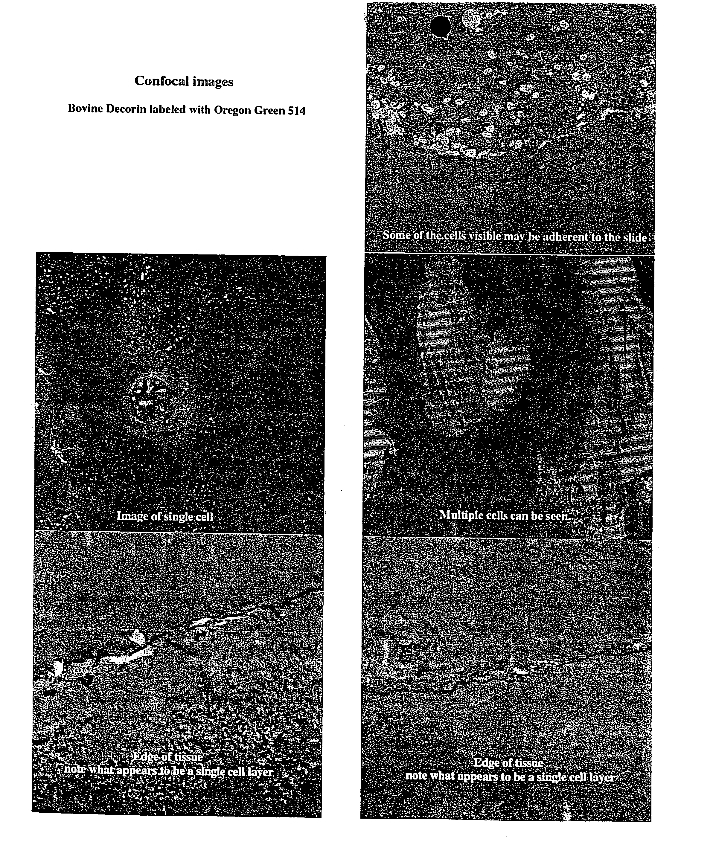
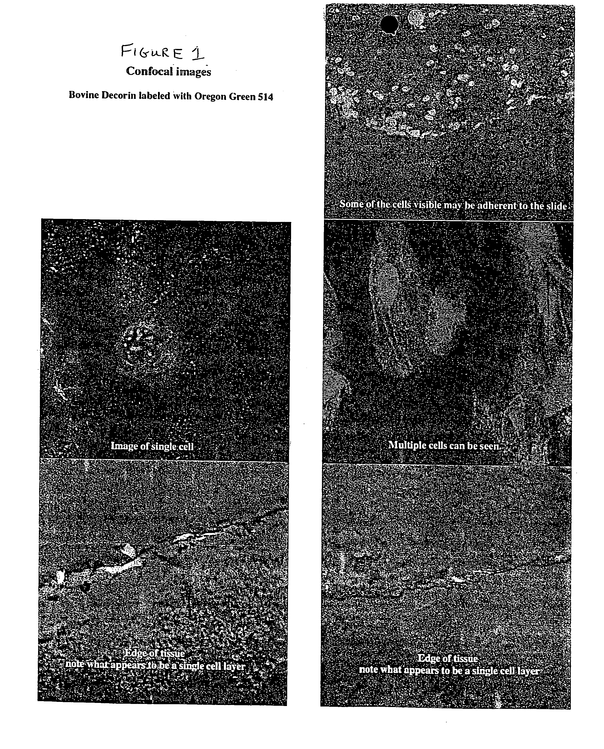
[0051] A series of ex vivo laboratory experiments were performed on enucleated porcine cornea to optimize the effects of stabilization. Enucleated porcine eyes were preserved in Optisol Solution and stored at 2-8° C. Treated eyes were examined visually and using biomicroscopy to monitor the effects on corneal epithelium, stroma, corneal clouding, corneal rigidity, anterior chamber response, and corneal thickness. Treated eyes underwent mechanical testing to evaluate changes in low and high modulus to evaluate the stress and strain under compressive force, as described in detail below. A decrease in low modulus indicated softening or a reduction in force needed to compress the collagenous network due to increased ability to force out fluids. In contrast, an increase in low modulus indicated stiffening. Any changes in the high modulus indicated damage to the collagen fibril network.

[0052] In the first phase of these ex vivo experiments, a total of 12 porcine eyes are prescreened. Enu...

example 2

[0054] The test eyes received both a molding treatment with a shaping lens followed by administration of decorin, an SLRP, having a concentration of 500 μg / ml buffered with 0.04M sodium phosphate to a pH of about 7.2. The solution was applied according to the following schedule: The mold was applied on Friday afternoon, and remained there until Monday morning when the molds were removed. After the mold removal, the eyes were then exposed to 100 μl of the decorin solution. The mold was reapplied onto each eye. Then each eye was placed back into Optisol solution. The application of decorin was repeated on Tuesday and on Wednesday.

[0055] The mold was reapplied to one eye. Subjective photographic assessment of that eye showed that the treatment with decorin with the mold reapplied resulted in a flat cornea with no rebound.

[0056] With respect to the second eye, after the eye was exposed to the decorin on Wednesday, the mold was not reapplied. This was done to determine if the eye would...

example 3

[0057] The test eyes received both molding treatment with shaping lens followed by administration of keratan sulfate, an SLRP, having a concentration of 100 μg / ml buffered with 0.04M disodium phosphate to a pH of about 7.2. The solution was applied according to the following schedule: The mold was applied on Friday afternoon, and remained there until Monday morning when the molds were removed. After the mold removal, the eyes were then exposed to 100 μl. The mold was reapplied onto each eye. Then each eye was placed back into Optisol solution. The application of keratan sulfate was repeated on Tuesday and on Wednesday.

[0058] The mold was reapplied to one eye. Subjective photographic assessment of that eye showed that the treatment with keratan sulfate with the mold reapplied resulted in a flat cornea with no rebound.

[0059] With respect to the second eye, after the eye was exposed to the keratan sulfate on Wednesday, the mold was not reapplied. This was done to determine if the eye...


PUM

PropertyMeasurementUnit
concentrationaaaaaaaaaa

Abstract

Two types of compositions having an eye-drop delivery system are used during or after an orthokeratology procedure to prevent or retard relaxation of corneal tissue back to the original anterior curvature of the cornea. Each composition functions independently from the others and is a different approach of preparing a stabilizing agent. The first composition is directed to a biologically compatible composition comprising fibril associated collagens with interrupted triple helices (FACITs) and / or small leucine-rich repeat proteoglycans (SLRPs). The fibril associated collagen family includes various types of collagens, such as type VI, type XX, type XII, and type XIV. The small leucine-rich repeat proteoglycans family includes decorin, keratocan, biglycan, epiphycan, lumican, mimecan, and fibromodulin. The second composition includes the enzyme found as a normal component of tissues, plasma, or epidermis, such as transglutaminase.

Description

BACKGROUND OF THE INVENTION [0001] 1. The Field of the Invention [0002] This invention relates to chemical compositions suitable for maintaining corneal curvature resulting from an orthokeratology procedure, and methods for making and applying the chemical compositions to a cornea for retaining the corneal curvature after undergoing orthokeratological therapy. [0003] 2. Description of the Related Art [0004] Structure and composition of a human cornea: The cornea is the first and most powerful refracting surface of the optical system of the eye. Production of a sharp image at the retinal receptors requires that the cornea be transparent and of appropriate refractive power. The refractive power of the cornea depends primarily on two factors: its curvature and its refractive index. When the cornea is misshapened or the axial length of the eye is too long or short, or the lens of the eye is functioning abnormally, various vision related problems, such as myopia, astigmatism, hyperopia, ...

Claims


Application Information

Patent Timeline
Patent Type & Authority Applications(United States)
IPC IPC(8): A61F2/14A61F9/00A61K38/17A61K38/39G02C7/04
CPCA61K38/1709G02C7/047A61K38/39
Inventor DEWOOLFSON, BRUCE H.DEVORE, DALE P.
Owner EUCLID SYST CORP
Learn More