Puncture-type endoscopic probe

a technology of endoscope and probe, which is applied in the direction of catheters, sensors, diagnostics, etc., can solve the problems of significant amount of data processing time, inability to obtain 3-dimensional tomographic images instantaneously, and inability to obtain 3-dimensional tomographic images

Inactive Publication Date: 2005-10-20
FUJI PHOTO OPTICAL CO LTD
View PDF3 Cites 107 Cited by
  • Summary
  • Abstract
  • Description
  • Claims
  • Application Information

AI Technical Summary

Benefits of technology

[0011] With the foregoing in view, it is an object of the present invention to provide a puncture-type endoscopic probe capable of carrying out both

Problems solved by technology

However, conventional confocal scanning endoscopes are only capable of obtaining optical slice images.
This requires a significant amount of time for data processing, where 3-dimensional tomographic images cannot be obtain

Method used


Image

  • Puncture-type endoscopic probe
  • Puncture-type endoscopic probe
  • Puncture-type endoscopic probe

Examples


Embodiment Construction

[0025] The embodiments of the puncture-type endoscopic probe of the present invention are described below with reference to the figures.

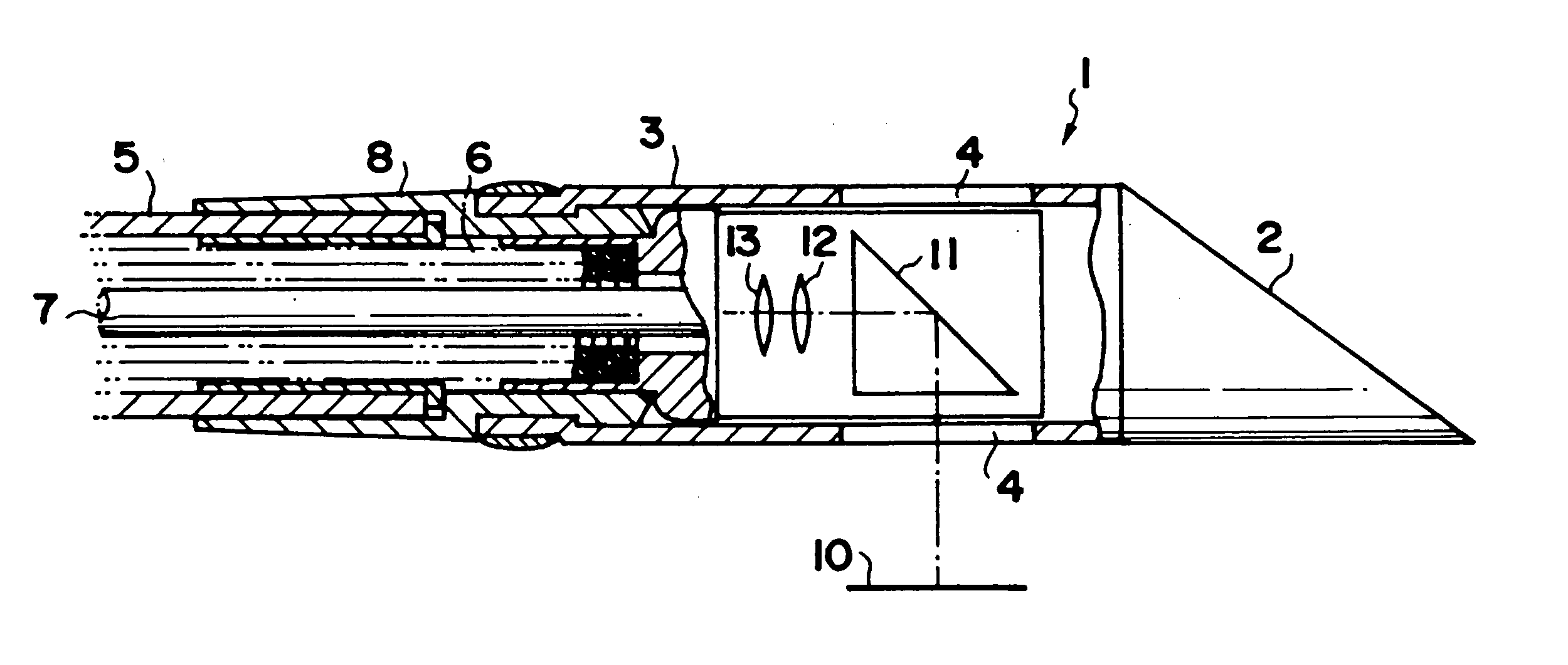
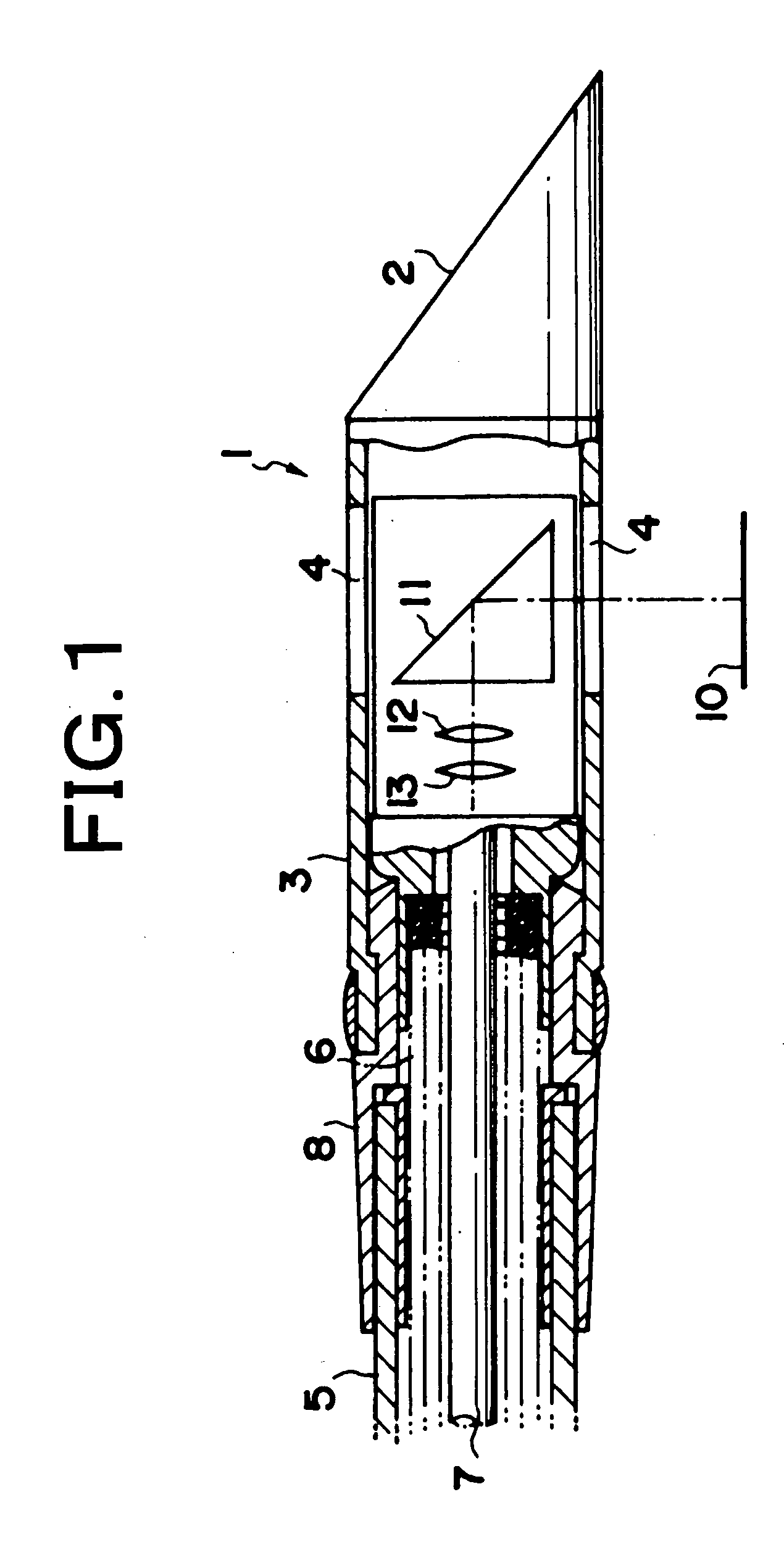
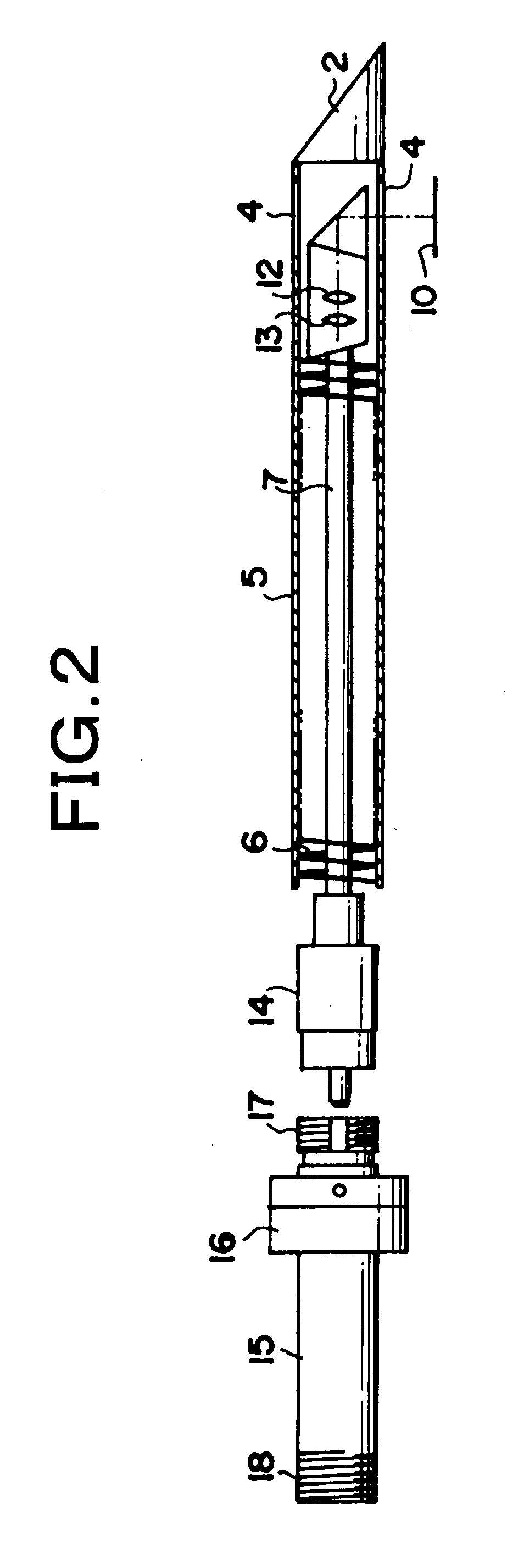
[0026]FIG. 1 and FIG. 2 are embodiments of the puncture-type endoscopic probe of the present invention. FIG. 1 is a schematic sectional view showing the tip of the probe and FIG. 2 is a schematic sectional view showing the probe and the rotation mechanism. FIG. 3 is a schematic diagram showing the implementation of inner tissue imaging using a puncture-type endoscopic probe related to the embodiments of the present invention.

[0027] In the puncture-type endoscopic probe 1, a protective section 3 which contains the object optical system which is attached to the tip of the flexible sheath 5 via the cylindrical connecting section 8 as shown in FIG. 1. A sectional roughly-triangular puncturing section 2 is attached at the tip of the protective section 3. The fiber bundle 7, whose outer circumference is sheathed in a helical spring 6, is contained withi...


PUM


Abstract

This puncture-type endoscopic probe of the invention comprises: an object optical system consisting of a fiber bundle which is composed of a plurality of optical fiber strands each having a light-emitting end which functions as a point light source, an imaging objective lens which focuses the light beams emitted from the light-emitting ends onto the suspected surface position and a light beam deflection member (prism) for deflecting light which is positioned between the light-emitting ends and the suspected surface position; and a puncturing section which is attached to the tip of the probe. The imaging objective lens is constituted by a micro lens array, which is positioned so that the light-emitting end positions and the suspected surface position are conjugated each other. The object optical system can be rotated around the rotational axis defined by the puncture direction.

Description

RELATED APPLICATIONS [0001] This application claims the priority of Japanese Patent Application No. 2004-094727 filed on Mar. 29, 2004, which is incorporated herein by reference. BACKGROUND OF THE INVENTION [0002] 1. Field of the Invention [0003] The present invention relates to a puncture-type endoscopic probe wherein the tip of the probe is inserted into the tissue in order to observe the same. [0004] 2. Description of the Prior Art [0005] Conventionally, confocal scanning endoscopes have been used as endoscopic devices wherein detailed tomographic images of the lesion area are obtained for the detailed observation of the same (For example, refer to Japanese Unexamined Patent Publication No. 2000-121961). [0006] Technology known as Optical Coherence Tomography (OCT) wherein tomographic images of the specimen are obtained using light interference is also being developed. [0007] However, conventional confocal scanning endoscopes are only capable of obtaining optical slice images. In...

Claims


Application Information

Patent Timeline
IPC IPC(8): G01N21/17A61B1/00A61B1/005A61B1/04A61B1/07A61B5/00A61B8/00G02B23/26
CPCA61B1/0008A61B1/00096A61B1/00101A61B1/00188A61B5/6852A61B1/042A61B1/07A61B5/0066A61B1/005
Inventor YAMATAKA, SHUUICHIFUJITA, HIROSHI
Owner FUJI PHOTO OPTICAL CO LTD
Why Eureka
Try Eureka