Nogo receptor antagonists

a nogo receptor and antagonist technology, applied in the field of neurobiology and molecular biology, to achieve the effect of inhibiting binding, inhibiting growth cone collapse, and decreasing the inhibition of neurite outgrowth

Active Publication Date: 2005-12-08
YALE UNIV
View PDF32 Cites 40 Cited by
  • Summary
  • Abstract
  • Description
  • Claims
  • Application Information

AI Technical Summary

Benefits of technology

[0010] The invention relates to soluble Nogo receptor-1 polypeptides and fusion proteins comprising them, and antibodies and antigenic fragments thereof directed against specific immunogenic regions of Nogo receptor-1. The invention also relates to immunogenic Nogo receptor-1 polypeptides that bind to the antibodies of the invention. The invention further relates to nucleic acids encoding the polypeptides o...

Problems solved by technology

When an elongation contacts a surface ...

Method used


Image

  • Nogo receptor antagonists
  • Nogo receptor antagonists
  • Nogo receptor antagonists

Examples


example 1

Production of Murine Monoclonal Anti-Nogo Receptor-1 Antibodies

[0156] Anti-Nogo receptor-1 antibodies that specifically bind an immunogenic Nogo receptor-1 polypeptide of the invention were made using the following methods and procedures.

Immunizations

[0157] Two immunization approaches were used:

1. COS-7 Cells or Cell Membranes Containing Nogo Receptor-1 (NogoR-1) As the Immunogen

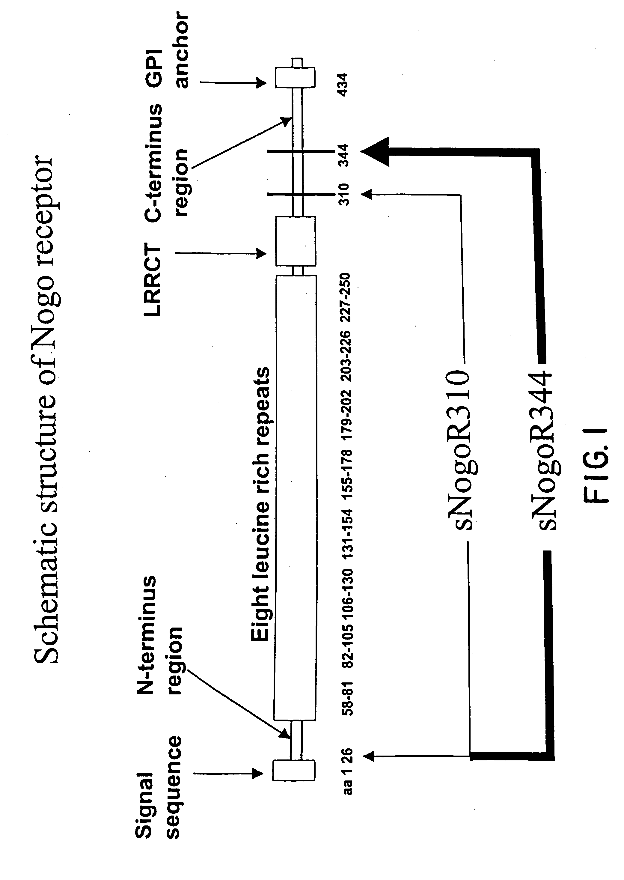
[0158] The rat Nogo receptor-1 gene (GenBank™ No. AF 462390) was subcloned into the mammalian expression vector pEAG1256 (Biogen®) that contained the CMV promotor and geneticin resistance gene for drug selection. The recombinant plasmid was transfected into COS-7 cells using Superfect (Qiagen®). Transfectants were selected using geneticin (Gibco™, 2 mg / ml), cloned and verified for surface expression of Nogo receptor-1 protein by FACS. COS-7 membranes were prepared from these cells according to procedures as described [Wang et al., J. Neurochem., 75:1155-1161 (2000)] with two washings, and stored at 1 ...

example 2

Production of Fab-Phage Anti-Nogo Receptor-1 Antibodies

[0170] Anti-Nogo receptor-1 Fab-phage antibodies that specifically bind an immunogenic Nogo receptor-1 polypeptide of the invention were also made by screening a Fab-phage library as follows.

[0171] The MorphoSys Fab-phage library HuCAL® GOLD was screened against recombinant rat soluble sNogoR310-Fc protein and COS7 cells expressing rat Nogo receptor-1. Fab-phages that specifically bound to Nogo receptor-1 were purified and characterized. The heavy chain of 14D5 is derived from the VH2 gene and the light chain is derived from the VK1 gene. The amino acid sequences of the CDRs of the heavy chain and light chain of one of these Fab-phages, 14D5, are shown in Table 4.

TABLE 4Amino Acid Sequence of CDRs of 14D5Amino Acid SequenceSEQ ID NO:Heavy Chain CDR1GFSLSTSGGSVG19Heavy Chain CDR2LIYSNDTKYYSTSLKT20Heavy Chain CDR3SRFWTGWYDV21Light Chain CDR1RASQNIAITLN22Light Chain CDR2LASSLQS23Light Chain CDR3QQYDNYPL24

[0172] 14D5 binds to ra...

example 3

Immunoprecipitation of Nogo Receptor-1 by Anti-Nogo Receptor-1 Monoclonal Antibodies

[0173] To perform the immunoprecipitation, 100 μl lysed cells or 50 μl PiPLC treated cells were mixed with 400 or 450 μl extraction buffer [10 mM Tris-HCl, pH 7.2, 0.5% Tween-20™, 0.2 mM PMSF] or RIPA buffer, respectively in the presence of 30 μl Protein A or G and 1-2 μg antibody. The mixture was incubated in a shaker at 4° C. for 16 hours.

[0174] Samples were spun gently to pellet the protein A or G coupled beads. The beads were washed three times with 1 ml wash buffer (10 mM Tris-HC1, pH 7.2, 0.1% Tween-20™). The final wash was performed using 10% of original wash buffer.

[0175] Beads were resuspended in 100 μl of 2× SDS with 10% beta-mercaptoethanol. Samples were incubated at room temperature before being run on a 4-20% Tris-Glycine gel for SDS-PAGE. As determined by SDS-PAGE gel analysis, monoclonal antibodies, 3G5 and 2F7, immunoprecipitate Nogo receptor-1.


PUM

PropertyMeasurementUnit
Solubility (mass)aaaaaaaaaa

Abstract

Disclosed are immunogenic Nogo receptor-1 polypeptides, Nogo receptor-1 antibodies, antigen-binding fragments thereof, soluble Nogo receptors and fusion proteins thereof and nucleic acids encoding the same. Also disclosed are compositions comprising, and methods for making and using, such Nogo receptor antibodies, antigen-binding fragments thereof, soluble Nogo receptors and fusion proteins thereof and nucleic acids encoding the same.

Description

FIELD OF THE INVENTION [0001] This invention relates to neurobiology and molecular biology. More particularly, this invention relates to immunogenic Nogo receptor-1 polypeptides, Nogo receptor-1 antibodies, antigen-binding fragments thereof, soluble Nogo receptors and fusion proteins thereof and nucleic acids encoding the same. This invention further relates to compositions comprising, and methods for making and using, such Nogo receptor antibodies, antigen-binding fragments thereof, immunogenic Nogo receptor-1 polypeptides, soluble Nogo receptors and fusion proteins thereof and nucleic acids encoding the same. BACKGROUND OF THE INVENTION [0002] Axons and dendrites of neurons are long cellular extensions from neurons. The distal tip of an extending axon or neurite comprises a specialized region, known as the growth cone. Growth cones sense the local environment and guide axonal growth toward the neuron's target cell. Growth cones respond to several environmental cues, for example, s...

Claims


Application Information

Patent Timeline
IPC IPC(8): A61K39/395A61K48/00A61P43/00C07H21/04C12N15/09C07KC07K5/00C07K14/00C07K14/47C07K14/705C07K16/00C07K16/18C07K16/28C07K16/46C07K19/00C12N1/15C12N1/19C12N1/21C12N5/06C12N5/10C12N15/11C12N15/85C12P21/02C12P21/08
CPCA61K2039/505C07K14/705C07K16/2863C07K2317/21C07K2317/55C07K2317/56C07K2317/565C07K2319/00C07K2319/30C07K2317/34Y10S530/809A61P21/02A61P25/00A61P25/14A61P25/16A61P25/28A61P43/00A61P9/10C07K7/08C12N15/11C07K14/47
Inventor LEE, DANIELPEPINSKY, R.LI, WEIWEIWORLEY, DANERELTON, JANESTRITTMATTER, STEPHENSAH, DINAHRABACCHI, SYLVIA
Owner YALE UNIV
Why Eureka
Try Eureka