
Apparatus and methods for occluding a hollow anatomical structure

a hollow tissue and anatomical structure technology, applied in the field of surgical methods and apparatus for occluding a hollow tissue structure, can solve the problems of atria quivering or beating too fast, normal, congestive heart failure, etc., and achieve the effect of positive attachment of the clamp to the tissu

Active Publication Date: 2005-12-15
ATRICURE
View PDF99 Cites 213 Cited by
  • Summary
  • Abstract
  • Description
  • Claims
  • Application Information

AI Technical Summary

Benefits of technology

[0015] In another aspect, the first and second clamping portions have complementary shapes in cross section such that the complementary shapes fit together in the clamping position. At least one of the first and second clamping portions may be convexly curved toward the other of the first and second clamping portions in cross section. This feature may assist with providing more uniform force distribution and / or more sealing force along the length of the clamp.
[0023] The present invention provides improved devices and methods for occlusion of hollow tissue such as the left atrial appendage. One advantage of various embodiments described herein is that the surgeon can open and close the clamp if needed to change the position of the clamp for a better result prior to release of the clamp onto the tissue. The configuration of the delivery and actuation device is such that the device can be used not only in an open surgical procedure, but in a minimally invasive surgical procedure during which, for example, the device is placed between or under the patient's ribs for access to the left atrial appendage. The implantable clamp has a geometry which traps appendage tissue within an annular opening thereby positively attaching the clamp to the tissue.

Problems solved by technology

However, recent medical research has uncovered some devastating complications including cardiomyopathy, congestive heart failure and stroke.
During atrial fibrillation, electrical signals from other parts of the heart disrupt the heart's normal rhythm and cause the atria to quiver or beat too fast.
This is fortunate, because a rapid ventricular heartbeat would be much more dangerous and potentially fatal.
The most significant danger is stroke.
During atrial fibrillation, the heart is not pumping normally or efficiently.
The blood begins to pool in the atria and this stagnation of blood can cause the blood to thicken and form clots.
When the heart experiences atrial fibrillation there may not be enough blood pumping to the brain or other organs.
This can cause dizziness, shortness of breath or organ failure.
Untreated atrial fibrillation will also weaken the heart due to phenomenon known as remodeling.
The fast abnormal rhythm in the atria causes electrical changes, and this can enlarge the heart.
For the prevention of blood clots, research has demonstrated that the anticoagulation warfarin (e.g., Coumadin) is effective in reducing the risk of blood clot formation and stroke but it does not totally eliminate the risk.
An anticoagulant such a warfarin interferes with the body's natural clotting mechanism.
While this pharmacological treatment may significantly reduce the risk of stroke, it also increases the risk of bleeding and may be inappropriate for many atrial fibrillation patients.
These clots can be slowly ejected from the left atrial appendage into the left atrium and left ventricle, and then released into the bloodstream thereby becoming an obstruction in the brain or other vascular structures.
Many surgeons are uncomfortable with making an opening in this wall without being able to repair it at the end of the procedure.
There are also issues of placing an occlusion device inside the heart.
If the occlusion device becomes detached within the heart, the result may be fatal.
When trying to place a flaccid loop around an irregular pedunculated structure, it can be difficult to make certain the loop is positioned at the base of the appendage.
When cinching the loop, it is very easy to over tighten the loop, and this can result in severing the delicate atrial appendage.
Even a partial tear can create problems in getting access to repair the tear.
This method of occlusion may not always seal the opening between the appendage interior and the atrium.
That is, there may still be a partial opening due to the way the appendage wall collapses during cinching of the loop.
Such a partial opening could still allow some flow into and out of the atrial appendage, leading to the problems mentioned above.
In addition, transforming the relatively flat structure of the appendage onto a round hard mass, as does a cinching method, could lead to other problems.
Due to the limited access, the ability to visualize the entire atrial appendage while placing the stapler in the correct location can be a problem.
It is very difficult to make certain the staple line makes a complete occlusion of the appendage.
Again, a partial occlusion of the appendage can still result in the formation and dislodgement of clots.

Method used


Image

  • Apparatus and methods for occluding a hollow anatomical structure
  • Apparatus and methods for occluding a hollow anatomical structure
  • Apparatus and methods for occluding a hollow anatomical structure

Examples


Embodiment Construction

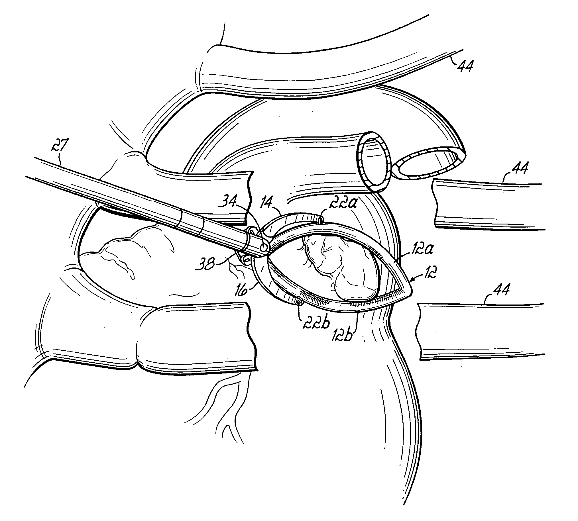
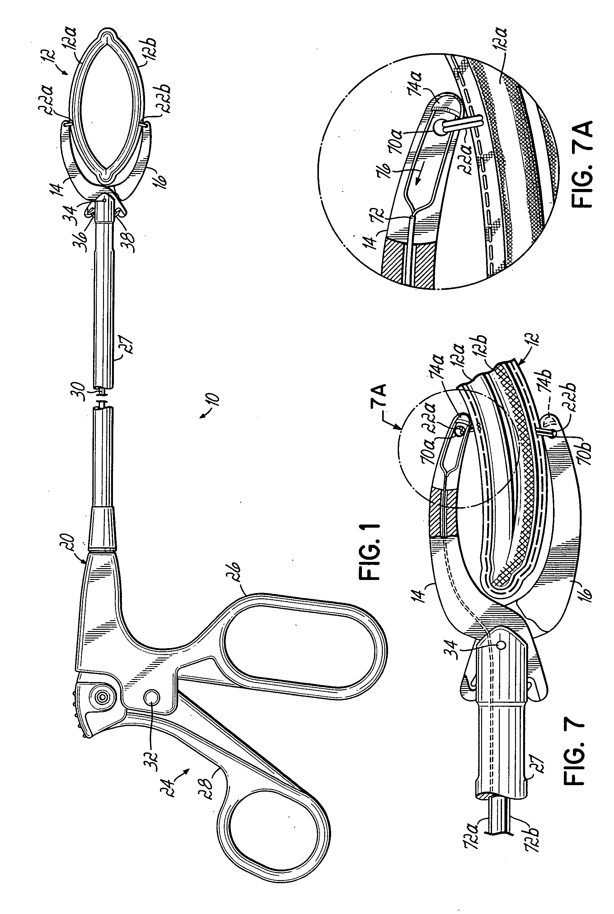
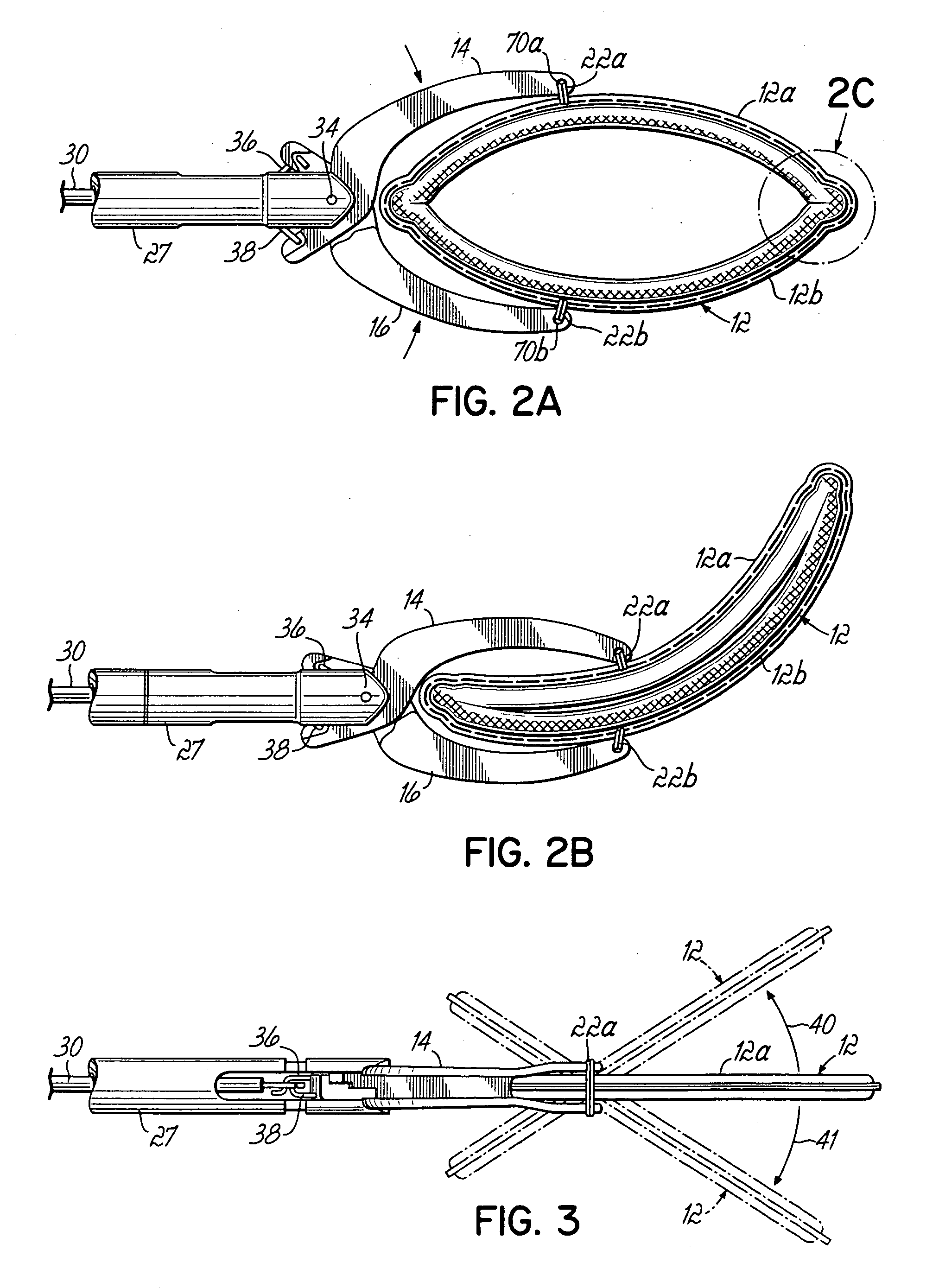
[0072] Referring initially to FIGS. 1, 2A, 2B and 3, a first embodiment of the invention includes an apparatus 10 comprising a clamp 12 having respective first and second clamping portions 12a, 12b secured between first and second jaws 14, 16. A delivery and actuation device 20 carries first and second jaws 14, 16 for actuating clamp 12 between open and closed or clamping positions as will be described further below. Clamp portions 12a, 12b are secured to jaws 14, 16 by respective sutures 22a, 22b. Delivery and actuation device 20 includes a pistol grip 24 having a stationary handle 26 coupled with an elongate jaw support member 27. A movable handle 28 is coupled with an actuating bar 30 and pivots with respect to stationary handle 26 at a pivot member 32. When movable handle 28 is depressed toward stationary handle 26, this action draws actuating bar 30 to the left as viewed in FIG. 1. Actuating bar 30 has a connecting portion 30a secured to respective rigid wire members 36, 38. Wi...


PUM


Abstract

A device for occluding a hollow anatomical structure includes a clamp having at least first and second clamping portions adapted to be placed on opposite sides of the anatomical structure. At least one of the first and second clamping portions is movable toward the other from an open position to a clamping or closed position to occlude the anatomical structure. The clamp has an annular shape configured to surround the hollow anatomical structure in the open position and a flattened shape in the clamping position configured to occlude the hollow interior of the anatomical structure. The clamp is preferably covered with fabric to promote tissue ingrowth. A clamp delivery and actuation device is provided for allowing the clamp to be applied in either an open surgical procedure or a minimally invasive procedure.

Description

FIELD OF THE INVENTION [0001] The present invention relates generally to surgical methods and apparatus for occluding a hollow tissue structure, such as when occluding vessels, or pedunculated structures such as an appendix, gall bladder or appendages on the heart. More specifically, the present invention relates to a method and device for occluding the left atrial appendage of the heart in either an open surgical procedure or minimally invasive procedure. BACKGROUND OF THE INVENTION [0002] Atrial fibrillation is a common cardiac rhythm disorder that affects more than two million people each year. Until relatively recently, atrial fibrillation was thought to be a nuisance arrhythmia with few consequences. However, recent medical research has uncovered some devastating complications including cardiomyopathy, congestive heart failure and stroke. [0003] During atrial fibrillation the upper part of the heart beats (quivers) faster than the rest of the heart. This phenomenon is due to th...

Claims


Application Information

Patent Timeline
IPC IPC(8): A61B17/08A61B17/10A61B17/12A61B17/122A61B17/128A61B17/28
CPCA61B17/12A61B17/122A61B2017/2825A61B17/1285A61B17/29A61B17/1227
Inventor COSGROVE, DELOS M.WILLIAMSON, WARREN P. IVBERKY, CRAIG B.
Owner ATRICURE
Learn More