Apparatus for imaging and treating a breast

Inactive Publication Date: 2006-01-12
TECHNISCAN
View PDF47 Cites 87 Cited by
  • Summary
  • Abstract
  • Description
  • Claims
  • Application Information

AI Technical Summary

Benefits of technology

[0021] It has been recognized that it would be advantageous to develop a method to facilitate further treatment of the breast or tumor after performing a scan or creating a tomography of the breast. In addition, it has been recognized that it would be advantageous to develop a method to facilitate biopsy, therapy and/or surgery after an ultrasound computer topography scan

Problems solved by technology

The current standard of care has significant problems, generating unacceptably high rates of false positive tests—between 8% and 10%—and upwards of 15% false negative tests.
The result is that many women suffer from unnecessary and invasive biopsies.
In addition, each year the U.S. healthcare system spends an estimated $2.1 billion on biopsies, which yield negative results more than 75% of the time.
When breast cancer cells reach the axillary (underarm) lymph nodes, they may continue to grow, often causing the lymph nodes in that area to swell.
Fibrocystic changes can cause breast swelling and pain.
Although widespread use of screening mammography has increased the number of breast cancers found before they cause any symptoms, some breast cancers are not found by mammography, either because the test was not done or because even under ideal conditions mammography cannot find every breast cancer.
It has difficulty imaging objects with low reflectiv

Method used


Image

  • Apparatus for imaging and treating a breast
  • Apparatus for imaging and treating a breast
  • Apparatus for imaging and treating a breast

Examples


Embodiment Construction

)

[0051] Reference will now be made to the exemplary embodiments illustrated in the drawings, and specific language will be used herein to describe the same. It will nevertheless be understood that no limitation of the scope of the invention is thereby intended. Alterations and further modifications of the inventive features illustrated herein, and additional applications of the principles of the inventions as illustrated herein, which would occur to one skilled in the relevant art and having possession of this disclosure, are to be considered within the scope of the invention.

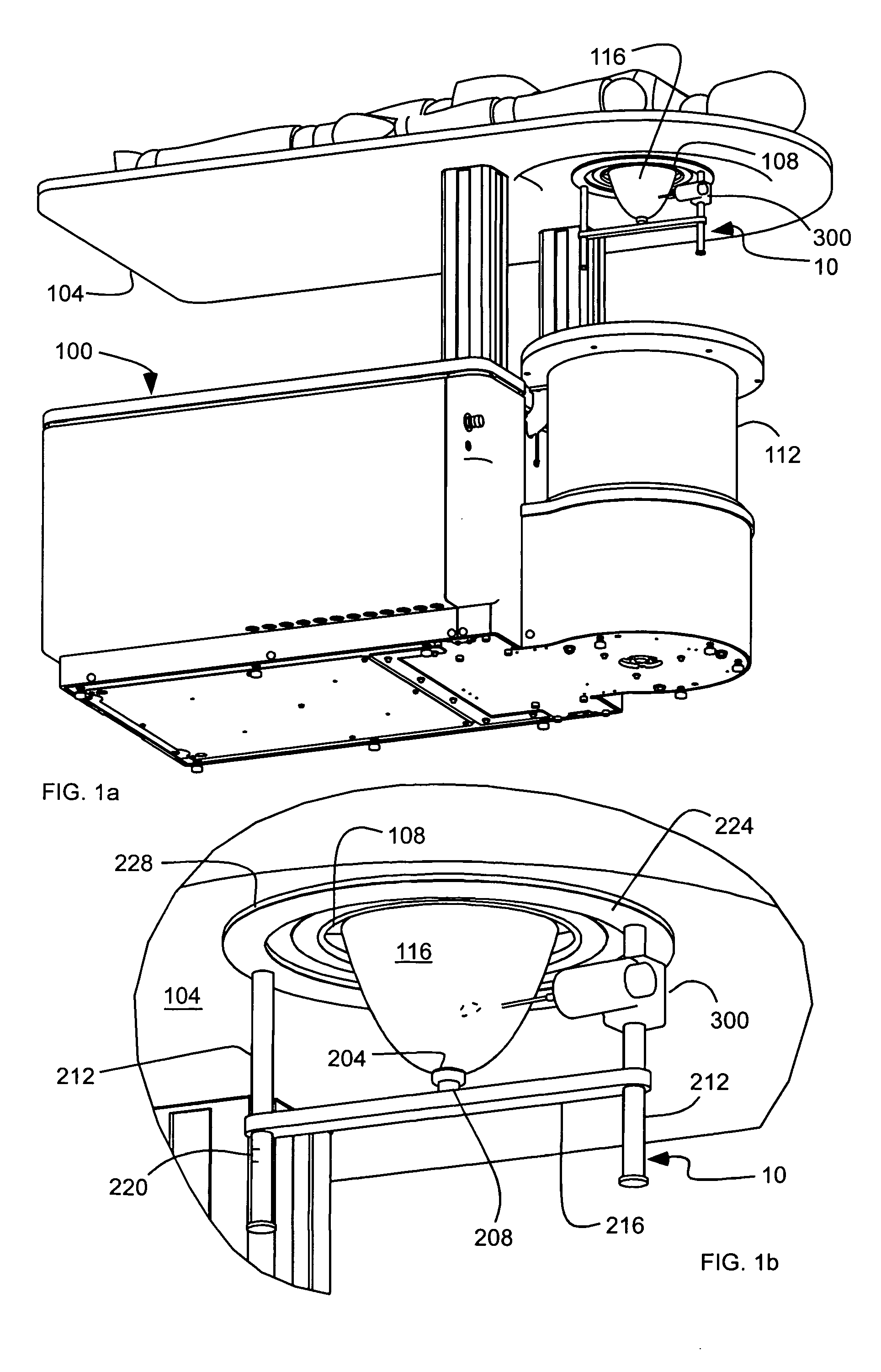
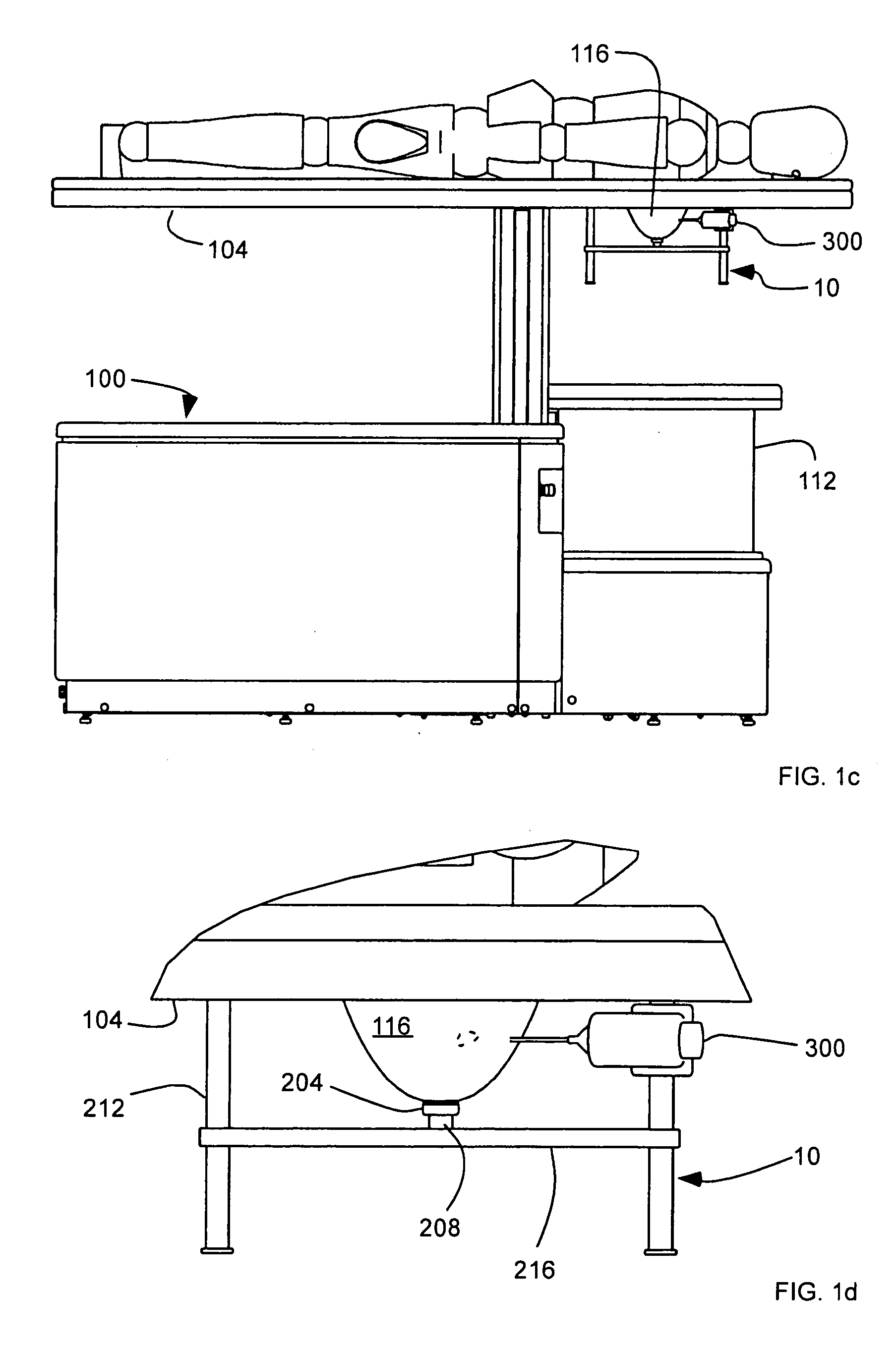
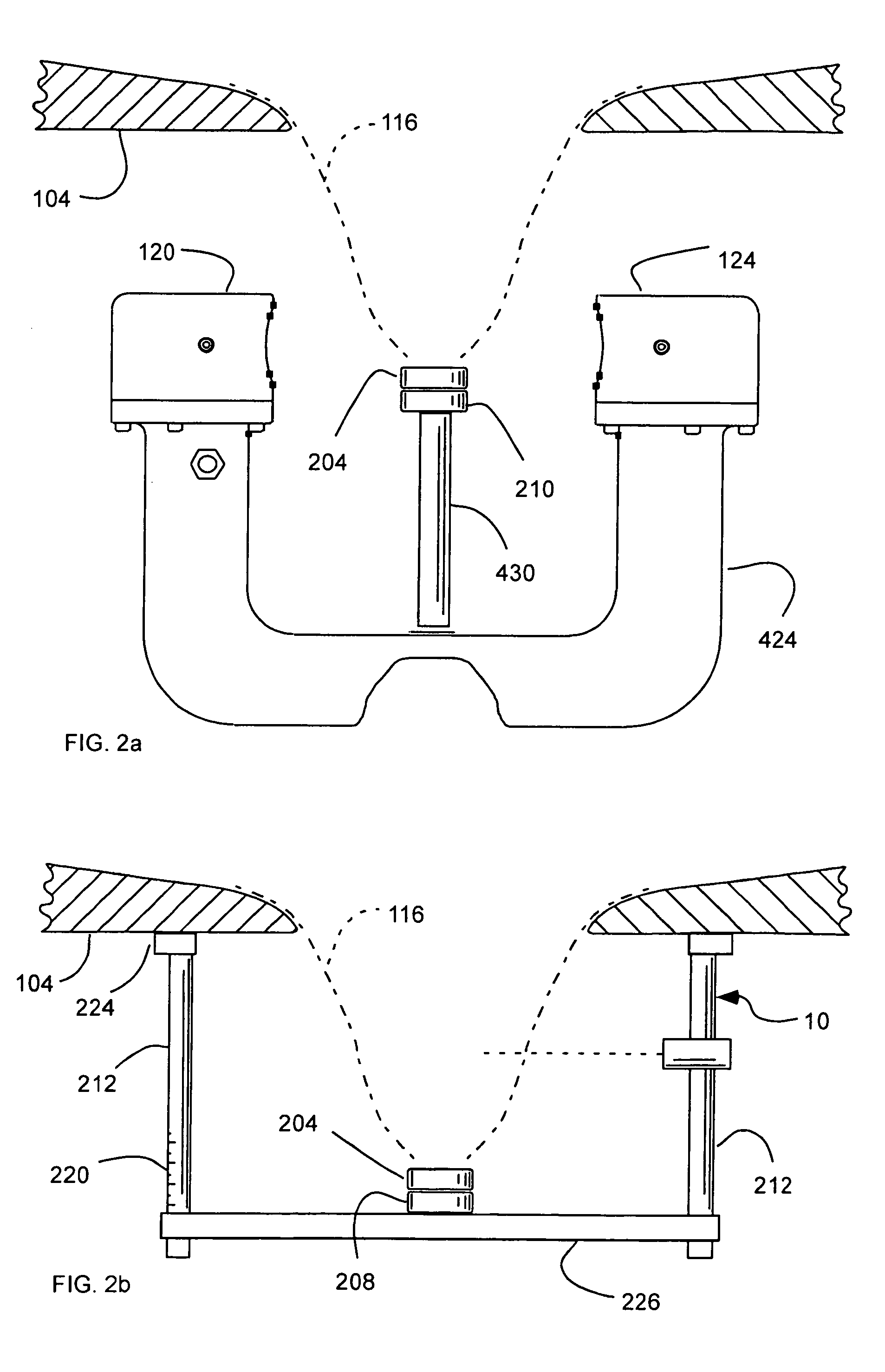
[0052] The present invention includes a method for further treating a tumor or a lesion of a breast while maintaining a position and a shape of the breast with respect to the chest wall of the patient as during scanning. Maintaining the position and the shape of the breast during further treatment allows for the tumor or legion to be more easily located, and allows three-dimensional image to be utilized to gui...


PUM


Abstract

A system for imaging and treating a breast of a patient includes a table configured to receive the patient thereon, having an aperture formed therein configured to receive the breast of the patient pendent therethrough and positionable over a bath configured to contain a medium. Means for transmitting and receiving ultrasound signals is disposed in the bath. A frame is disposable out of the bath, and includes means for securing the breast to the frame to substantially maintain a position and a shape of the breast out of the bath as in the bath.

Description

PRIORITY CLAIM [0001] Priority of U.S. Provisional Patent Application Ser. No. 60 / 580,416, filed Jun. 16, 2004, is claimed, and which is herein incorporated by reference. This application is a continuation-in-part of U.S. patent application Ser. No. 10 / 821,407, filed Apr. 8, 2004, which is herein incorporated by reference.RELATED APPLICATION(S) [0002] This application is related to U.S. patent application Ser. No. ______, filed Jun. 15, 2005, as TNW docket no. 23473.NP, entitled “Method for Imaging and Treating a Breast,” which is herein incorporated by reference. BACKGROUND [0003] 1. Field of the Invention [0004] The present invention relates generally to a method for maintaining the position and the shape of a patient's breast during scanning and treatment, such as biopsy, therapy and / or surgery. [0005] 2. Related Art [0006] Other than skin cancer, breast cancer is the most common cancer among women, and is the second leading cause of cancer death in women, after lung cancer. Acco...

Claims


Application Information

Patent Timeline
IPC IPC(8): A61B5/05
CPCA61B5/415A61B5/418A61B5/4312A61B8/0825A61B2019/5276A61B19/201A61B2017/008A61B2019/205A61B8/406A61B90/17A61B90/11A61B2090/378
Inventor HANOVER, BARRY K.JOHNSON, STEVEN A.ROBINSON, DAVIDWISKIN, JAMESBORUP, DAVID
Owner TECHNISCAN
Why Eureka
Try Eureka