Process for detecting PrP using a macrocyclic adjuvant ligand

a macrocyclic and adjuvant ligand technology, applied in the field of process for detecting prp using a macrocyclic adjuvant ligand, can solve the problems of slowing down the development of epidemiological models, difficult infected animals' detection of prpsup>sc/sup>, and poor recognition of human physiopathology

Inactive Publication Date: 2006-01-26
AGENCE FRE DE SECURITE SANITAIRE DES ALIMENT +3
View PDF2 Cites 5 Cited by
  • Summary
  • Abstract
  • Description
  • Claims
  • Application Information

AI Technical Summary

Problems solved by technology

The delayed appearance after an incubation period of 2 to 6 years and the slow development of symptoms in cattle infected with ESB has considerably slowed the development of epidemiological models.
Detecting the protein pathogen PrPsc is difficult in infected animals that are otherwise healthy before the development of the disease and especially in the blood and urine of diseased animals.
In fact, as the presence of the protein pathogen PrPsc in the blood and the lymphoid liquids shows well before the cerebral disturbance, and thus well before the possibility of detecting the neurological signs suggestive of a clinically declared prion disease, the physiopathology in humans is poorly recognized and, not being able to carry out experimental infections as in sheep, the absence of a detection test in the blood or another biological fluids does not allow it to be studied and to thus prevent human to human transmission by blood donation or to treat infected persons before the cerebral lesions have begun; and
However, those methods constantly come up against the difficulty of identifying PrPsc in a reliable manner when it is in a low quantity in a biological sample and especially in biological fluids.

Method used


Image

  • Process for detecting PrP using a macrocyclic adjuvant ligand
  • Process for detecting PrP using a macrocyclic adjuvant ligand
  • Process for detecting PrP using a macrocyclic adjuvant ligand

Examples


example 1

Preparation of Macrocyclic Adjuvant Ligands

1.1 Preparation of p-sulfonato-3,7-(2-carboxy-methyloxy)-calix-[6]-arene (called AML 1)

[0098] This macrocyclic adjuvant ligand AML1 has the general formula V:

[0099] This ligand was synthesized as follows: A suspension of calyx[6]arene in acetonitrile in the presence of a base equivalent (K2CO3) was agitated on reflux. After 30 minutes, 0.5 equivalent of alkylating agent (ethyl bromoacetate) was added to the reaction mixture, that was maintained on reflux and under agitation 24 hours. After cooling of the suspension the solvent was evaporated under reduced pressure and the resulting solid was dissolved in 200 ml CH2Cl2 and washed twice with a solution of hydrochloric acid (1M) and once with demineralized water. After having dried on magnesium sulfate (MgSO4) the organic solvent was evaporated under reduced pressure and permits the obtention of a white powder. This powder was purified on a silica chromatography column (eluent: CH2Cl2: he...

example 2

Detection of PrP Using AML1 not Coupled to a Solid Support

2.1 Preparation of Samples

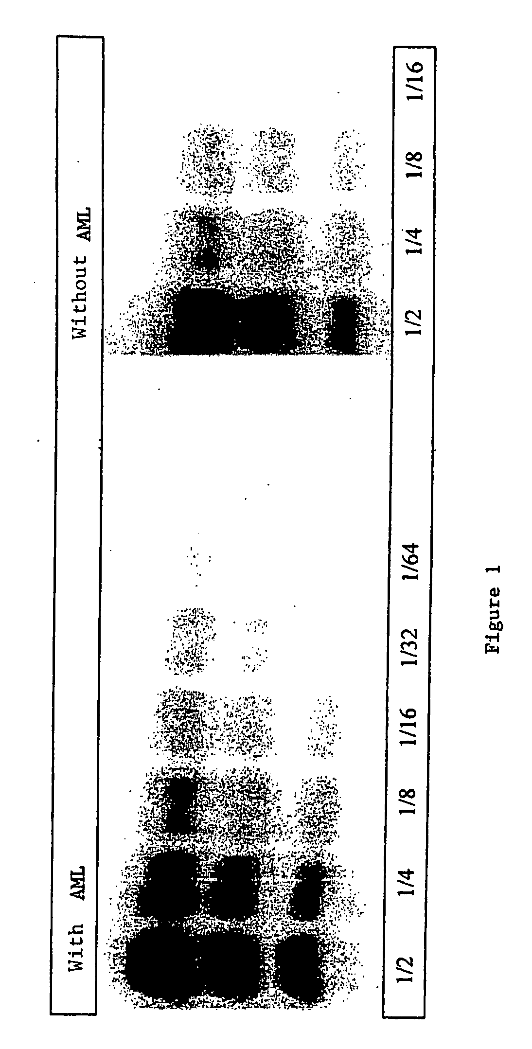
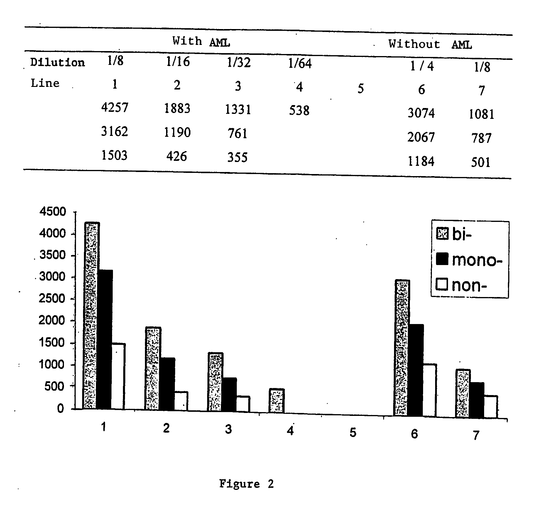
[0112] The samples that were used for the examples of FIGS. 1 and 2 were prepared as follows:

[0113] Sample without AML: 0.5 g brain tissue was crushed in 4.5 ml 5% glucose solution to obtain a suspension with 10% weight / volume. 1 μg proteinase K (Boehringer) in 10 μl was added per 100 μl brain homogenate at 10% in glucose at 5% (equivalent to 10 mg of brain). The solution was placed under vortex and incubated at 37° C. for another hour. After the addition of 100 μl denaturing Laemmli buffer, the mixture was heated 5 minutes at 100° C., centrifuged at 12000 G for 5 nm, and the supernatants recovered to make them migrate on SDS PAGE.

[0114] Sample with AML: 0.5 g brain tissue was crushed in 4.5 ml 5% glucose solution to obtain a suspension with 10% weight / volume. 1.5 μg proteinase K (Boehringer) in 10 μl was added per 100 μl brain homogenate at 10% in glucose at 5% (equivalent to 10 mg of brain). T...

example 3

Detection of Recombinant PrP Using the C6S Graft on a Solid Support

3.1 Detection of Bovine Recombinant PrP on Plates

a) Protocol

[0127] The protocol used in this experiment was from a classic ELISA.

[0128] According to Example 1, grafting of activated amine plates (NH S) was performed for 6 hours with a range of sulfonated calix-6-arenes (C6S) diluted in a solution of PBS 50 mM (saline phosphate buffer). The plates were then rinsed with distilled water before being saturated with a solution of PBST (PBS Tween) (0.05% milk 5%) for 1 hour at ambient temperature. After rinsing, a solution of recombinant bovine protein prion (PrP) diluted to a concentration of 0.25 μg / ml was deposited in PBST 0.05%. It was incubated one hour at ambient temperature. The plate was rinsed again three times. It was then incubated with a solution of anti-PrP antibodies marked with peroxidase diluted at 0.5 μg / ml in PBST 0.05% for one hour at ambient temperature. The antibody used recognizes the region def...


PUM

PropertyMeasurementUnit
concentrationaaaaaaaaaa
temperatureaaaaaaaaaa
pHaaaaaaaaaa

Abstract

A process for detecting the forms of the prion pathogens responsible for subacute, transmissible, spongiform encephalopathies, including a macrocyclic adjuvant ligand (AML), free or bound to a support, that is added to a biological sample capable of containing PrPsc, the resulting suspension then being reacted with an anti-PrPsc antibody, and the presence of PrP is then detected.

Description

RELATED APPLICATION [0001] This is a continuation of International Application No. PCT / FR2003 / 003857, with an international filing date of Dec. 19, 2003 (WO 2004 / 059322 A1, published Jul. 15, 2004), which is based on French Patent Application No. 02 / 16383, filed Dec. 20, 2002.FIELD OF THE INVENTION [0002] This invention relates to a process for detecting the forms of prion pathogens responsible for subacute, transmissible, spongiform encephalopathies. BACKGROUND [0003] The native or normal prion protein, designated PrP or PrPc, for the cellular prion protein is a glycoprotein broadly expressed in the lymphoid and neuronal cells of mammals. [0004] Conformational changes of PrPc result in the appearance and the propagation of the protein pathogen PrPc, that is resistant to the proteinase K. This protein pathogen can be indifferently called PrPsc or PrPres. Accumulation of PrPsc in the organs of animals is at the origin of numerous diseases and especially trembling in small ruminants, ...

Claims


Application Information

Patent Timeline
Patent Type & Authority Applications(United States)
IPC IPC(8): G01N33/53G01N33/68
CPCG01N33/6896G01N2800/2828Y10S436/823Y10S530/807Y10S435/961
Inventor MOUSSA, ALYCOLEMAN, ANTHONY WILLIAMSHAHGALDIAN, PATRICKSILVA, ERIC DAMARTIN, AMBROISELAZAR, ADINA NICOLETALECLERE, EDWIGEDUPIN, MARILYNEPERRON, HERVE
Owner AGENCE FRE DE SECURITE SANITAIRE DES ALIMENT
Why Eureka
Try Eureka