Endoscopic system for treating inside of body cavity

a technology of endoscopic system and body cavity, which is applied in the field of endoscopic system, can solve the problems of difficult positioning of balloon catheter, difficult to clearly see the markings, and difficult to dissect only the lumen wall to be separated from the organ, and achieve the effect of perforating the lumen wall reliably and safely

Inactive Publication Date: 2006-02-02
APOLLO ENDOSURGERY INC
View PDF10 Cites 171 Cited by
  • Summary
  • Abstract
  • Description
  • Claims
  • Application Information

AI Technical Summary

Benefits of technology

[0016] The present invention has been made in view of the above-described circumstances. It is an object of the present invention to provide an endoscopic system for perforating a lumen wall reliably and safely and a treatment device applicable thereto.

Problems solved by technology

In this system, it is difficult to dissect only the lumen wall to be separated from the organ adjacent to the lumen wall when perforating the lumen wall.
However it is difficult to position this balloon catheter at a required location under the endoscope.
However when treatment is carried out by using this balloon catheter, the balloon is loaded on the markings, thus making it difficult to clearly see the markings.
This high-frequency catheter is believed to have the possibility of the balloon slipping during dilation.
Because an electrode always protrudes, proper insertion properties relevant to a physiological wall cannot be obtained.

Method used


Image

  • Endoscopic system for treating inside of body cavity
  • Endoscopic system for treating inside of body cavity
  • Endoscopic system for treating inside of body cavity

Examples


first embodiment

[0061]FIG. 1 to FIG. 10 each show an endoscopic system for treating the inside of a body cavity according to a first embodiment of the present invention.

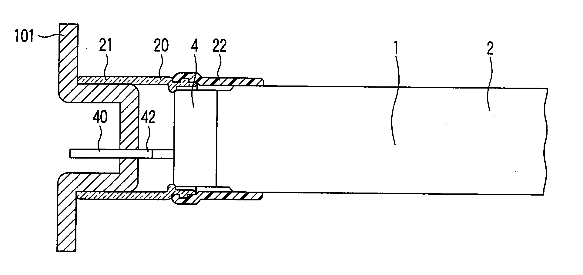
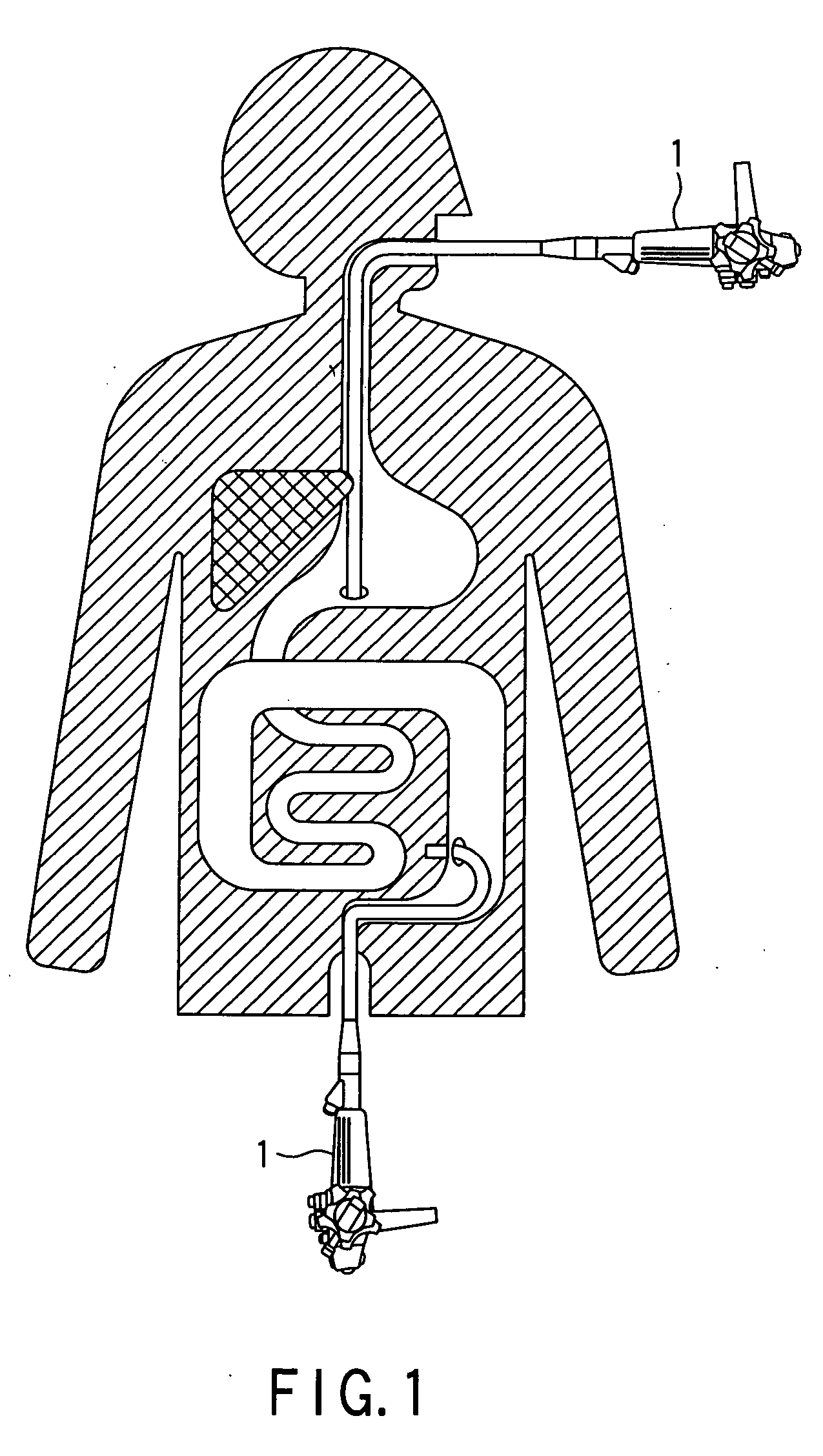
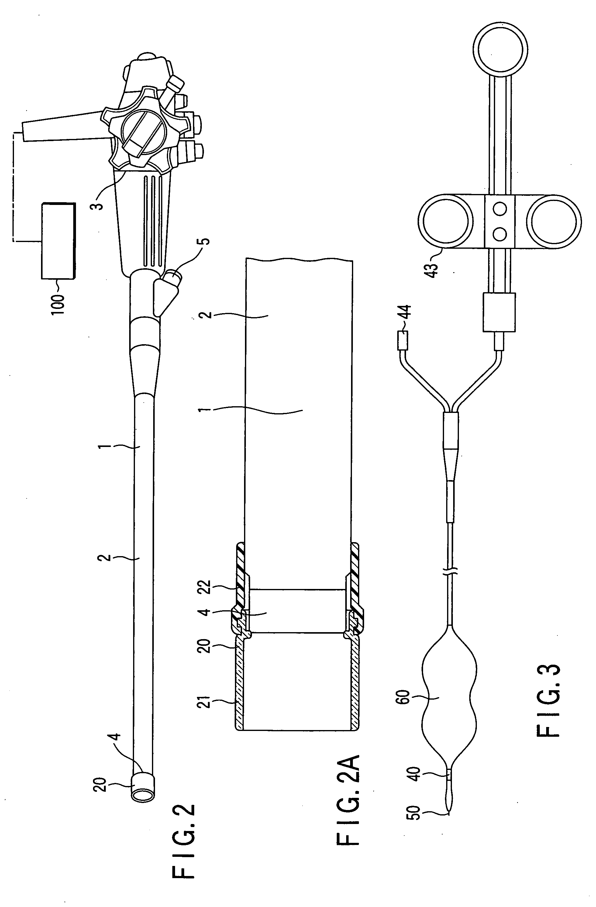
[0062] As shown in FIG. 1 and FIG. 2, an endoscopic system for treating the inside of a body cavity according to the present embodiment comprises the endoscope 1 inserted into a body through a natural opening of a human body. Further, a system according to the present embodiment comprises: a transparent cap 20 mounted on a distal end portion of an endoscope 1 as shown in FIG. 2A; and an opening treatment device 40 inserted into a body via the endoscope 1 as shown in FIG. 3.

[0063] This endoscope 1 comprises an endoscopic insert portion 2 to be inserted into a body; an endoscope distal end portion 4 that is at a distal end of an endoscope insert portion; and an endoscope manipulating portion 3 for manipulating an endoscope insert portion. This endoscope manipulating portion 3 is connected to an endoscope main body or a suction unit ...

second embodiment

[0087]FIG. 17 and FIG. 18 show a second embodiment of the present invention. In a variety of embodiments described below, like elements similar to those according to the first embodiment are designated by like reference numerals. A detailed description is omitted here.

[0088] An endoscopic system for treating the inside of a body cavity according to the present embodiment is composed of an over-tube 30, an endoscope 1 inserted into this over-tube 30; and an opening treatment device 40 inserted into this endoscope 1.

[0089] The over-tube 30 consists of a tubular over-tube sheath 31; and a proximal portion 32 disposed at a proximal end of the over-tube sheath 31. A suction port 33 communicating with the inside of the over-tube 30 is provided at this proximal portion 32.

[0090] The over-tube sheath 31 has a hollow structure whose cross section is circular, for example, and is formed of a polymeric resin material such as polytetrafluoroethylene (PTFE), expanded polytetrafluoroethylene (...

third embodiment

[0097]FIG. 20 shows a distal end portion 41 of an opening treatment device 40 according to the system of a third embodiment of the present invention.

[0098] A high-frequency surgical knife 50 according to the present embodiment can be removed from an opening treatment device sheath 42. This sheath 42 is reduced in inner diameter of a distal end portion 41 as compared with that of the above-described embodiment, and a shoulder portion is formed. A diameter dilution portion for housing a sheath side stopper in the above-described embodiment is omitted here. When the high-frequency surgical knife 50 is inserted into the opening treatment device sheath 42, a wire side stopper 53 securely fitted to the high-frequency knife 50 abuts against a shoulder portion formed in inner hole of the distal end portion 41. In this manner, the sliding of the high-frequency knife 50 to the distal end direction is restricted.

[0099] In this embodiment, the sheath 42 itself restricts the sliding of the hig...


PUM


Abstract

There is provided an endoscopic inserting system. The system includes an endoscope which is inserted into a lumen inside a body through a natural opening of a human body, an opening member which forms an opening for inserting the endoscope into a thoracic cavity or an abdominal cavity from the lumen inside the body at a wall portion of the lumen, and a retracting member which, when forming the opening, retracts the wall portion of the lumen.

Description

CROSS-REFERENCE TO RELATED APPLICATIONS [0001] This application claims the benefit of U.S. Provisional Application No. 60 / 365,266, filed Mar. 18, 2002.BACKGROUND OF THE INVENTION [0002] The present invention relates to an endoscopic system for observing and / or treating the inside of the abdominal cavity and / or thoracic cavity by inserting the system into the human body, and a treatment device applicable thereto. [0003] In general, there are a variety of systems for inserting an endoscope through a natural opening in the human body, dissecting a lumen, and then, treating the inside of the abdominal cavity. [0004] For example, in U.S. Pat. No. 5,297,536, a treatment system as shown in FIG. 34 is disclosed. This system is composed of a dissecting device for perforating a lumen wall; an endoscope insert member for inserting an endoscope, a tube, an endoscope, and a pneumoperitoneum device for deflating the abdominal cavity; and a closing device. [0005] When surgery of the inside of the ...

Claims


Application Information

Patent Timeline
Patent Type & Authority Applications(United States)
IPC IPC(8): A61B1/00A61M29/00A61B17/28A61B17/00A61B17/02A61B17/30A61B17/34A61B18/14
CPCA61B17/00234A61B17/0218A61B17/29A61B17/3478A61B18/1482A61B2018/1407A61B2017/00296A61B2017/306A61B2017/3445A61B2017/3488A61B2017/00278
Inventor SUZUKI, KEITATSUKAGOSHI, TSUYOSHISEKINE, RYUTAKALLOO, ANTHONY NICHOLASKANTSEVOY, SERGEY VENIAMINOVICH
Owner APOLLO ENDOSURGERY INC
Why Eureka
Try Eureka