Ultrasonic cellular tissue screening system

a cellular tissue and ultrasonic technology, applied in ultrasonic/sonic/infrasonic image/data processing, instruments, applications, etc., can solve the problems of misrepresentation of overall findings, process error, and haphazard coverage of scanned tissue, so as to achieve reliably screened and easy removal

Inactive Publication Date: 2006-02-16
SONOCINE INC
View PDF23 Cites 8 Cited by
  • Summary
  • Abstract
  • Description
  • Claims
  • Application Information

AI Technical Summary

Benefits of technology

[0013] The present invention is directed to an improved system of ultrasonic scanning and diagnostics of breast tissue. A sequence of cross-sectional ultrasonic images of tissue are generated. To create the appropriate environment for such scanning, a fabric covering adapted for holding the breast tissue in place accommodates an ultrasonic coupling agent to absorb the agent for conducting ultrasonic energy with minimal interference. The covering may be preimpregnated, may include a pattern usable by an optical measurement sensor and may be constructed for easy removal. A pad which is ultrasonically conductive may be placed beneath the fabric covering to cover the breast nipple.

Problems solved by technology

Because the operator must choose a few frames from the large number generated during the scan, the process is open to error.
The operator may fail to select an image of an important finding, or may select an image that misrepresents the overall findings.
In addition, since the operator is manipulating the probe by hand, and the speed of the probe over the tissue cannot be correlated with the image capture rate of the probe, the coverage of the scanned tissue is somewhat haphazard.
As a result, the operator does not record a series of images that represent a contiguous and complete set of images for the entire scanned tissue.
Nor does the manual operation of the probe allow for entirely uniform coverage of the tissue, even if multiple passes are used.
Unfortunately, this analog method is not matched to the digital sonographic recording of individual frames.
Consequently, there is a great loss of detail that prevents the evaluation of individual frames, which limits the usefulness of the videotape for diagnosing tissue anomalies.
In addition, the use of separate videotapes for individual patients is expensive, and creates a storage problem because of the bulkiness of the tapes.
The interpreting physician has no way to vary the speed of playback or to vary the size of the images.
Nor can the physician vary the inherent contrast and brightness of the images, only the monitor settings.
These difficulties lengthen the review time and prevent optimum viewing.
Both of these methods are imperfect.
Physical examination, whether performed by the woman herself or by a physician or other health care provider, usually cannot detect cancers smaller than ½ inch in diameter.
Mammography is unable to detect as many as 30 percent of cancers smaller than ½ inch.
Mammograms also use radiation and necessitate painful compression of the breasts, which discourage women from having routine mammograms.
When using ultrasound as a screening method for the entire breast, however, malignancies are usually difficult to pick out of the background tissue.
In the past there have been two schemes to use ultrasound for breast screening, but they failed to gain acceptance due to their unacceptably low success rate in finding cancers.
This method had the disadvantage of being somewhat haphazard in breast coverage.
The variable speed of manual motion does not allow the tissue to be uniformly imaged because the speed is not synchronized to the frame capture rate of the ultrasound probe.
Videotaping also results in a degradation of the images for the reasons described above.
To date, no method has been developed to uniformly and reliably use ultrasound probes to create a contiguous and complete set of scan images for an entire area of cellular tissue, such as a human breast.

Method used


Image

  • Ultrasonic cellular tissue screening system
  • Ultrasonic cellular tissue screening system
  • Ultrasonic cellular tissue screening system

Examples


Embodiment Construction

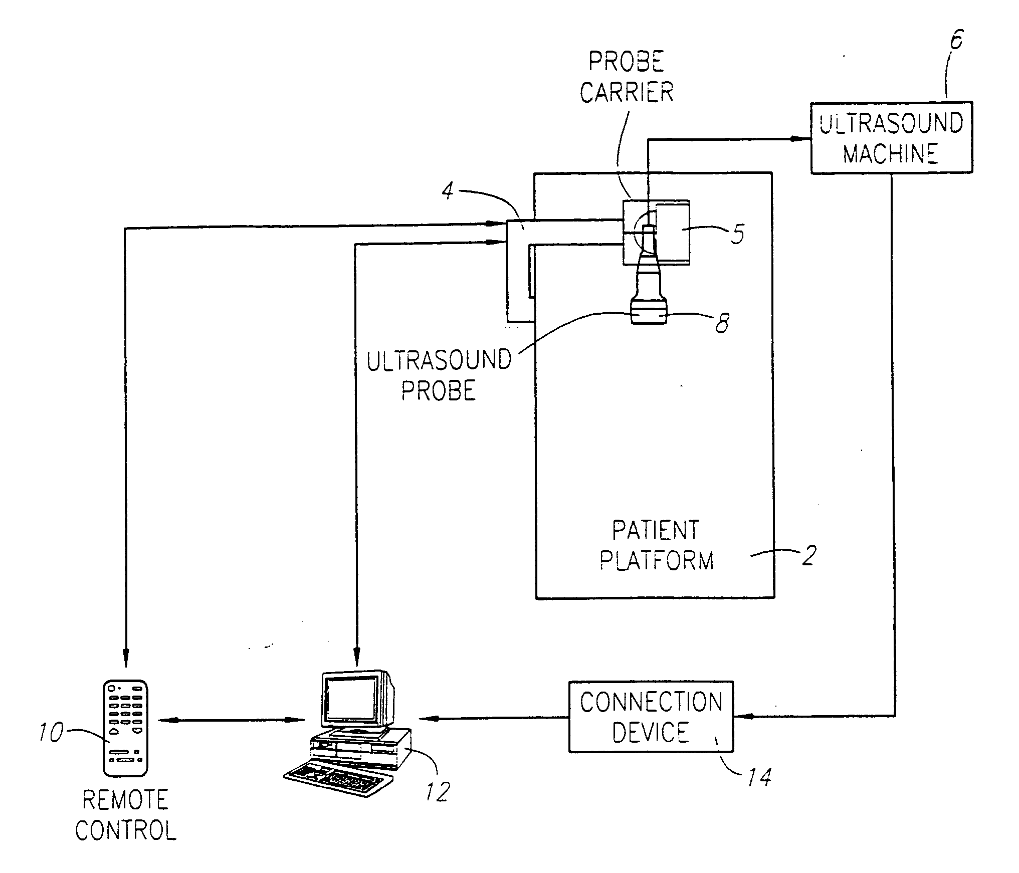
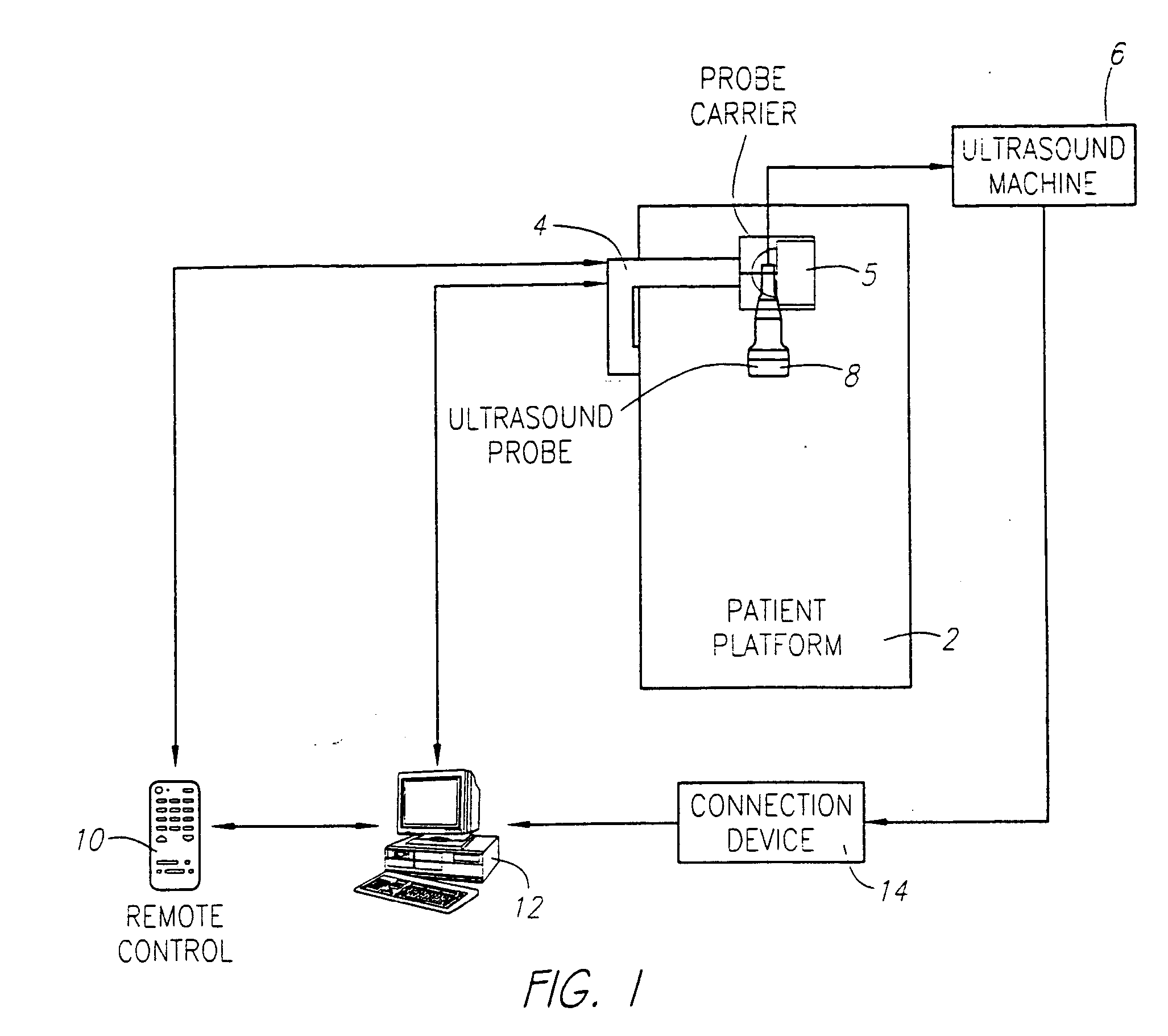
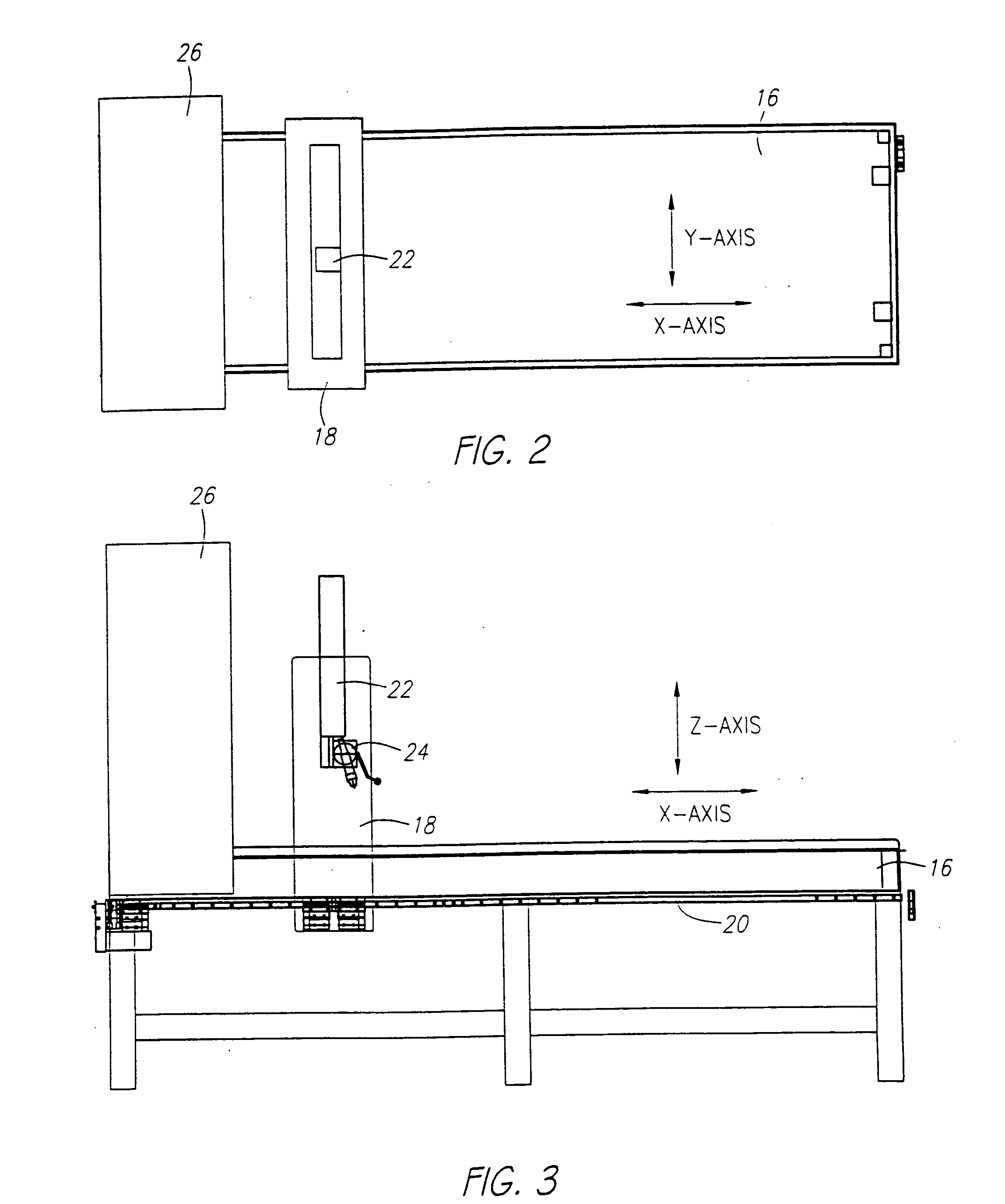
[0031] As shown in FIG. 1, a preferred embodiment is comprised of a patient platform 2 to steady the patient and provide a base for the support member 4, the probe carrier 5 connected with the support member 4 that is capable of translational movement to guide the probe across the tissue to be scanned, a standard medical ultrasound device 6 with an associated probe 8, a remote control device 10 that operates the probe carrier 4, a standard computer 12, a connection device 14 between the ultrasound device 6 and the computer 12, and a viewing program that obtains images from the ultrasound device and converts them into images compatible with the viewing program and displays the images. The medical ultrasound scanning device 6 with associated probe 8, computer 12, and connection device 14 are commercially available.

[0032] The mechanical carrier 4 holding the ultrasound probe 8 can be connected with the ultrasound scanner 6. Synchronization between the probe holder mechanical carrier 4...


PUM


Abstract

Ultrasonic scanning and diagnostics for cellular tissue are disclosed. An ultrasonic probe is moved across cellular tissue at a rate that is synchronized with the image capture rate of the ultrasonic scanner, to achieve a contiguous and complete set of scan images of the tissue. The probe can be held in a single position as it is moved across the tissue, or it can be dynamically adjusted during the scan to provide optimal contact with the scanned tissue. The image data are captured and converted to a format that is easily stored and compatible with a viewer. The viewer allows playback of the scanned images in a manner that is optimized for screening for cancers and other anomalies. A location function allows the user to select a point of interest on an individual scan image, and choose another known reference point, and the function calculates and provides the distance from the reference point to the point of interest in three dimensions. The system can be used for virtually any tissue, but can also be optimized for breast cancer screening. A pad of different density may be placed over the nipple to provide a reference point that is visible in the scan images. The breast may be covered with a fabric that is constructed in such a manner to hold the breast in place and reduce ultrasonic scanning errors, as well as holding the pad in place. The location function described above will use the nipple pad as a reference point from which to measure any detected cancers or other anomalies.

Description

PRIORITY [0001] This is a divisional application of U.S. patent application Ser. No. 09 / 687,128, filed Oct. 13, 2000 in the name of Kevin M. Kelly et al., for which priority under 35 U.S.C. 120 is claimed. The disclosure of U.S. patent application Ser. No. 09 / 687,128 is incorporated herein by reference.BACKGROUND OF THE INVENTION [0002] 1. Field of the Invention [0003] The field of the present invention is ultrasonic scanning and diagnostics for cellular tissue. [0004] 2. Background of the Invention [0005] Ultrasonic probes have been used for scanning cellular tissue for many years. Presently, any medical ultrasound examination, whether of the heart, pelvis, abdomen, soft tissues or any other system, is usually displayed as a number of individual frames or pictures from a study performed in a dynamic movie like manner. The usefulness of the scan, however, is dependent on the skill of the operator, who manipulates the probe by hand while watching the scan images on a monitor to ident...

Claims


Application Information

Patent Timeline
Patent Type & Authority Applications(United States)
IPC IPC(8): A61B8/14A61B8/00A61B8/08G01S15/89
CPCA61B8/0825A61B8/406A61B8/4209A61B8/565A61B8/488A61B8/54B82Y15/00A61B8/4281A61B8/4245A61B8/4218A61B8/085A61B8/145A61B8/4254A61B8/4444A61B8/463A61B8/5223
Inventor KELLY, KEVIN M.ROYCE, ROGER
Owner SONOCINE INC
Why Eureka
Try Eureka