Ultrasonic endoscope and ultrasonic endoscopic apparatus

an endoscope and ultrasonic technology, applied in the field of ultrasonic endoscopes and ultrasonic endoscopes, can solve the problems of shortening the scanning time (frame period) for obtaining images for one frame, and reducing the frame rate, so as to improve the frame rate, reduce the influence of crosstalk between plural ultrasonic beams, and improve the image quality of ultrasonic tomographic images

Inactive Publication Date: 2006-03-16
FUJIFILM CORP +1
View PDF24 Cites 9 Cited by
  • Summary
  • Abstract
  • Description
  • Claims
  • Application Information

AI Technical Summary

Benefits of technology

[0014] According to the present invention, the frame rate can be improved by simultaneously transmitting the same number of ultrasonic beams at a certain angle interval in each of plural angle regions formed by dividing a scanning angle region by a predetermined angle. Further, in the case where the angle interval of the simultaneously transmitted ultrasonic beams is set t

Problems solved by technology

However, according to the mechanical radial scanning method, compared to a scanning method of performing scanning with viewing angle of 90° using a convex ultrasonic transducer array in an external ultrasonic imaging apparatus, in the case where conditions of s

Method used


Image

  • Ultrasonic endoscope and ultrasonic endoscopic apparatus
  • Ultrasonic endoscope and ultrasonic endoscopic apparatus
  • Ultrasonic endoscope and ultrasonic endoscopic apparatus

Examples


Embodiment Construction

[0026] Hereinafter, preferred embodiments of the present invention will be described in detail by referring to the drawings. The same reference numbers will be assigned to the same component elements and the description thereof will be omitted.

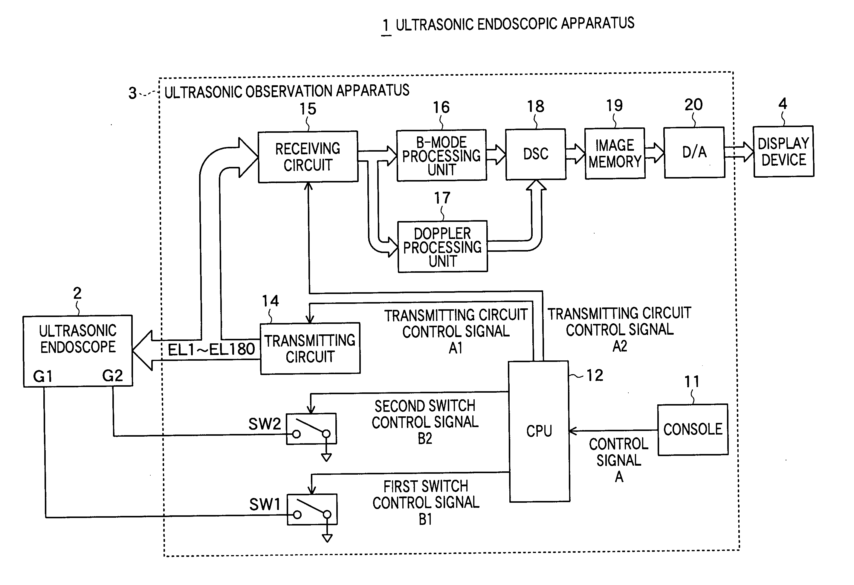
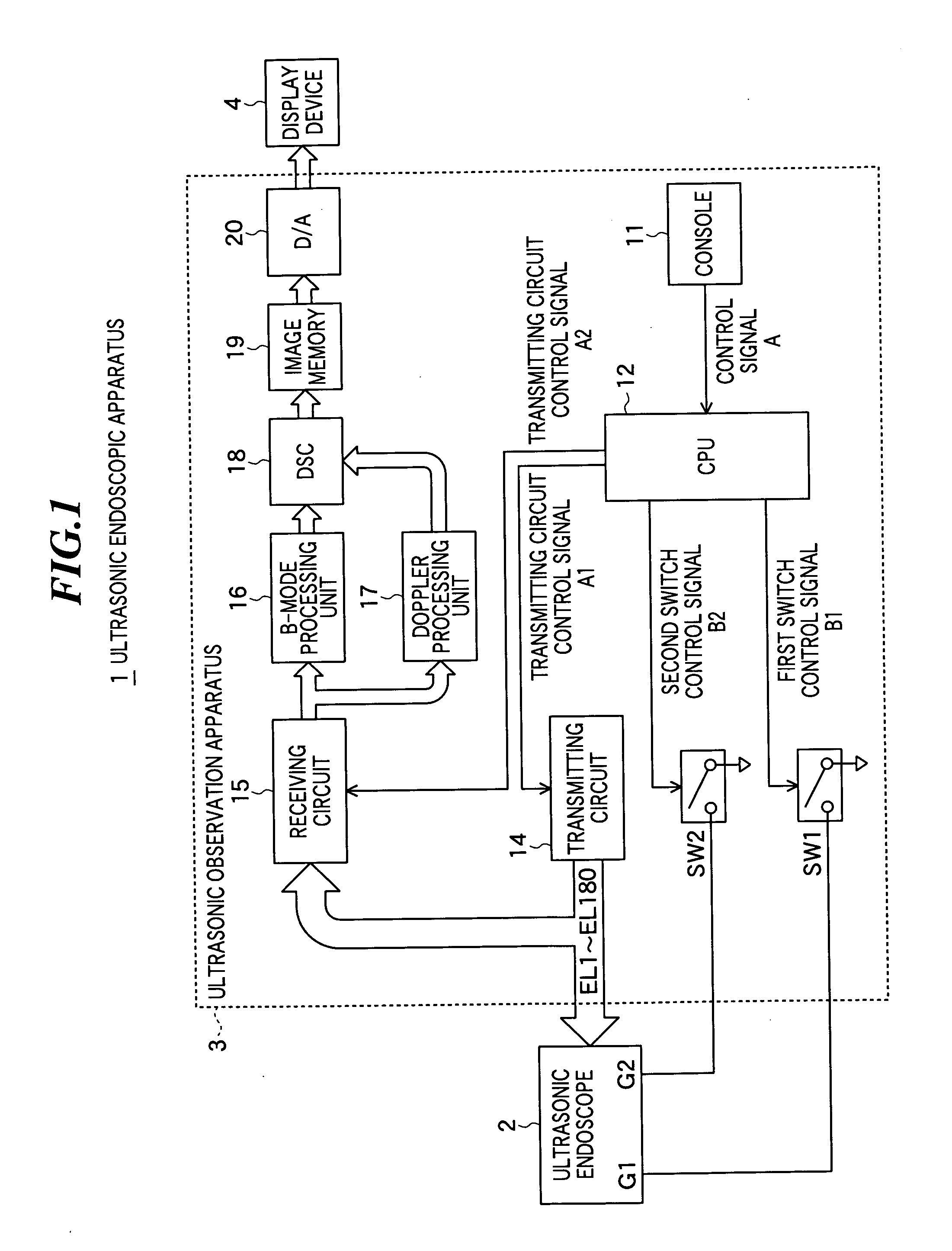
[0027] As shown in FIG. 1, an ultrasonic endoscope apparatus 1 according to one embodiment of the present invention includes an electronic radial ultrasonic endoscope 2, an ultrasonic observation apparatus 3 to which the ultrasonic endoscope 2 is connectable, a display device 4 connected to the ultrasonic observation apparatus 3.

[0028] The ultrasonic observation apparatus 3 includes a console 11, a CPU (central processing unit) 12, first and second switches SW1 and SW2, a transmitting circuit 14, a receiving circuit 15, a B-mode processing unit 16, a Doppler processing unit 17, a digital scan converter (DSC) 18, an image memory 19, and a digital / analog converter (D / A converter) 20.

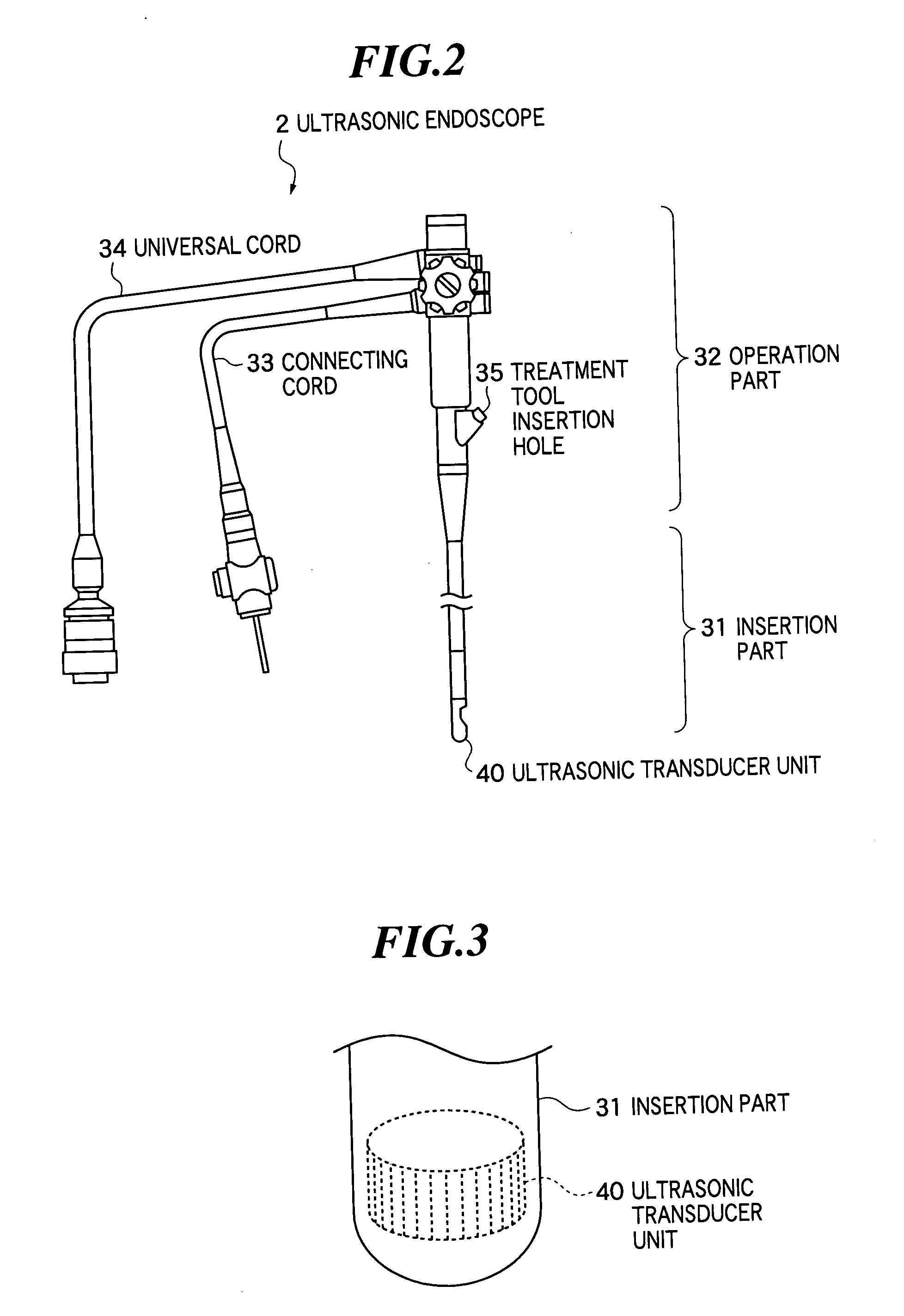
[0029] As shown in FIG. 2, the ultrasonic endoscope 2 includ...


PUM


Abstract

In an ultrasonic endoscope capable of being inserted into a body of a patient and imaging ultrasonic tomographic images, a frame rate in electronic radial scanning operation is improved. The ultrasonic endoscope includes: an ultrasonic transducer unit including plural ultrasonic transducers, each having a first electrode and a second electrode, for transmitting and receiving ultrasonic waves; a plurality of first interconnections each connected to the first electrodes of predetermined ultrasonic transducers; and a plurality of second interconnections each connected to the second electrodes of ultrasonic transducers having the first electrodes not connected to each other; wherein the ultrasonic transducer unit performs scanning operation by simultaneously transmitting the same number of ultrasonic beams in each of plural angle regions when selectively supplied with drive signals via the plurality of first interconnections and the plurality of second interconnections.

Description

BACKGROUND OF THE INVENTION [0001] 1. Field of the Invention [0002] The present invention relates to an ultrasonic endoscope capable of being inserted into a body of a patient and imaging ultrasonic tomographic images for medical diagnoses and an ultrasonic endoscopic apparatus including such an endoscope. [0003] 2. Description of a Related Art [0004] Recent years, an ultrasonic endoscope has been used for medical diagnoses by inserting it into a body of a patient and imaging ultrasonic tomographic images. In such an ultrasonic endoscope, generally, a mechanical radial scanning method of performing scanning with viewing angle of 360° by mechanically rotating an array including plural ultrasonic transducers (ultrasonic vibrators) that transmit and receive ultrasonic waves has been adopted. [0005] However, according to the mechanical radial scanning method, compared to a scanning method of performing scanning with viewing angle of 90° using a convex ultrasonic transducer array in an e...

Claims


Application Information

Patent Timeline
IPC IPC(8): A61B8/14A61B8/12
CPCA61B8/12A61B8/145A61B8/4461G01S15/8979A61B8/4488G01S15/8922A61B8/4483
Inventor SATOH, YOSHIAKITSUJITA, KAZUHIRO
Owner FUJIFILM CORP
Why Eureka
Try Eureka