

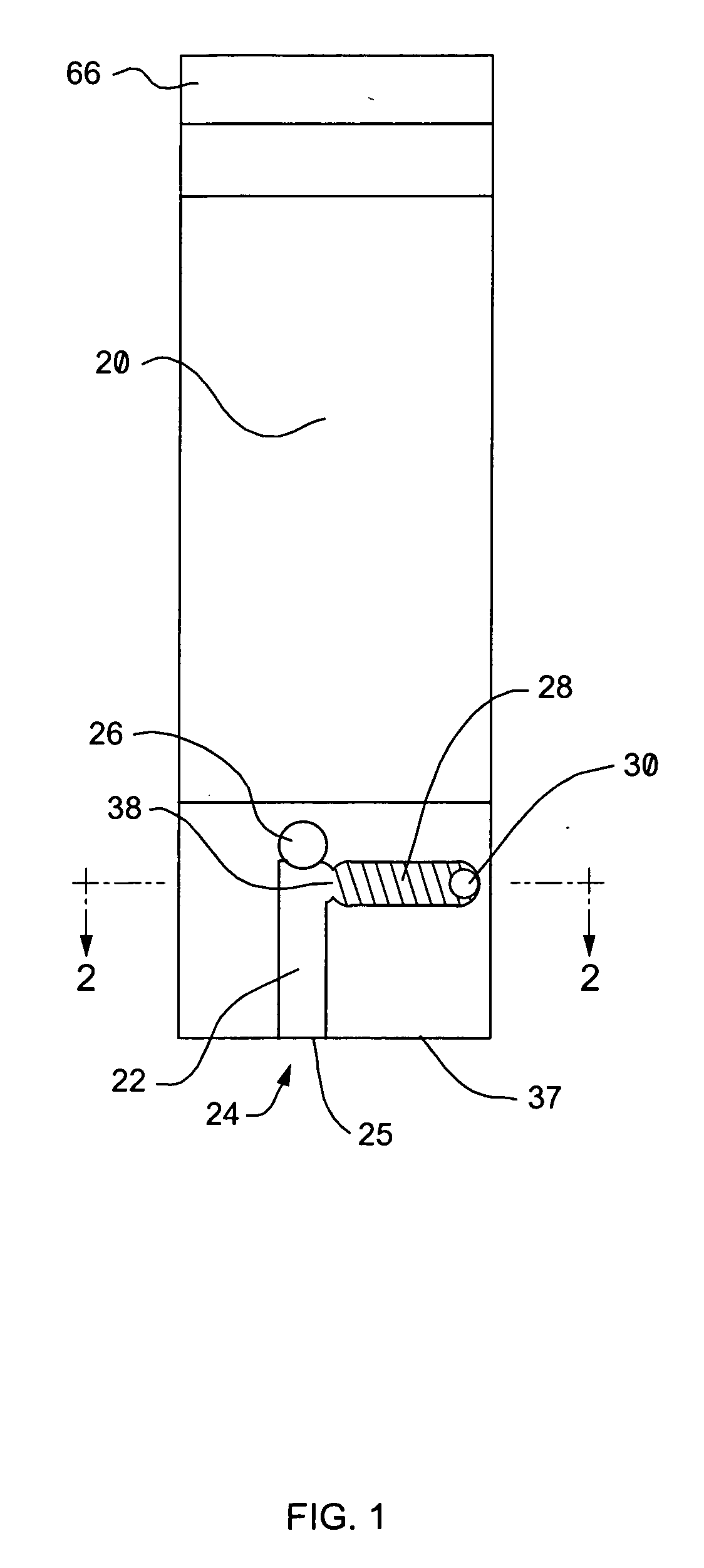
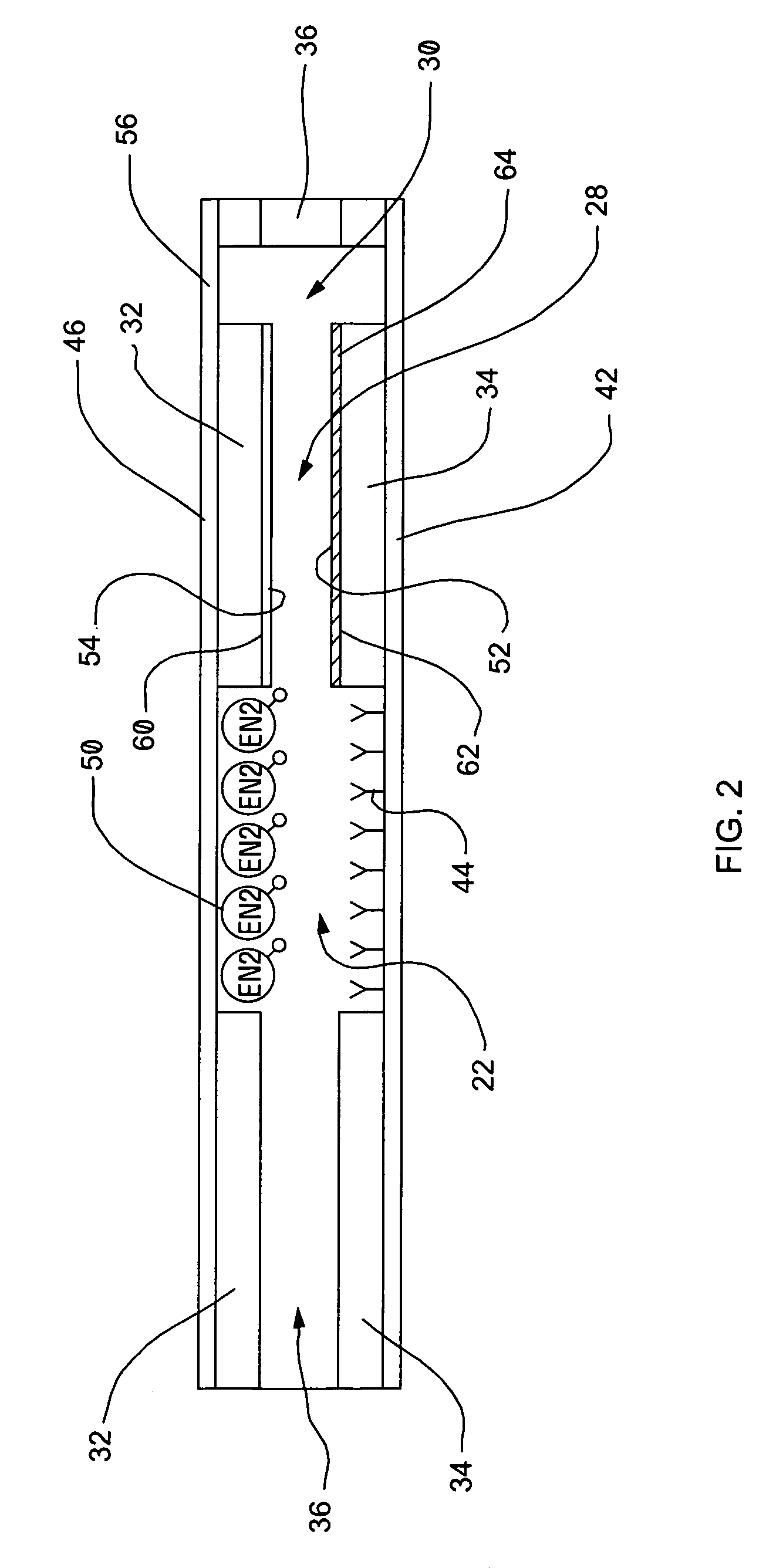
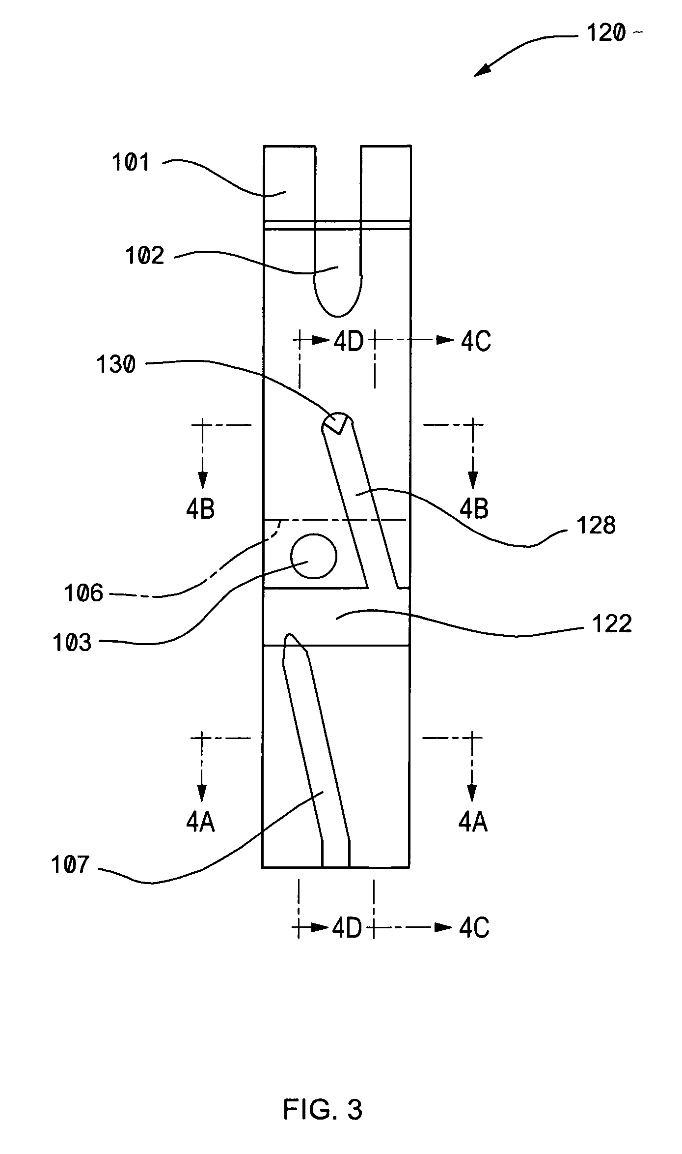
Biosensor apparatus and methods of use
a biosensor and apparatus technology, applied in the field of biomedical sensors, can solve the problems of generating biohazardous liquid waste, steps introducing complexity into the assay procedure, and methods involving expensive equipmen
- Summary
- Abstract
- Description
- Claims
- Application Information
AI Technical Summary
Benefits of technology
Problems solved by technology
Method used
Image

Examples
example
[0070] An exemplary immunosensor assay for human C Reactive protein in whole blood using magnetic beads coated with Human C reactive protein was performed.
[0071] C reactive protein (CRP) was covalently attached to 1.5 micron Carboxylated BioMag magnetic beads (Cat no BM570; Bangs Laboratories, Indianapolis In, USA). 21.9 mg of beads (1 ml) were washed 4 times with 50 mM MES (Morpholinoethanesulphonic acid (Sigma-Aldrich, St Louis, Mo. USA) buffer pH 5.2 by incubating with this buffer and using a magnet to concentrate the beads on the side of the tube and after 2 min remove the buffer with a transfer pipette. After the fourth wash, the beads were suspended in a final volume of 0.34 ml 50 mM MES. 40 ul of 100 mg / ml EDAC (Sigma, St Louis, Mo. USA) was added and after 5 minutes 450 ug of CRP in Phosphate buffered saline (Hytest, Turku Finland) was added. Beads were allowed to incubate for a further 30 min at room temperature. Unbound CRP was removed by using a magnet to concentrate the...
PUM
| Property | Measurement | Unit |
|---|---|---|
| distance | aaaaa | aaaaa |
| distance | aaaaa | aaaaa |
| distance | aaaaa | aaaaa |
Abstract
Description
Claims
Application Information