Biodegradable pericardia constraint system and method

a pericardia constraint and biodegradable technology, applied in the field of biodegradable pericardia constraint system and method, can solve the problems of depressed ventricular function, high wall stress within and around the infarct, and abnormal ventricular wall motion

Inactive Publication Date: 2006-06-22
G&L CONSULTING
View PDF25 Cites 16 Cited by
  • Summary
  • Abstract
  • Description
  • Claims
  • Application Information

AI Technical Summary

Problems solved by technology

This is often due to clotting in a coronary blood vessel.
Within seconds of a myocardial infarction, the under-perfused myocardial cells no longer contract, leading to abnormal ventricular wall motion, high wall stresses within and surrounding the infarct, and depressed ventricular function.
These high stresses eventually kill or severely depress function in the still viable myocardial cells.
This results in a wave of dysfunctional tissue spreading out from the original myocardial infarct region.
During the early period after MI the infarcted region is particularly vulnerable to distorting forces.
Therefore late enlargement is due to complex alterations in LV architecture involving both infarcted and non-infarcted zones.
Reopening the occluded artery within hours of the initial occlusion can decrease tissue death, and thereby decrease the total magnitude of infarct expansion, extension, and ventricular remodeling.
These treatments are effective but clearly not satisfactory alone.
In many cases, patients arrive at the appropriately equipped hospital too late for these acute therapies.
In other cases, their best efforts fail to reopen blood vessels sufficiently to arrest expansion of the infarct.
These therapies are also associated with considerable risk to the patient and high cost.

Method used


Image

  • Biodegradable pericardia constraint system and method
  • Biodegradable pericardia constraint system and method
  • Biodegradable pericardia constraint system and method

Examples


Embodiment Construction

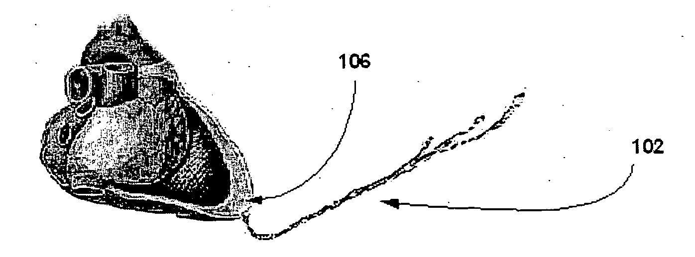
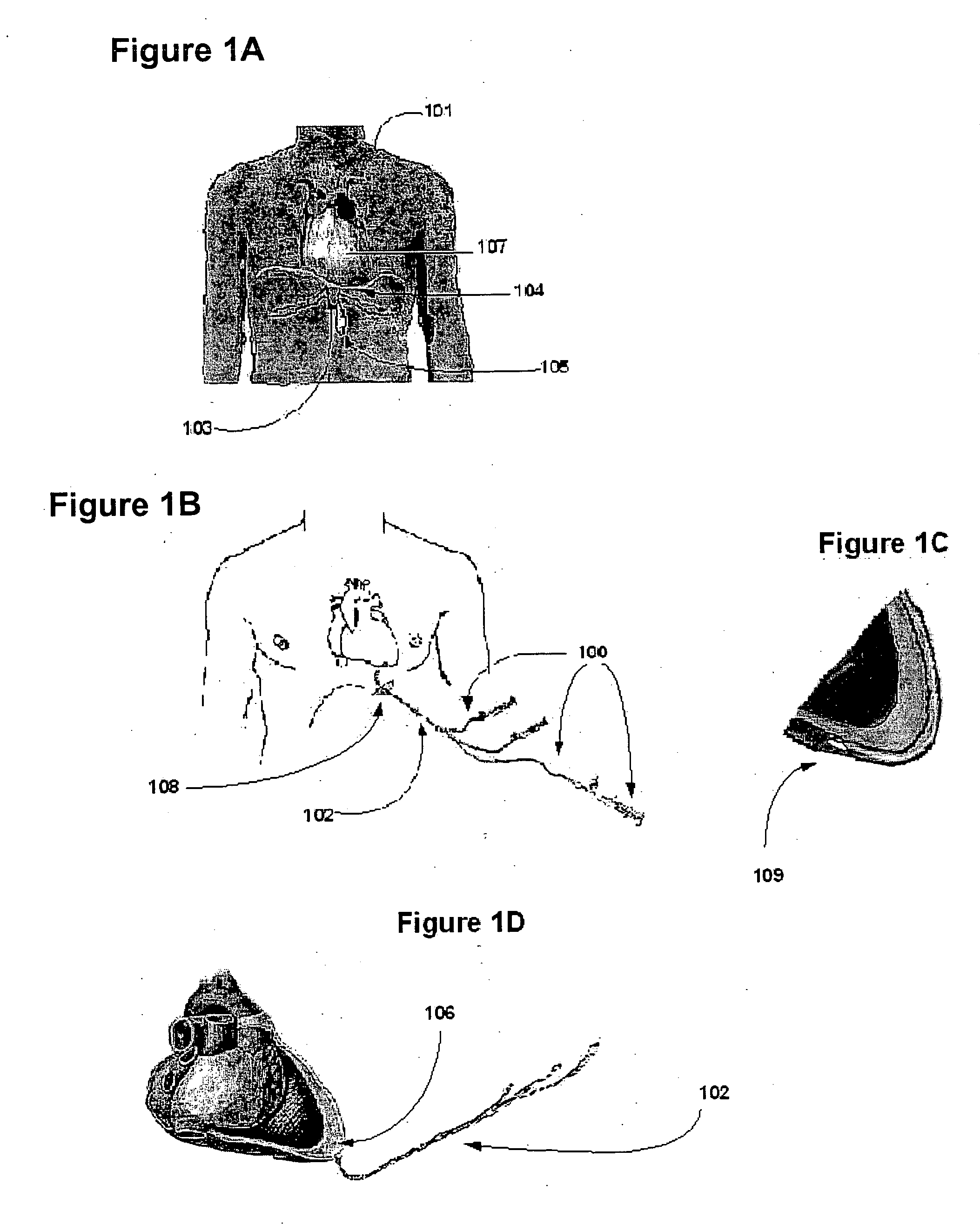
[0023]FIG. 1A, B, C, D illustrates the treatment of a patient 101 with the system 100 for injection of BES into the pericardial sac of the heart. The distal end of injection cannula 102 is partially inserted into the pericardial sac of the heart 107. Cannula 102 crosses the patient's skin in the xiphoid area 103 via subxiphoidal incision 105. The diaphragm 104 is incised during surgery down to the pericardial surface. Through this incision 108, the pericardium 107 may be easily visualized and a small incision or a puncture 109 is made in pericardium to accommodate a cannula insertion. The distal tip of the cannula 102 has an opening and is in fluid communication with the pericardial (also called intrapericardial) space 106. The proximal end of the cannula 102 is connected to the delivery system 100 containing the biodegradable viscoelastic substance such as sterile cross linked collagen gel, balloon inflation media such as saline, and tissue sealant such as BioGlu.

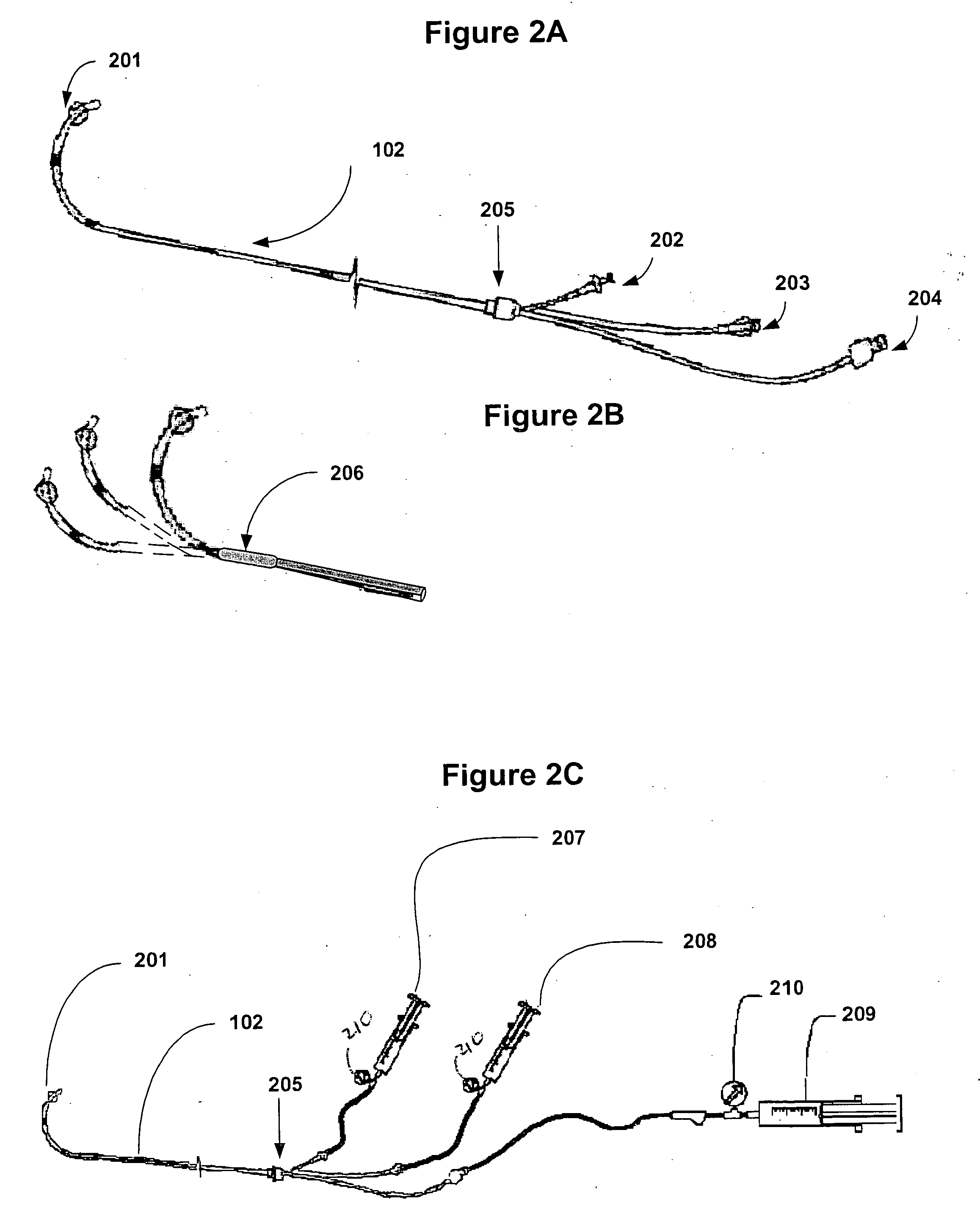
[0024]FIGS. 2 A, ...


PUM


Abstract

A system has been developed for injecting a biodegradable pericardial constraint including: a biodegradable viscoelastic substance (BES); an external injection container for the BES; a cannula having a distal section adapted to be inserted into a pericardial sac of a mammalian heart and a proximal section connectable to the external injection container; wherein BES from the injection container is injected into the pericardial sac through the cannula.

Description

RELATED APPLICATIONS [0001] This application is a continuation-in-part (CIP) application of U.S. patent application Ser. No. 10 / 808,397, entitled “Method and System To Treat and Prevent Myocardial Infarct Expansion” filed Mar. 25, 2004, which claims priority under 35 U.S.C. §119(e) to U.S. Provisional application 60 / 457,246, filed Mar. 26, 2003, and this application also claims priority under 35 U.S.C. §119(e) to U.S. Provisional Patent Application Ser. No. 60 / 628,923, entitled “Biodegradable Pericardial Constraint”, filed Nov. 19, 2004, the entirety of all of these related applications are incorporated by reference herein. BACKGROUND OF INVENTION [0002] A Myocardial Infarction (MI) or heart attack, occurs when the blood supply to some part of the heart muscle (myocardium) is abruptly stopped. This is often due to clotting in a coronary blood vessel. Blood supplying the heart muscle comes entirely from two coronary arteries, both lying along the outside surface of the heart. If one ...

Claims


Application Information

Patent Timeline
Patent Type & Authority Applications(United States)
IPC IPC(8): A61M1/00
CPCA61B17/00491A61B2017/00247A61B2018/00392A61M25/0084
Inventor CHERNOMORSKY, ARYGELFAND, MARKLEVIN, HOWARD R.
Owner G&L CONSULTING
Why Eureka
Try Eureka