High-resolution three-dimensional imaging of mammalian hearts

a three-dimensional imaging and heart technology, applied in image enhancement, instruments, image data processing, etc., can solve the problems of limiting the type of information that can be gained, the final specimen may not reflect well, and the information regarding the cell context in the organ is los

Inactive Publication Date: 2017-04-20
UNIV OF WASHINGTON
View PDF1 Cites 7 Cited by
  • Summary
  • Abstract
  • Description
  • Claims
  • Application Information

AI Technical Summary

Benefits of technology

[0007]The invention provides a method of preparing a tissue sample for high-resolution three-dimensional tissue imaging. In one embodiment, the method comprises embedding a fixed tissue sample in an embedding medium; sectioning the embedded tissue into sections of 20-1000 μm thickness; staining the sectioned tissue; mounting the stained 20-1000 μm thick tissue sections on a coverslip and glass slide: and cl

Problems solved by technology

However, the limitations of traditional imaging techniques restrict the type of information that can be gained.
However, these proteases can also damage the cells, and this processing increases the time from living tissue to preserved specimen, meaning the final specimen may not well reflect what occurs in vivo.
Though this technique is well suited for studying single cells in detail, information regarding the cells' context in the organ is lost.
Tissue sections can offer a view of the cell in situ, but have their own limitations.
Cryosectioning frequently utilizes fresh tissue that is cryopreserved, with the resulting sections typically being post-fixed with alcohol and/or acetone, though samples may also be fixed with formalin prior to cryopreservation (3), Cryosections are generally thin (>10 μm); thicker sections can be obtained but section quality is often compromised (rolling, ruffling, or cracking).
A major limitation of using frozen sections is that the cryopreservation process can disrupt the morphology o

Method used


Image

  • High-resolution three-dimensional imaging of mammalian hearts
  • High-resolution three-dimensional imaging of mammalian hearts
  • High-resolution three-dimensional imaging of mammalian hearts

Examples


example 1

lution Three-Dimensional Cardiac Imaging

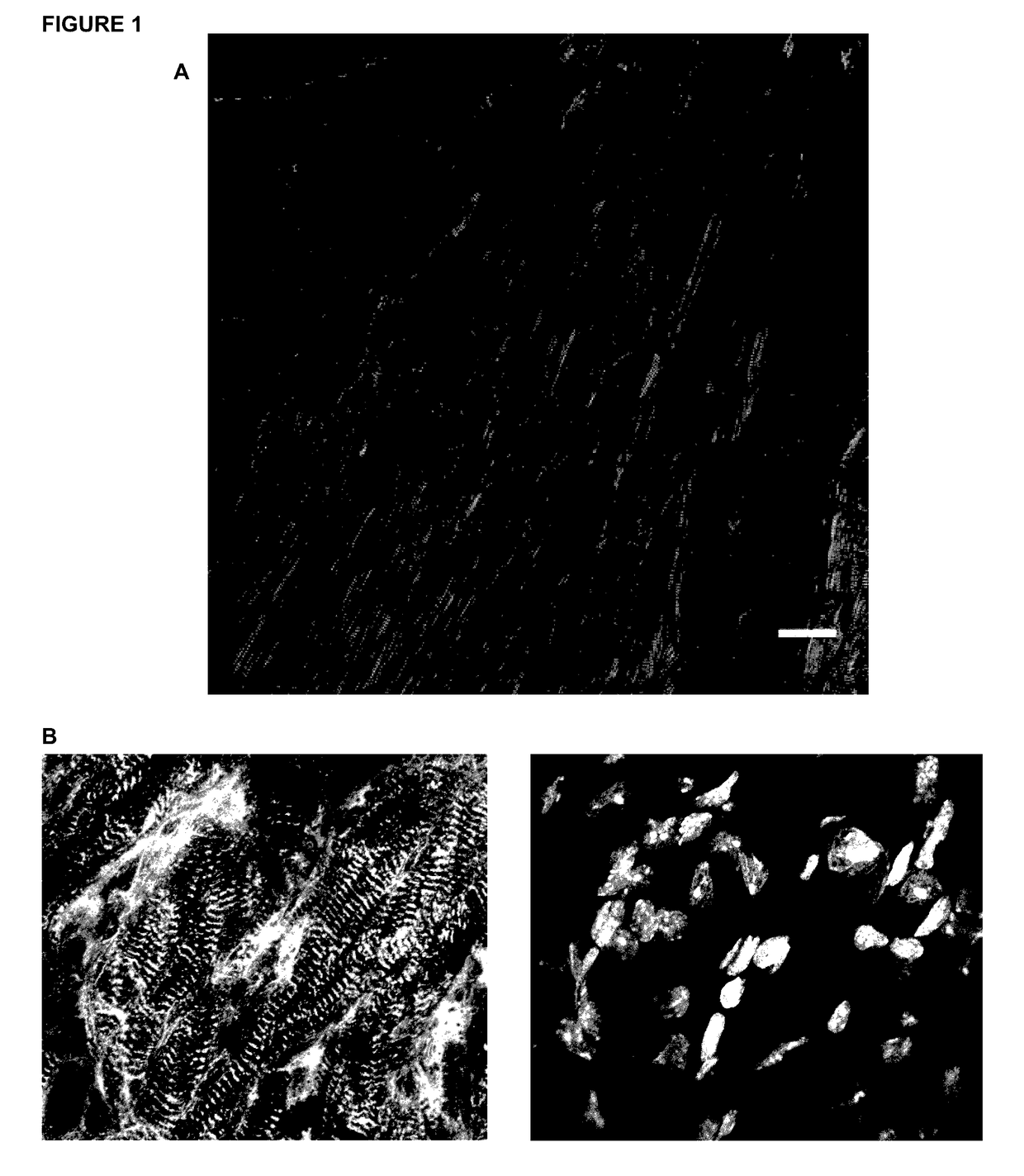
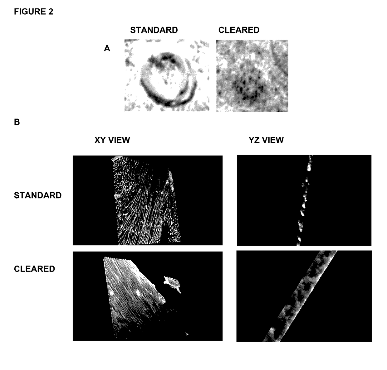
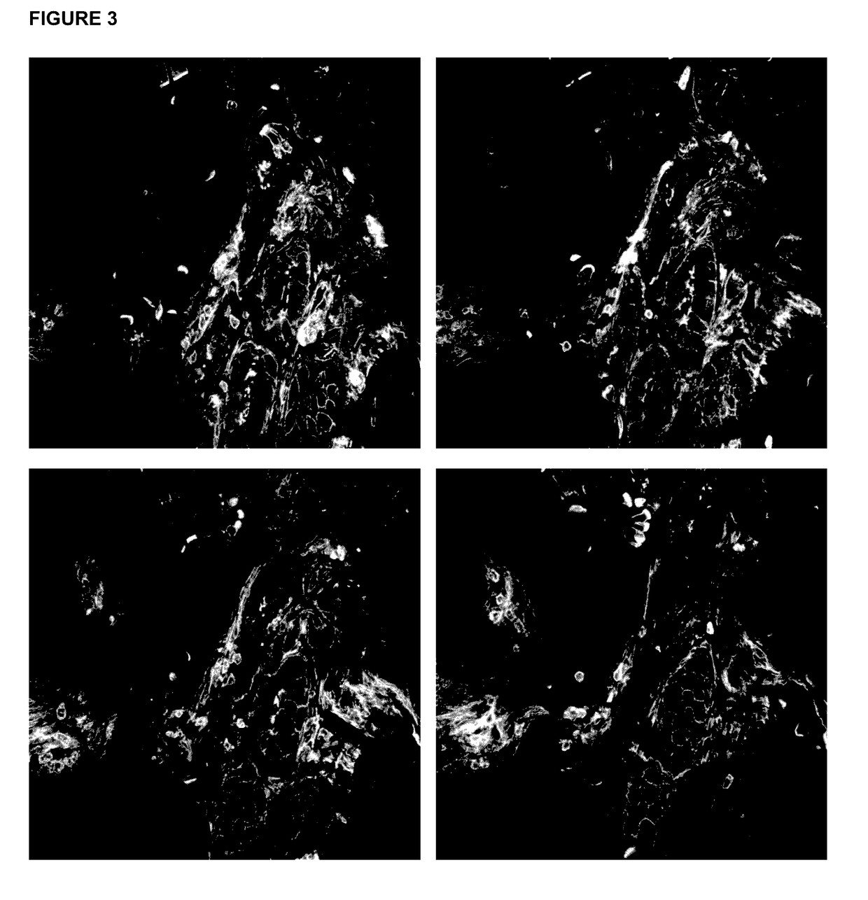
[0068]In this work we established a sample preparation method that utilizes vibratorne sectioning, tissue clearing, and confocal microscopy techniques. We generated 3D images of adult mammalian hearts with 0.2 μm resolution in xy and 0.3 μm resolution in yz / xz, spanning several cardiac myocytes of thickness, in native tissue. We used this innovation to assess fibrosis and sarcomere disarray in human ischemic heart disease samples and to obtain volumetric quantification of mitotic activity, apoptosis, and chromatin subnuclear structure in adult mouse cardiac myocytes,

[0069]We followed established conventional protocols for imaging paraffin sections and cryosections, Hearts for paraffin sections were fixed with 4% PFA and sectioned at 4 μm thiCkneSS. Fresh hearts were frozen in OCT medium, cryosectioned at 10 μm thickness, and fixed with 100% methanol prior to staining, For High-Resolution Three-Dimensional Cardiac Imaging, we tested various fix...

example 2

rt Preparation

[0070]Obtain human tissue. We used IRB approved LVAD core from patient with ischemic heart disease, keeping fresh sample in a cold, high potassium buffer to keep the myocytes arrested, such as KB buffer.

[0071]Transfer tissue to glass plate, with scalpel, trim heart piece into a cube, ˜3 mm×3 mm×3 mm, keeping sample on ice. *Note, larger pieces do not section well.

[0072]Fix tissue (Choose fix appropriate for downstream applications, other fixatives may be used):

[0073]Transferring sample to 4% paraformaldehyde in PBS, precooled to 4 C. Incubate overnight at 4 C.

OR

[0074]100% methanol (MeOH), precooled to −20 C. Incubate at −20 C for 30 min to 1 hr. In a stepwise manner, incubate the sample in precooled 80% MeOH / 20% PBS, 60% MeOH / 40% PBS, with each incubation for 30 minutes at −20 C.

[0075]Wash 3× with PBS, keep in PBS at 4 C until embedding.

example 3

rt Preparation

[0076]Euthanize animal as approved on animal protocol, according to government and insfitute guidelines, we used isoflurane overdose. Collect heart in cold KB buffer.

[0077](Optional, but recommended) Cannulate aorta with blunt 26 g needle, secure with suture:

[0078]Perfuse 4 ml KB buffer, precooled at 4 C, 2 mL / min to wash the blood out of the coronary vessels.

[0079]Perfuse Fixative:

[0080]4 mL 4% PFA, 4 C, 2 mL / min

OR

[0081]2 mL 100% MeOH, −20 C, 1 mL / min

[0082]Fix Tissue:

[0083]Transfer sample to 4% paraformaldehyde in PBS, precooled to 4 C. Incubate 16 hrs at 4 C.

[0084]100% methanol (MeOH), precooled to −20 C. Incubate at −20 C for 30 min to 1 hr. In a stepwise manner, incubate the sample in precooled 80% MeOH / 120% PBS, 60% MeOH / 40% PBS, with each incubation for 30 minutes at −20 C.

[0085]Wash 3' with PBS, keep in PBS at 4 C until embedding.


PUM


Abstract

Methods and materials for high-resolution three-dimensional imaging of mammalian tissues, such as heart and intestine, are described. Both methods of tissue preparation for imaging and methods of imaging are described, as well as kits comprising materials and media for use in the methods.

Description

[0001]This application claims benefit of U.S. provisional patent application No. 62 / 244,105, filed Oct. 20, 2015, the entire contents of which are incorporated by reference into this application.TECHNICAL FIELD OF THE INVENTION[0002]The present invention relates to methods and materials for high-resolution three-dimensional imaging of mammalian tissues, such as heart and intestine. Both methods of tissue preparation for imaging and methods of imaging are descrbed, as well as kits comprising materials and media for use in the methods.BACKGROUND OF THE INVENTION[0003]Microscopy has long been appreciated as a methodology to directly study cells. However, the limitations of traditional imaging techniques restrict the type of information that can be gained. To be compatible with high resolution imaging, hearts are either dissociated into single cells or are sectioned into thin slices; resulting in specimens that are eventually mounted on glass slides for viewing. Each method (dissociatin...

Claims


Application Information

Patent Timeline
IPC IPC(8): G01N1/30G06T3/40G01N33/483G06T7/00G01N1/36G01N1/28
CPCG01N1/30G01N1/36G01N1/286G06T2207/30004G06T7/0012G06T3/4053G01N2001/305G01N33/4833G01N1/06G01N1/312
Inventor MACLELLAN, W. ROBBEL-NACHEF, DANNY
Owner UNIV OF WASHINGTON
Why Eureka
Try Eureka