
Image-based computational mechanical analysis and indexing for cardiovascular diseases

Active Publication Date: 2006-07-06
WORCESTER POLYTECHNIC INSTITUTE
View PDF0 Cites 96 Cited by
  • Summary
  • Abstract
  • Description
  • Claims
  • Application Information

AI Technical Summary

Benefits of technology

[0010] Advantageously, Applicant's invention employs a three-dimensional model of an organ or biological system that includes fluid-structure interactions (e.g., blood-vessel interactions) and the structure of the organ or biological system. The three-dimensional models of the invention further include various components of the organ or biological system, e.g., various plaque components for atherosclerotic plaques. Therefore, the invention can accurately model mechanical distribution(s), such as stress / strain distribution(s), in the organ or biological system under investigation. Also, Applicant's invention can identify mechanical biological index (indices) to quantify the degree of the disease status, which can help physicians in decision-making processes.

Problems solved by technology

Image-based clinical diagnosis in the art generally lacks mechanical analysis.
Current technology for diagnosis of, for example, cardiovascular diseases (e.g., carotid plaque rupture, coronary plaque rupture and aneurism rupture), does not include an accurate and reliable computational mechanical analysis.
Some 2D MRI-based models and 3D Structure-only or fluid-only models known in the art are not generally adequate for decision-making purposes.

Method used


Image

  • Image-based computational mechanical analysis and indexing for cardiovascular diseases
  • Image-based computational mechanical analysis and indexing for cardiovascular diseases
  • Image-based computational mechanical analysis and indexing for cardiovascular diseases

Examples


example 1

Unsteady Stress Variations in Human Atherosclerotic Plaques and Sensitivity Analysis Using 3D MRI-Based Fluid-Structure Interactions (FSI) Models

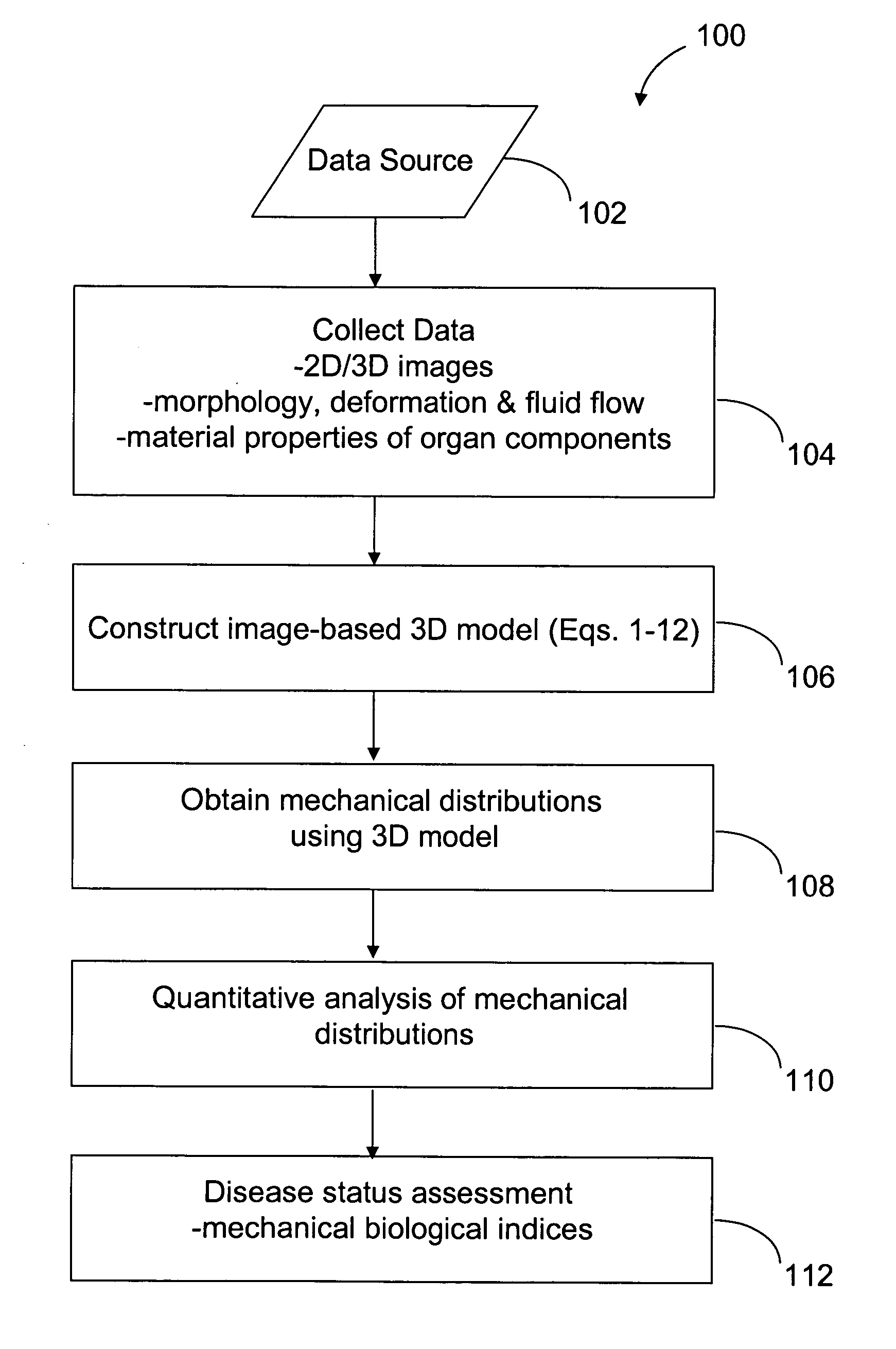
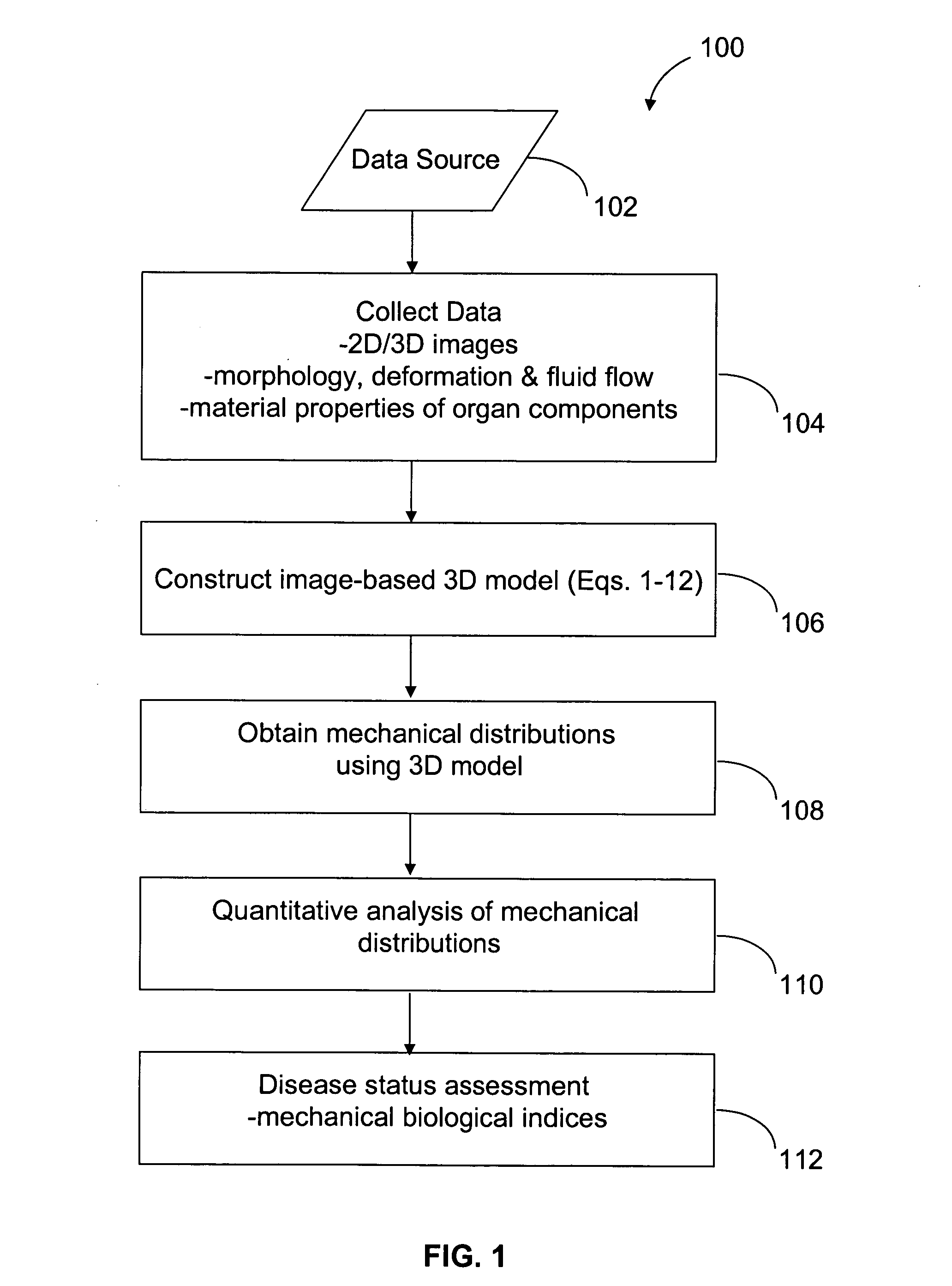
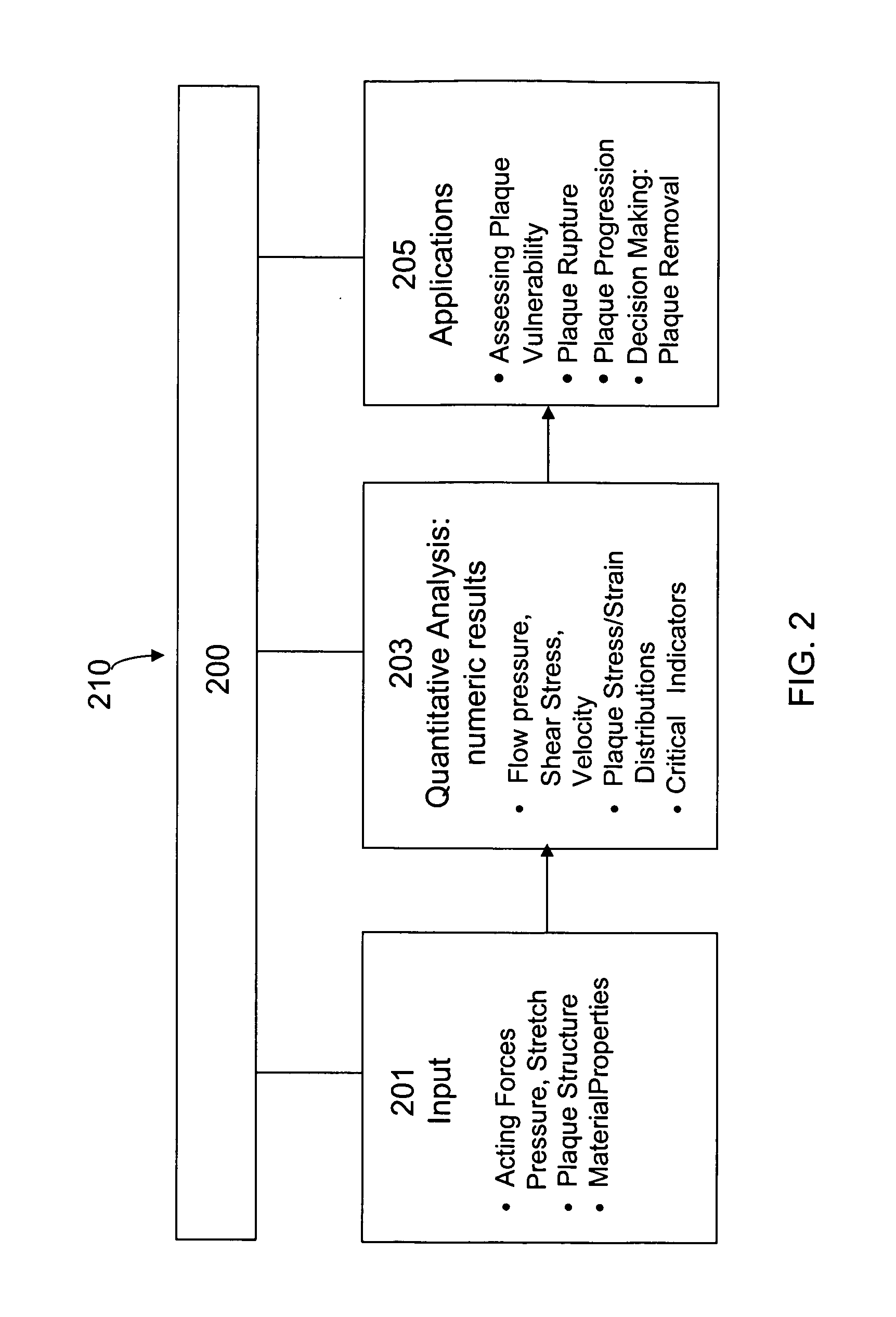
[0072] This example illustrates methods and computer systems 100, 210 of the invention for providing assessment for plaque vulnerability to rupture.

[0073] An unsteady three-dimensional (3D) MRI-based computational model for human atherosclerotic plaques with multi-component plaque structure and fluid-structure interactions (FSI) was introduced to perform mechanical analysis and identify critical flow and stress / strain conditions which might be related to plaque rupture.

[0074] Also, in this example, “local” maximal stress / strain values and their variations were investigated. A “critical site tracking” (CST) approach was used to concentrate on plaque stress / strain behaviors at well-selected critical locations which include locations of local maxima, locations of very thin cap and any sites of special interest. Stress / strain distributions a...

example 2

Local Maximal Stress Hypothesis and Computational Plaque Vulnerability Index (CPVI) for Atherosclerotic Plaque Assessment

[0093] In Example 1, a 3D MRI-based computational model with multi-component plaque structure and fluid-structure interactions (FSI) was introduced to perform mechanical analysis for human atherosclerotic plaques and identify critical flow and stress / strain conditions which may be related to plaque rupture. In this Example 2, a “local maximal stress hypothesis” and a stress-based computational plaque vulnerability index (CPVI) were used to assess plaque vulnerability. Local maximal stress results tracked at critical sites in the plaque were be used to determine CPVI values. A semi-quantitative histopathological method was used to classify plaques into different grades, according to factors which were generally known to correlate with plaque vulnerability. This histopathological classification was used as the “gold standard” to validate the computational indices o...

example 3

3D Image-Based Computational Modeling for Patient-Specific Mechanical Analysis of Human Hear Right Ventricles

[0119] In this example, the MRI techniques and computational modeling were combined to build a patient-specific 3D computational model with fluid-structure interactions (FSI) for the human right ventricle (RV). Right ventricular dysfunction is one of the more common causes of heart failure in patients with congenital heart defects.

[0120] Due to the complexity of the motion of a human right ventricle and related blood flow and structure stress / strain behaviors, some simplifications were made in the modeling so that proper measurements could be obtained and the model could be solved to obtain flow and stress / strain information. Based on the solutions, mechanical analysis could be performed to analyze cardiac functions of the right ventricle. The patient-specific 3D computational modeling techniques of the invention can be further employed in computer-aided cardiac surgery pla...


PUM


Abstract

A method and a computer system, for providing an assessment for disease status of a disease, such as a cardiovascular disease, employ construction of image-based 3D computational model of an organ representative of the disease status; computationally obtaining a certain mechanical distribution using the 3D-organ model; and computational, quantitative analysis of the mechanical distribution to provide an assessment for disease status of a disease. The image-based 3D computational model includes a fluid-structure interaction and multiple components within the organ.

Description

RELATED APPLICATION [0001] This application claims the benefit of U.S. Provisional Application No. 60 / 635,021 filed on Dec. 10, 2004. The entire teachings of this provisional application are incorporated herein by reference.GOVERNMENT SUPPORT [0002] The invention was supported, in whole or in part, by a grant DMS-0072873 from the National Science Foundation and a grant NIH / NIBIB, 1 R01 EB004759-01 from the National Institute of Health. The Government has certain rights in the invention.BACKGROUND OF THE INVENTION [0003] Cardiovascular disease (CVD) is the No. 1 killer in the United States and responsible for 40% of all deaths in 2000. More than 60% of all myocardial infarction is caused by rupture of a vulnerable plaque. A large number of victims of the disease who are apparently healthy die suddenly without prior symptoms. About 95 percent of sudden cardiac arrest victims die before reaching a hospital. About 250,000 people a year die of coronary artery disease (CAD) without being ...

Claims


Application Information

Patent Timeline
IPC IPC(8): G06G7/48G06F19/00
CPCG06F19/321G06F19/3437G16H30/40G16H50/50Y02A90/10
Inventor TANG, DALIN
Owner WORCESTER POLYTECHNIC INSTITUTE
Learn More