Complete airway stabilization system and method

a complete airway and stabilization technology, applied in the field of endotracheal intubation, can solve the problems of inadvertent unintentional movement, and multiple severe pulmonary difficulties, and achieve the effect of preventing unintentional head movement and preventing malpositioning of the endotracheal tub

Active Publication Date: 2006-08-10
SECURISYN MEDICAL
View PDF82 Cites 30 Cited by
  • Summary
  • Abstract
  • Description
  • Claims
  • Application Information

AI Technical Summary

Benefits of technology

[0012] The present invention provides a multi-pronged approach to preventing malpositioning of the endotracheal tube and maintaining the endotracheal tube in the correct and proper position for intubation. A new and improved endotracheal tube and faceplate interact with one another to prevent relative movement, even under conditions where bodily fluids cover the tracheal tube. A new and improved stabilization collar fits around the neck and prevents unintentional movement of the head and neck while simultaneously interacting with the faceplate to assure that the endotracheal tube is maintained in the proper position while the head and neck are stabilized against unintentional movement. In contrast to known prior art arrangements, the present invention provides a complete system and method for positioning, maintaining and stabilizing the endotracheal tube.
[0015] In addition to the endotracheal tube, another form of the apparatus includes a faceplate which contacts the face of the patient at the mouth. A variably-sized orifice is formed in the faceplate. The orifice expands in size to accept the endotracheal tube and the projections, and the orifice contracts in size to constrict within a selected recess and contact the projections adjacent to the selected recess and create a barrier which limits longitudinal movement of the endotracheal tube relative to the faceplate at the selected recess.

Problems solved by technology

An endotracheal tube may also become necessary when a patient's own respiratory drive is either ineffective or absent due to trauma, disease, drugs or anesthesia, thus requiring mechanical ventilation through the endotracheal tube.
The failure to ventilate the other lung can lead to multiple severe pulmonary difficulties.
If the tube moves completely out of its proper position in the trachea and into the esophagus, the patient will become hypoxic due to the lack of ventilation to the lungs, typically resulting in life-threatening brain injury within several minutes.
Even after the endotracheal tube has been positioned correctly, subsequent movement of the patient can lead to inadvertent mal-positioning of the endotracheal tube.
Such unintentional movement is not uncommon, because the patient frequently needs to be moved, particularly when the patient is moved from an out-of-hospital setting to an emergency department of a hospital.
Even repositioning an intubated patient in a hospital bed may cause unintentional movement of the endotracheal tube.
Inadvertent pulling on, or other excessive movement of the air conduit, may transfer movement to the endotracheal tube, thereby moving the endotracheal tube and possibly inadvertently repositioning it from its proper position.
While the use of the umbilical tape in this manner provides some benefit, the restraint available from the umbilical tape usually diminishes because the tape becomes covered and saturated with blood, saliva, or other bodily fluids, thereby diminishing the frictional restraint of the tape around the endotracheal tube.
One of the problems with many of these auxiliary mechanical contact devices is that the gripping action becomes less effective when the endotracheal tube becomes wet from bodily fluids, thereby substantially diminishing or eliminating any restraint provided by these devices.
Restricting the cross-sectional size of the air passageway decreases the ventilatory efficiency of the tube, thereby decreasing the respiratory airflow.
Insufficient airflow can lead to hypoxemia, and increased pressure can lead to barotrauma in the lungs.
Resistance to expiratory airflow leads to multiple adverse effects within the lungs.
Impairing a patient's ventilations may result in serious medical effects, particularly with patients whose functions are already compromised.

Method used


Image

  • Complete airway stabilization system and method
  • Complete airway stabilization system and method
  • Complete airway stabilization system and method

Examples


Embodiment Construction

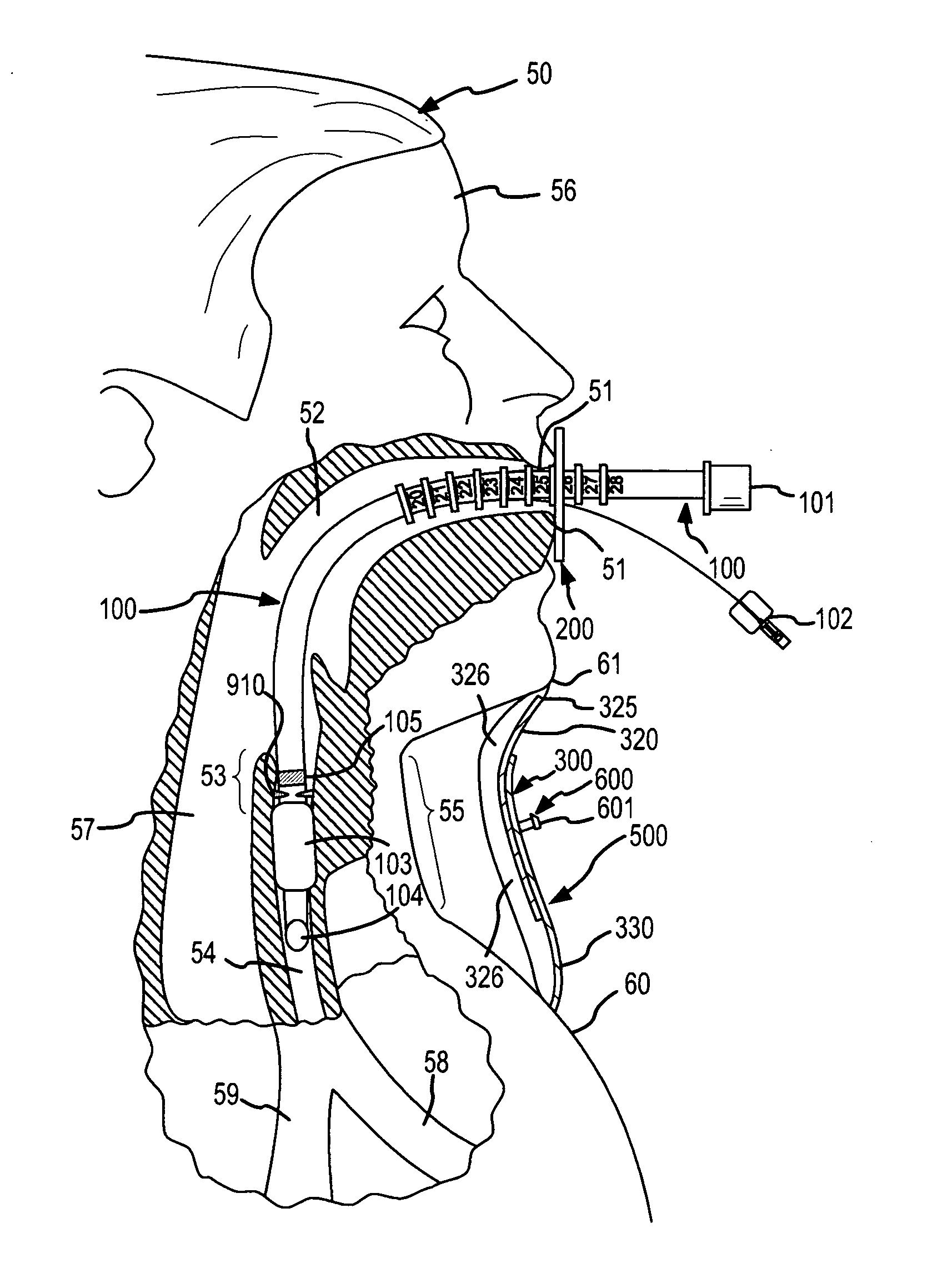
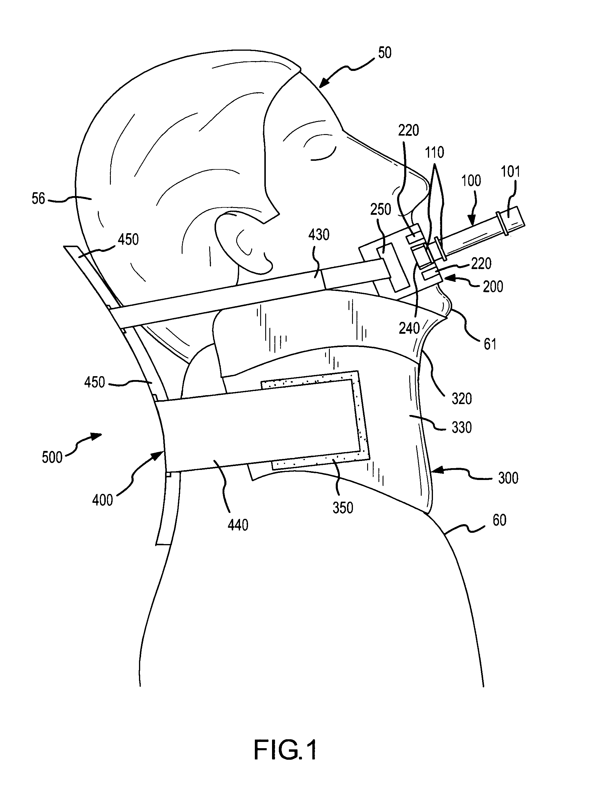
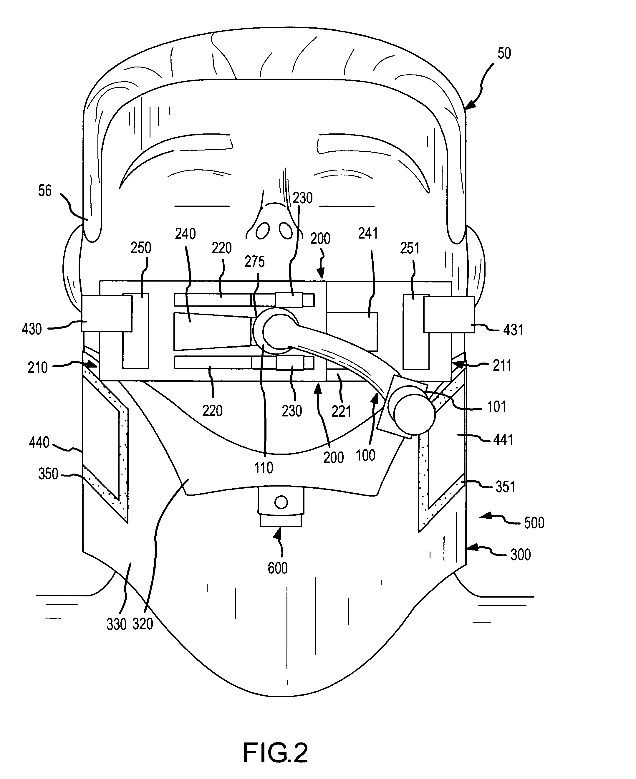
[0040] A complete airway stabilization system which incorporates the present invention is shown in FIGS. 1, 2 and 3. The complete airway stabilization system is used to intubate a patient 50 under conditions where natural respiration is impossible or severely compromised. The airway stabilization system establishes an air passageway to the lungs for respiration of the patient 50, while eliminating many of the risks and difficulties associated with intubation with an endotracheal tube.

[0041] The airway stabilization system comprises an endotracheal tube 100, a faceplate 200 and a stabilization collar 500. The endotracheal tube 100 is inserted through the mouth 51, throat 52 and larynx 53 of the patient 50 (FIG. 3), and into the trachea 54 by using conventional intubation procedures. The faceplate 200 fits over the patient's mouth 51 and interacts with the endotracheal tube 100 to restrain the endotracheal tube 100 relative to the faceplate 200. The stabilization collar 500 fits arou...


PUM


Abstract

An endotracheal airway is established using an endotracheal tube having an exterior retention structure defined by a series of longitudinally spaced recesses separated by projections. A variably-sized orifice in a faceplate constricts within a recess and contacts the projections to create a barrier against movement of the endotracheal tube. The faceplate is retained on the mouth by connection to a stabilization collar which surrounds the neck of the patient to restrain against flexion and extension which could reposition the endotracheal tube from its desired position.

Description

CROSS REFERENCE TO RELATED APPLICATION [0001] This application claims the benefit of U.S. Provisional Application US60 / 593,702, filed Feb. 7, 2005.FIELD OF THE INVENTION [0002] This invention relates to endotracheal intubation, and more particularly to a new and improved system and method which make use of an endotracheal tube, a faceplate, and a head and neck stabilization collar, all of which cooperatively interact to provide improved stabilization and maintenance of an airway into the lungs for respiration. BACKGROUND OF THE INVENTION [0003] Endotracheal intubation is a medical procedure used to place an endotracheal tube (artificial airway) into a patient's airway. The endotracheal tube becomes necessary when a patient is unable to maintain a patent airway on their own due to unconsciousness, trauma, disease, drugs or anesthesia. An endotracheal tube may also become necessary when a patient's own respiratory drive is either ineffective or absent due to trauma, disease, drugs or ...

Claims


Application Information

Patent Timeline
Patent Type & Authority Applications(United States)
IPC IPC(8): A62B9/06A61M16/00
CPCA61F5/055A61M16/0488A61M16/0497A61M2205/32A61M16/0493
Inventor KANOWITZ, ARTHUR
Owner SECURISYN MEDICAL
Why Eureka
Try Eureka