Quantitative non-invasive method for detecting degree of malignancy in tumors and application thereof

a tumor and non-invasive technology, applied in the field of tumor hemodynamic parameters measurement and non-invasive measurement of tumors, can solve the problems of insufficient credibility, limited suitability of aforementioned detection, and many tumors, and achieves low resistance area, high resistance area, and high ratio of neovascular blood vessels

Inactive Publication Date: 2006-10-26
IND TECH RES INST +1
View PDF6 Cites 33 Cited by
  • Summary
  • Abstract
  • Description
  • Claims
  • Application Information

AI Technical Summary

Benefits of technology

[0032] In general, tumor contains both cooped arteries (high resistance area) and neovascular blood vessels (low resistance area). The higher the ratio of neovascular blood vessels, namely the higher the ratio of low resistance area, and the higher the chance of malignant tumor. Consequently, the Angiogenesis Index (A...

Problems solved by technology

The outcomes of aforementioned detection are limited to suitability when compared with the results of invasive tumor sampling with blood vessel staining or with those of cell culture.
This is due to many insufficient assumptions on detection of images and lack of precise quantification indexes, which results not enough credibility.
People skilled in the art understand that arterial vessels in tumors are either difficult to locate or many vessels are found all at once.
And the differences among vessels are quite significant which result in problems for carrying out the abovementioned method.
Even though the abovementioned methods provide a preliminary quantitative tool for tumor classification, tumors containing soft tissue and blood vessels are highly v...

Method used


Image

  • Quantitative non-invasive method for detecting degree of malignancy in tumors and application thereof
  • Quantitative non-invasive method for detecting degree of malignancy in tumors and application thereof
  • Quantitative non-invasive method for detecting degree of malignancy in tumors and application thereof

Examples


example 1

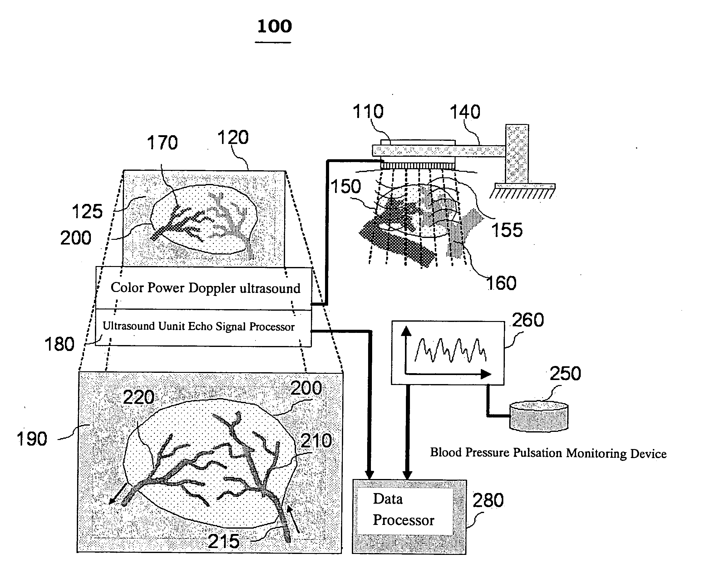
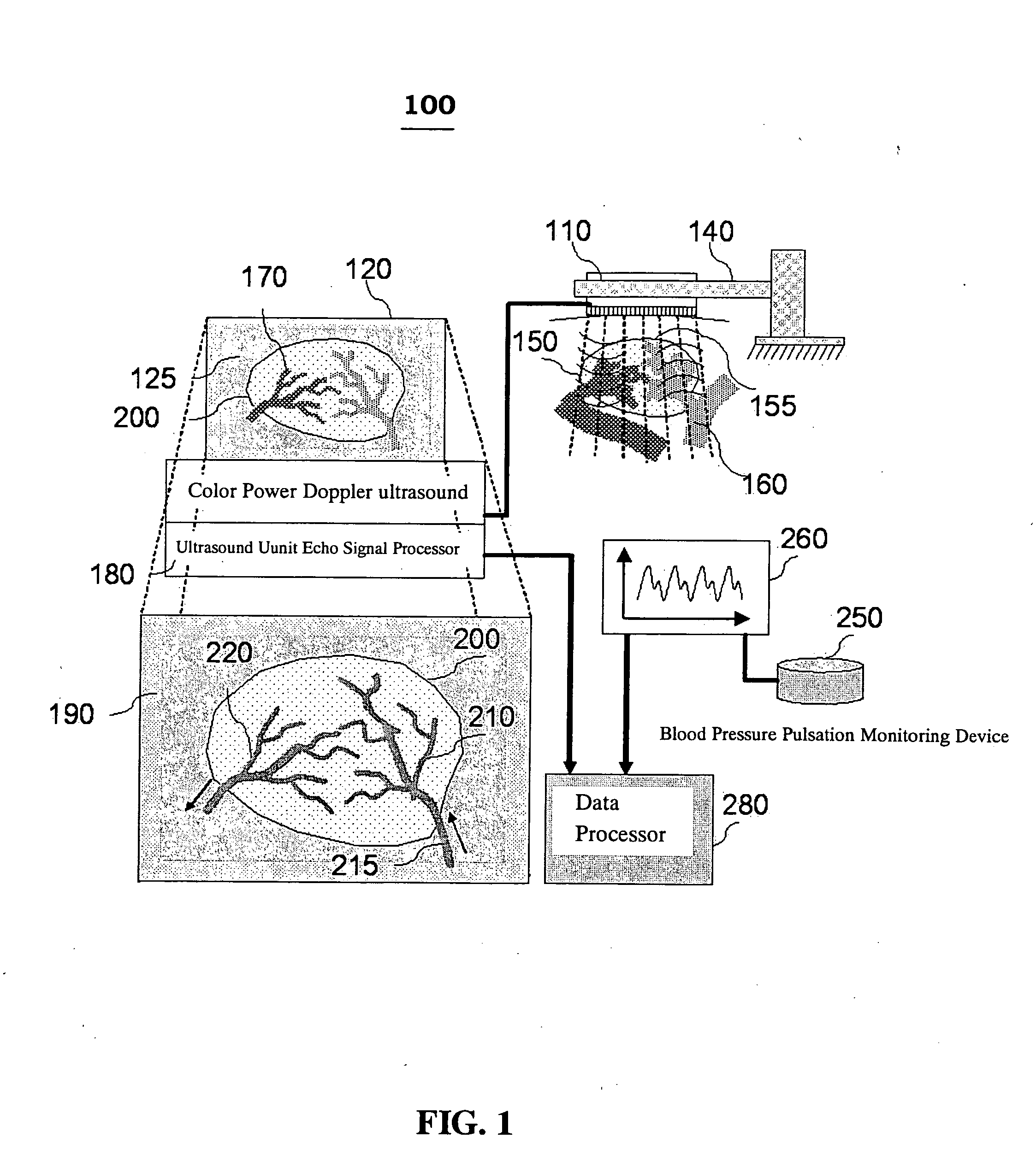
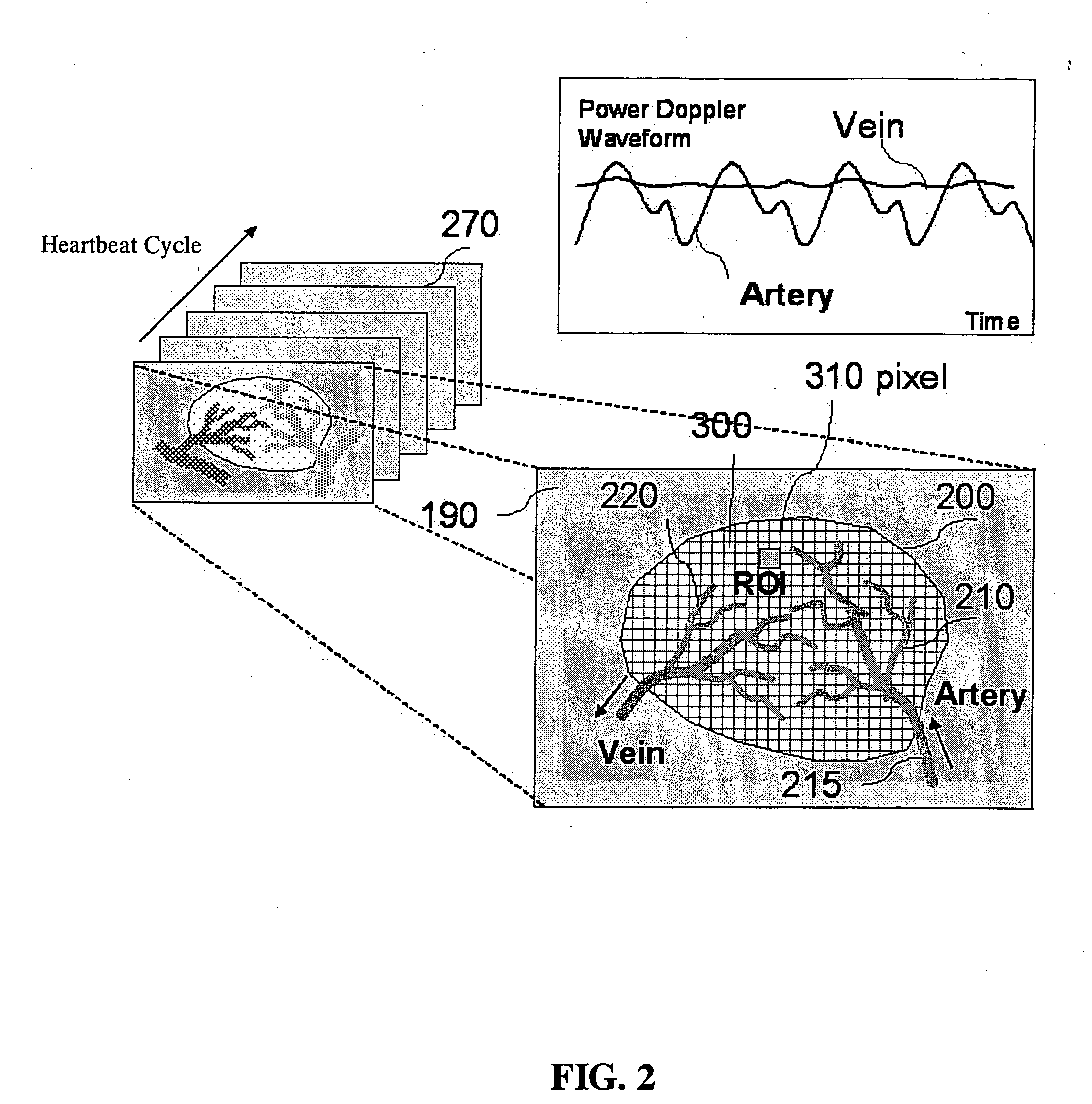
[0061]FIG. 4 is an embodiment showing a diagnosed result using the method of the present invention in breast tumor detection. The sequential Power Doppler images of the breast tumor are shown in the bottom. Middle square represents the margin of region of interest (ROI) and cross-section of blood vessel which is contoured by Power Doppler ultrasound. Upper right corner shows the results expressing arterial vessel nests and venous vessel nests in the tumor ROI cross-section distinguished with arrows by Power Doppler variance waveform analysis (upper right corner). The present invention uses the flow ultrasounic imaging analysis to study the interaction between tumor and pressure of supplying artery, to distinguish the PDVI arterial and venous regions in tumor, and to effectively calculate the degree of angiogenesis in tumor. The neovascular density ratio in tumor detected with ultrasound can be helpful in disease subtype classification, in stratifying patients for either enhance the ...

example 2

[0062]FIG. 5 is an embodiment showing a diagnosed result using the method of the present invention in kidney blood perfusion detection. The vessel pixels contributed to tumor differential vascularity index (TDVI) area are also shown. (A) represents the ultrasonic Power Doppler image during maximal systolic stage; (B) shows the Power Doppler pixels in tumor region which are higher than energy threshold during maximal systolic stage and lower than energy threshold during maximal diastolic stage; and (C) shows the difference of TDVI area in red scale. The pixel location calculated with TDVI represents a group of blood vessels with low perfusion (the reflection signals of blood flow in the location will appear in systolic phase but disappear in diastolic phase). New blood vessels of tumor have small vessel diameters, low perfusion, thin and soft wall, which have made TDVI a good indicator for the area ratio of new blood vessels. TDVI of the invention can further be used to represent the...

example 3

[0063] Thyroid cancer can be divided into 4 groups according to the tissue malignancy, which are described respectively below: [0064] (1) Normal: tissues are sampled from another normal side of a thyroid tumor; [0065] (2) Nodule Goitar (NG): patients usually require no operative intervention but sonography detection; [0066] (3) Follicular Adenoma: lots of blood vessels proliferated in patients, the distinction between an adenoma versus a carcinoma is difficult, thyroidectomy is performed since carcinoma may be rendered from adenoma; [0067] (4) Papillary thyroid carcinoma (PTC): tumor of malignancy.

[0068] A total of 53 patients with tumor are scanned with Doppler ultrasound in the tumor regions non-invasively. The CDVI, FMBV, PDVI indices from common use and the TDVI index in the present invention are analyzed to determine the degree of abnormality and aggressiveness of the cancer cells

[0069] The tumor tissue sections are collected invasively and stained for endothelial cell CD34 a...


PUM


Abstract

Disclosed is a method for detecting the degree of malignancy in tumors noninvasively, which comprises the steps of: using a Power Doppler ultrasound unit to scan a tumor and capture sequential color imagines in a complete heartbeat cycle, and choosing an area of interest (AREA_ROI) from the images; labeling pixels reflecting signals of bloodflow in the imagines during one heartbeat cycle to contour an area of tumor blood vessels (AREA_vessel); calculating a difference of PDVI between maximal systolic pressure and diastolic pressure during the heartbeat cycle to obtain tumor differential vascularity index (TDVI), in which PDVI is the ratio obtained by dividing pixels of AREA_vessel by a total area in the section of AREA_ROI; and determining the degree of malignancy by the TDVI. The method of the present invention can be applied to monitor the response of tumor to clinical treatment.

Description

BACKGROUND OF THE INVENTION [0001] 1.Field of the Invention [0002] The present invention relates to a method for assessment of tumors, especially relates to a novel method of non-invasive measurement of tumor hemodynamic parameters to detect the degree of malignancy in tumors. [0003] 2. The Prior Arts [0004] Tumor blood flow plays an important role for tumor growth and metastasis from the view of tumor metabolism. It's also crucial to the selection of optimal therapy and a marker for response to therapy. Jain reviewed the current knowledge of physical parameters for normal, benign and malignant neoplastic tissues in Cancer Research, volume 48(10): 2641-58, 1988. Non-invasive techniques for assessment of tumors like magnetic resonance imaging (MRI), ultrasound B-Mode Image are known for imaging tumor sizes, structures of blood vessels in tumors, vascular network of tumor, integrity of tumor surface, resistance indices such as pulsed index (PI) and resistance index (RI), and so on. Th...

Claims


Application Information

Patent Timeline
IPC IPC(8): A61B8/00
CPCA61B5/021A61B8/08A61B8/04
Inventor SHAU, YIO-WHAHSIEH, FON-JOUCHANG, KING-JENPAO, SUN-HUACHEN, CHIUNG-NEIN
Owner IND TECH RES INST
Why Eureka
Try Eureka