Shape-memory port-access tube

a technology of memory port and access tube, which is applied in the field of memory port access tube, can solve the problems of rescheduled surgery, large administrative, professional and emotional cost, and difficulty in rescheduling surgery, so as to facilitate the withdrawal of the tube, reduce the diameter of the tube, and improve the ability to travers

Inactive Publication Date: 2006-11-09
NAPIER BRADFORD
View PDF31 Cites 25 Cited by
  • Summary
  • Abstract
  • Description
  • Claims
  • Application Information

AI Technical Summary

Benefits of technology

[0023] A NaPier tube is typically used in conjunction with an endoscope with an outside diameter small enough to be inserted in the axial lumen of an unactivated NaPier tube. For instance, a 1.8 mm (e.g., for pediatric endoscopy) to 3.8 mm (e.g., for adult endoscopy) outside diameter bronchoscope is used with NaPier endotracheal tubes having a slightly larger axial lumen diameter than the bronchoscope's outside diameter. Before insertion of the NaPier tube into a passage, the operator inserts the endoscope into the axial lumen of the NaPier tube until the distal end of the endoscope is aligned with the distal end of the NaPier tube. The operator inserts the NaPier tube into the proximal opening of the passage, manipulates the navigational controls on the proximal end of the endoscope based on the images of the passage provided by the endoscope, and progressively feeds the tube into the passage until the distal ends of the endoscope and NaPier tube, still aligned, reach the desired location (“target”) in the passage. The NaPier tube is activated and the endoscope is then typically withdrawn. The activated NaPier tube is used for examination, diagnosis, anesthesia, surgery, therapy, or other procedure, and after completion of the procedure(s), the operator withdraws the NaPier tube from the passage by pulling on the proximal end. Pulling on the proximal end of the tube generates an elongating tensile force that causes the shape-memory element to reduce its diameter, which facilitates withdrawal of the tube.
[0024] Various embodiments of the NaPier tube are adapted for use in specific passages. For instance, a NaPier ET tube can be made with an inflatable cuff near the distal end; when inflated by a pressurized gas or fluid introduced into a longitudinal “inflation duct” between the proximal end and the cuff, the cuff expands to provide an airtight, tracheal seal around the activated tube. A NaPier ET tube with an inflatable cuff in the distal portion of the tube is called an “inflatable cuff” NaPier ET tube. Alternatively, rather than using inflation to activate a cuff, the cuff can be formed by imparting a larger memorized diameter to a distal portion of a round shape-memory element; when the tube is activated, such distal portion expands until the inner wall of the trachea prevents further expansion, thereby providing a substantially airtight seal. A NaPier ET tube with such a larger memorized diameter in the distal portion of the tube is called an “integral cuff” NaPier ET tube. A NaPier ET tube without a cuff is called a “cuffless” NaPier ET tube. A NaPier ET tube can also be made with a longitudinal duct (“dispensing duct”) between a hub at the proximal end and a point near the distal end for administration of nebulized anesthetic and / or medicament. A NaPier ET tube typically has an unactivated axial lumen diameter of less than 4 mm, is used with a bronchoscope with outside diameter slightly smaller than the axial lumen diameter of the unactivated NaPier ET tube, and upon activation the tube (in an embodiment for use in adults) expands to an axial lumen diameter of about 7 mm and outside diameter of about 8 mm. After activation of a NaPier ET tube, the lumen of the NaPier ET tube (i.e., the airway) does not collapse when a ventilator is attached to the proximal end of the NaPier ET tube.
[0025] The NaPier ET tube enables much less traumatic placement of an endotracheal tube as a result of using a smaller diameter, unactivated tube, and especially when placed using direct (endoscopic) vision. The NaPier ET tube provides improved ability to traverse limited airways in young children and infants, or secondary to tumors, infection, or abnormal anatomy. For nasotracheal intubation, if a NaPier ET tube is used, there is no advancement of a large endotracheal tube through the nose before or after placement of the bronchoscope in the trachea. The NaPier ET tube is less traumatic to the nasal passage and provides a safer deployment; there is greatly reduced risk of bleeding and of blood obstructing the airway during placement. An unactivated NaPier ET tube is much easier to place than a standard endotracheal tube during oratracheal or, especially, during nasotracheal intubation. This means that placement in a limited passage can be done by an operator with less training than an otolaryngologist, anesthetist, pulmonologist, or other specialist. For instance, an emergency medical technician or combat medic could use a NaPier ET tube rather than perform a tracheostomy, which permanently scars the patient's throat. The NaPier ET tube satisfies the demand for a lower risk apparatus and method of endotracheal intubation, especially as an alternative to tracheostomy and to avoid the cost of failed intubations.
[0026] The NaPier tube can be selected to expand to the exact outer diameter of a standard endotracheal tube, but the thinner walls of the expandable endotracheal tube provide a larger lumen (the wall of the NaPier ET tube is typically less than half the thickness, but with no compromise in strength, compared with a standard endotracheal tube); this represents a significant advance in the art of endotracheal tubes. The larger tube lumen provided by the NaPier ET tube has reduced resistance to respiratory airflow, and the increased lumen area provides more space for insertion of instruments through the tube after the tube is activated, compared with standard endotracheal tubes.

Problems solved by technology

Endotracheal intubation can be problematic when a patient has a limited airway secondary to congenital problems, anatomic blockage (e.g., large tonsils; redundant pharyngeal mucosa; prolapse of the tongue base; tumors of larynx, pharynx or hypopharynx; post radiation edema of the pharynx), infective processes (e.g., epiglottitis, retropharyngeal abscess, or prevertebral abscess), medical conditions (e.g., obesity, general anesthesia by IV), or existing medical devices (e.g., neurosurgical stability devices, cervical collars, and halo devices).
Rescheduled surgeries incur a large administrative, professional, and emotional cost.
Moreover, cases of asphyxia after a failed intubation still occur.
Even in non-obese patients, general anesthesia induced by intravenous injection can cause the pharyngeal, hypopharyngeal, and laryngeal regions to become flaccid, which makes intubation (for ventilation and for delivery of nebulized anesthetic) problematic.
The passage from the nose or mouth to the trachea may be small, limited, or tortuous.
Placement of a traditional, rigid, oral endotracheal tube, such as commonly available endotracheal tubes made by MALLINCKRODT®, SHERIDAN®, RUSCH®, or PORTEX® (collectively called, “traditional endotracheal tubes”), in small, limited, or tortuous passages may be difficult or dangerous; the heightened risk in some cases leaves no alternative but a tracheostomy.
Traditional endotracheal tubes have an average wall thickness of 1 to 2 mm, which means that about 2 to 4 mm of the overall diameter of a tube is consumed by the structure and is unavailable for ventilation.
Such additional apparatus or means adds complexity and potential points of failure to intubation, and does not solve the problem of guidance of a port-access tube through limited passages.
The prior art lacks any enabling disclosure of a radially self-expanding endotracheal tube or port-access tube.
Self-expanding stents require very complicated, multi-element, catheter-based activation systems designed to inhibit forward movement of the catheter tip and to inhibit “jumping” of the stent while a proximal lever or grip is squeezed to retract the jacket surrounding the stent.
The existing art of expanding endotracheal tubes has not solved this problem.

Method used


Image

  • Shape-memory port-access tube
  • Shape-memory port-access tube
  • Shape-memory port-access tube

Examples


Embodiment Construction

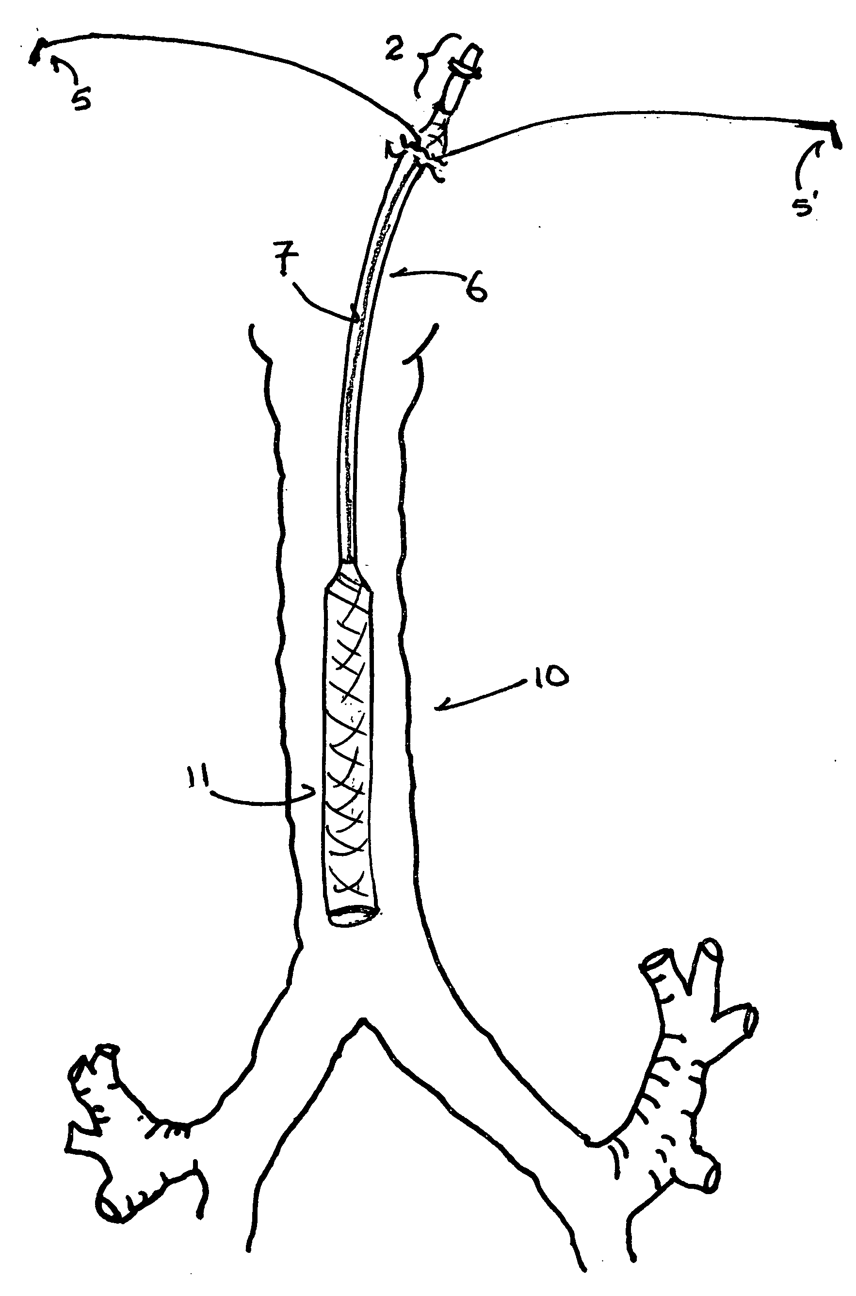
[0038] An embodiment of the NaPier tube with a round lumen after activation (“round-lumen embodiment’) is a circumferentially (i.e., radially about the longitudinal axis) self-expanding port-access tube for insertion through a port into a body passage. A basic, unactivated embodiment of a NaPier tube comprises a compressed, round, tubular, shape-memory element (“frame”) in circumferential association with a thin, expandable, elastomeric or fabric wall material and contained in a removable sheath. The removable sheath maintains the shape-memory element in a compressed state; the sheath extends around the exterior surface of the full-length of a plain tube or from the distal end to the hub, defined below, of a hubbed tube.

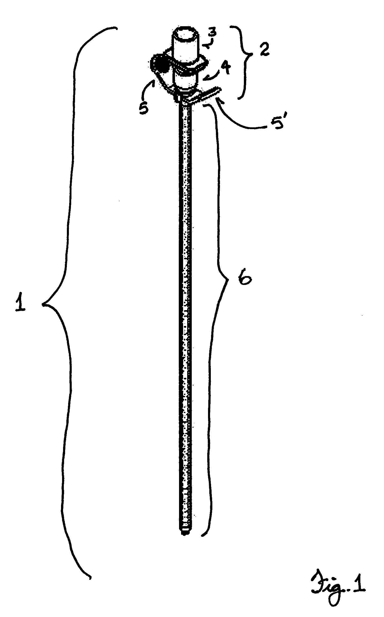
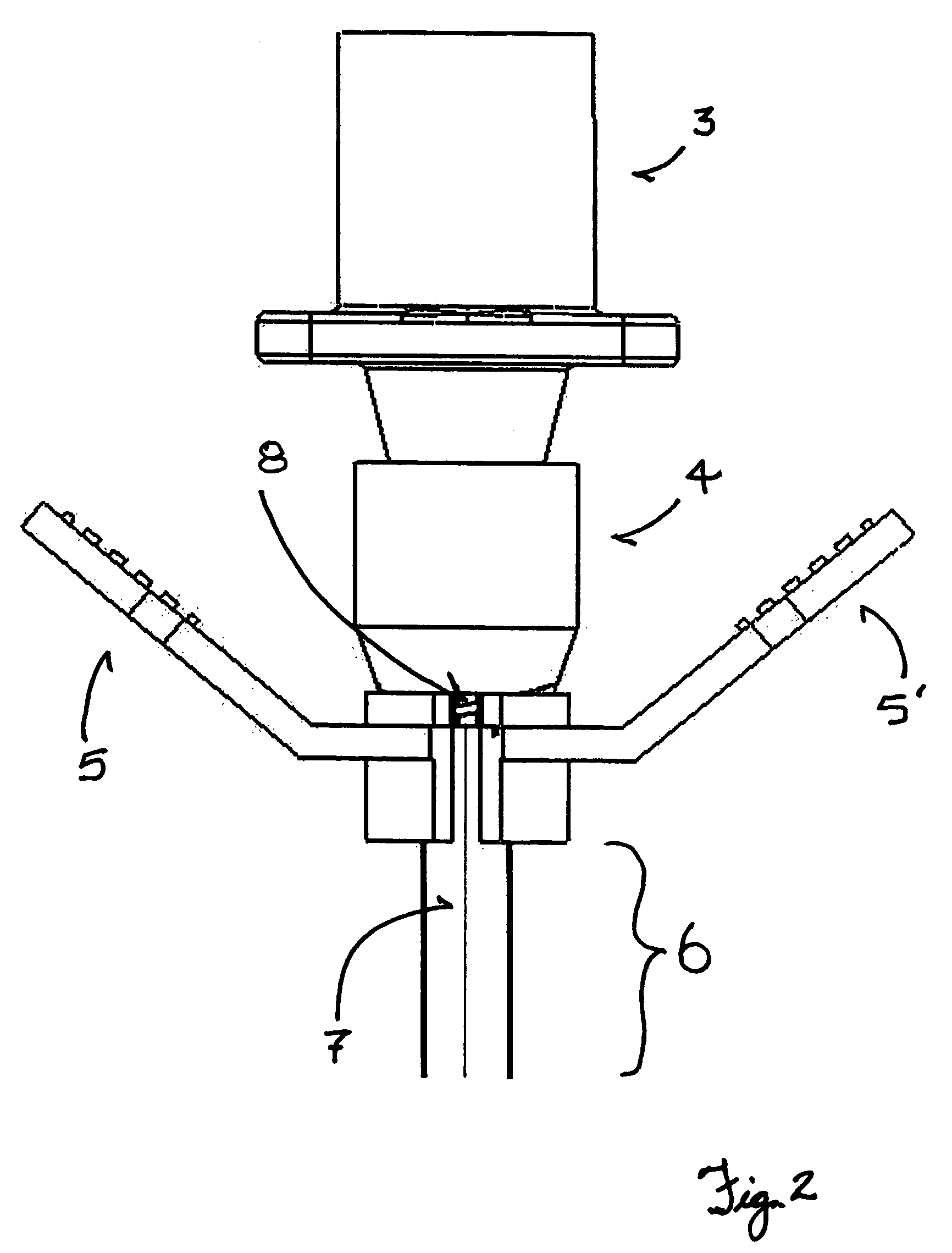
[0039]FIG. 1 shows an unactivated, hubbed NaPier tube (1) with a preferred embodiment of a hub (2) and a tear-away sheath. A fitting (3) is solidly affixed to a sleeve (4) that has external threads on its distal portion. The fitting (3) and sleeve (4) form the hub (...


PUM


Abstract

The NaPier tube is a self-expanding port-access tube for insertion into body passages. A NaPier tube is typically used with an endoscope disposed within the lumen of an unactivated tube to enable visual navigation to place the tube in a passage. A NaPier tube comprises a shape-memory element, wall material, and a removable sheath. A tear-away, removable sheath maintains the shape-memory element in a compressed state. Upon placement of the distal end of a NaPier tube in the target location in a passage, the sheath is ruptured and removed through the port and the shape-memory element expands to its memorized geometries. Embodiments of the NaPier tube are adapted by length and memorized dimensions for endotracheal intubation and for port-access procedures, such as endoscopic surgery.

Description

BACKGROUND OF THE INVENTION [0001] 1. Technical Field [0002] This invention relates to shape-memory tubes that are inserted, while compressed and sheathed, through a natural or surgical port into a natural or surgical passage in humans and animals; after placement in a passage, the compressing sheath is removed and the shape-memory tube of the invention expands to its memorized diameter. In an endotracheal embodiment of the invention, after placement of a shape-memory tube from the mouth or nose of a patient to the patient's trachea, the tube is expanded to its memorized diameter and the lumen of the expanded tube is used to facilitate respiration, observation, medication (including anesthesia), surgery, and / or therapy. Navigation of a shape-memory tube during insertion and placement is typically guided by an endoscope disposed in the lumen of the tube. The tube is easily removed after use. [0003] 2. Description of the Related Art [0004] A primary object (i.e., technical problem to ...

Claims


Application Information

Patent Timeline
Patent Type & Authority Applications(United States)
IPC IPC(8): A61F2/20A61F2/04
CPCA61B1/0058A61B1/267A61B17/3439A61B2017/00867A61M16/0404A61M16/04A61M16/0434A61M16/0463A61F2/95A61M16/0459A61M16/0486A61M16/0481
Inventor NAPIER, BRADFORD
Owner NAPIER BRADFORD
Why Eureka
Try Eureka