Invasive chemometry

a chemometric analysis and in vivo technology, applied in the field of in vivo chemometric analysis, can solve the problems of patient discomfort, sample loss or misidentification, and inconvenience of sample collection, so as to enhance the coherence of image and improve the rigidity and durability of conduits

Active Publication Date: 2006-12-07
CHEMIMAGE TECH LLC
View PDF79 Cites 53 Cited by
  • Summary
  • Abstract
  • Description
  • Claims
  • Application Information

AI Technical Summary

Benefits of technology

[0007] The invention relates to method of assessing one or more components of an animal tissue (e.g., a human tissue), either in vitro or in vivo. The method includes assessing multiple irradiated regions of an animal body to identify at least one region that exhibits a first optical characteristic of the tissue. A second optical characteristic of the component (e.g., a Raman shift characteristic of the component) is assessed for at least one identified region (and preferably for multiple regions identified as exhibiting the first optical characteristic). In this method, interfering signals from the same or a different component in a tissue other than the tissue of interest can be reduced or avoided. In some embodiments, one or more confirmatory optical characteristics of the tissue can be assessed at the identified regions to confirm that the individual identified regions correspond to the tissue of interest. The second optical characteristic of the component can, if desired, be assessed only at identified regions that exhibit the confirmatory optical characteristic.
[0010] In one embodiment, the optical conduits are rigidly fixed relative to one another (e.g., the optical conduits are fused or enclosed within an optically clear jacket). Such fixation of conduits to one another can improve the rigidity and durability of the conduits and can enhance the coherence of the image produced using the conduits, in coherent imaging systems. Significantly, the collection axes of the optical conduits need not necessarily be substantially parallel, as in an imaging device. At least in embodiments in which a coherent image of the assessed body regions is not required, there need not be any particular relationship among the collection axes of the conduits. The axes can be substantially parallel, in dispersed parallel bundles, or irregularly arranged. Because the first and second optical characteristics are collected using the same optical fiber for any particular body location, there need be no particular geometric arrangement among the conduits.

Problems solved by technology

Such invasive techniques have disadvantages including discomfort to the patient during sample collection, inconvenience of sample collection, and the possibility that collected samples can be lost or misidentified.
Discomfort and inconvenience are magnified in situations in which frequent or regular sample collection is required, such as with blood glucose determinations for diabetic patients.
However, each of these has certain disadvantages and limitations.

Method used


Image

  • Invasive chemometry
  • Invasive chemometry
  • Invasive chemometry

Examples


example 1

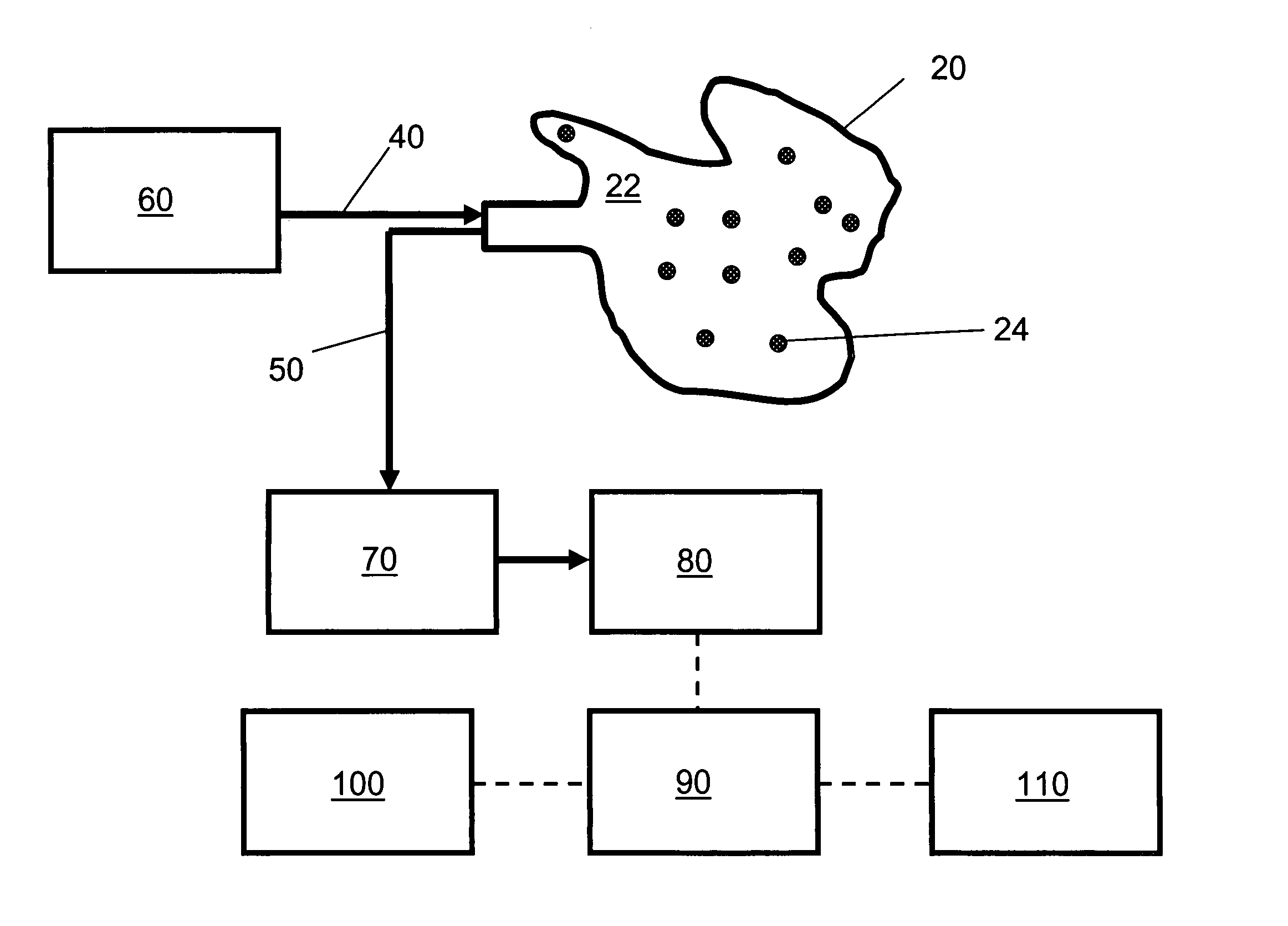
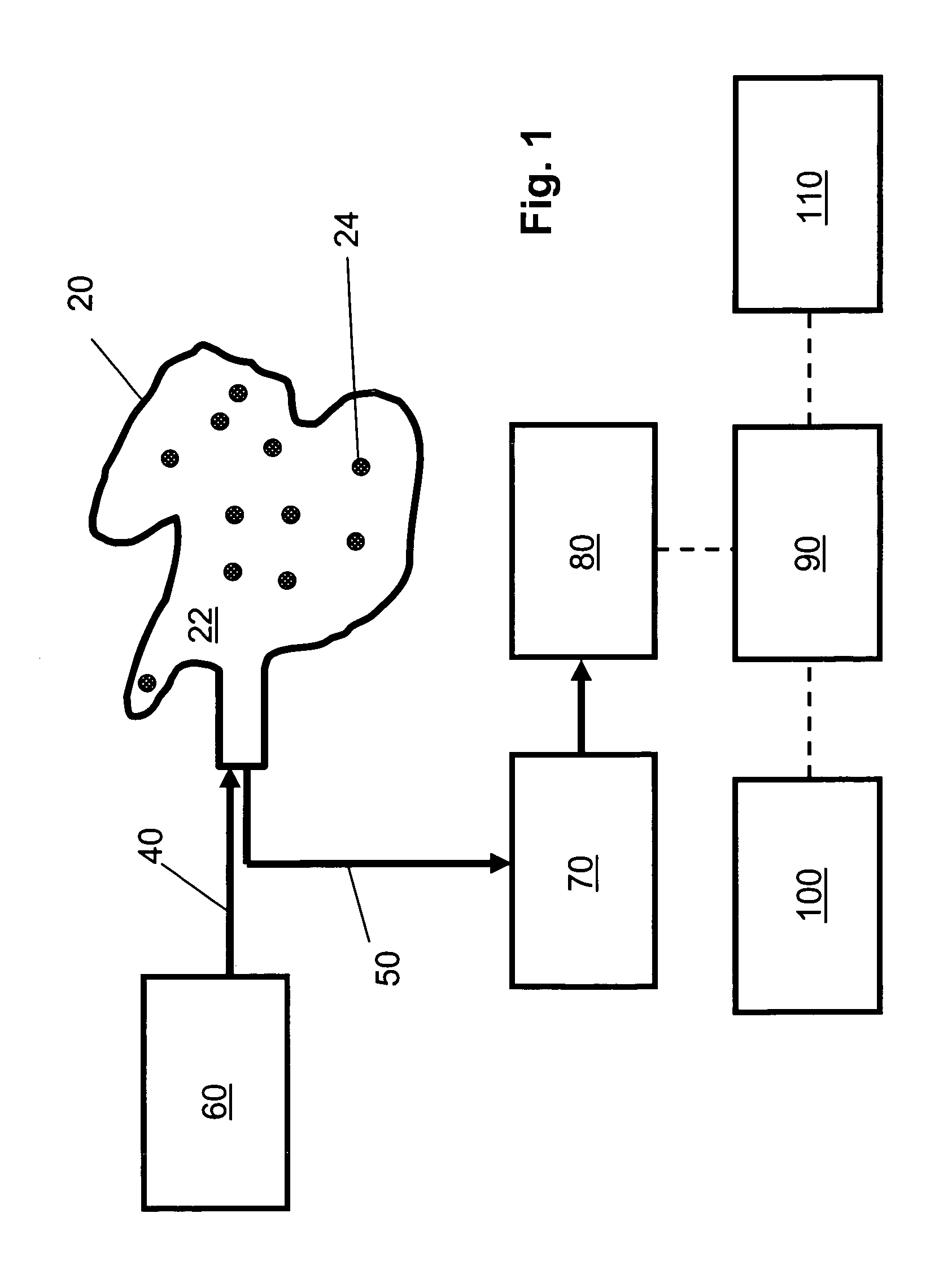
[0128]FIG. 1 is a schematic diagram of an embodiment of the non-invasive chemometric analysis device described herein. In FIG. 1, a probe 20 has a surface 22 that can be applied to a vascularized surface of an animal. Multiple holes 24 extend through the surface, through which radiation can pass (e.g., through air, a lens, or one or more optical fibers in the hole). In the embodiment shown in FIG. 1, optical fibers for illumination 40 are optically coupled with a radiation source 60 and pass through the holes 24 in the probe 20 to illuminate whatever lies adjacent the surface 22. Optical fibers 50 for collecting light reflected, emitted, or scattered from whatever lies adjacent the surface 22 are optically coupled to a detector 80, optionally by way of an optical element 70 such as a tunable filter or an interferometer. The detector 80 assesses light transmitted thereto by the optical fibers 50 to determine an optical property corresponding to discrete regions of space adjacent the ...

example 2

[0132] Blood Glucose Determination

[0133] The invention described herein provides an integrated method to perform a non-invasive, rapid measurement of the body chemistry of a conscious or unconscious person. The method involves using a small multipoint probe inserted under the tongue or into another vascularized tissue-lined body surface or cavity. The probe can perform several measurements simultaneously (e.g., under computer control) at several points without a need to move the probe. Several features of the probe, where and how it is located and the laser excitation wavelength used, can be routinely optimized for Raman scattering assessment of glucose and other blood components. Known spectral unmixing and other software algorithms can be applied to the acquired Raman data and enhance the selectivity and sensitivity of the methods for detection and quantification of analytes. Such enhancements can improve the accuracy and reduce the time between sampling and production of an anal...

example 3

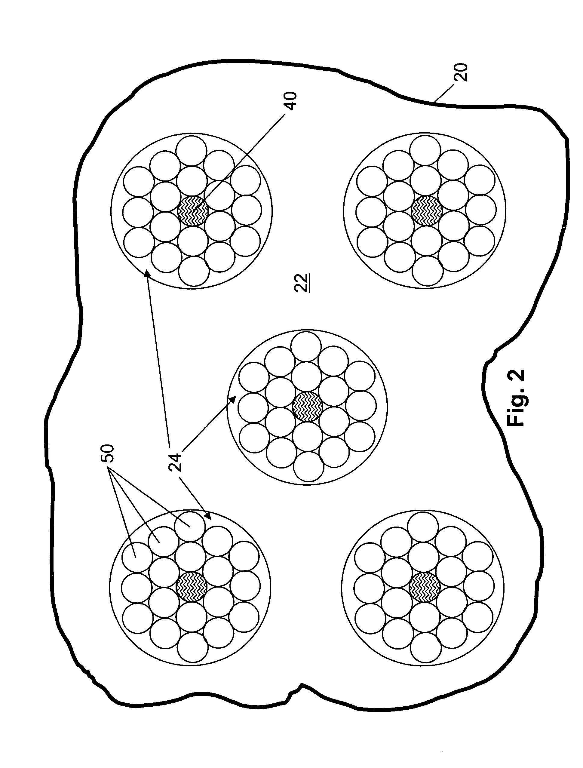
[0138] Assessment of a Tissue Component in One or More Tissues

[0139] The devices described herein assess optical properties at multiple body locations. A plurality of tissue types can therefore be assessed simultaneously using the devices, the number of assessable tissues depending on the number of tissue types that are contacted by or in close opposition to a radiation-collecting optical conduit of the device. Careful placement of an optical probe described herein by a skilled operator can increase or decrease the likelihood that a particular tissue type will be contacted by the probe. Furthermore, if desired, probes can be constructed to maximize the likelihood that the probe will contact a tissue of interest when the probe is applied to or inserted into the body of an animal.

[0140]FIG. 6 illustrates assessment of one or more tissue components in multiple tissue types. The device used in the analysis illustrated in FIG. 6 includes an elongate probe 20 that, in FIG. 6, is emplace...


PUM


Abstract

The invention relates to methods and devices for assessing one or more components of a selected tissue in an animal. The present invention permits non-invasive assessment of tissue components in a body structure containing multiple tissue types by assessing multiple regions of the animal's body for an optical characteristic of the tissue of interest and separately assessing one or more optical (e.g., Raman or NIR) characteristics of the tissue component for one or more regions that exhibit the optical characteristic of the tissue of interest.

Description

CROSS-REFERENCES TO RELATED APPLICATIONS [0001] This application is a continuation-in-part of co-pending U.S. application Ser. No. 11 / 146,458, which was filed on 7 Jun. 2005, the entire contents of which are incorporated herein by reference.BACKGROUND OF THE INVENTION [0002] The invention relates generally to the field of in vivo chemometric analyses of chemical components of cells, tissues, or organs in a living organism. [0003] Analyses of the chemical composition of blood and other tissues are among the most commonly performed medical diagnostic techniques. Typically, such analyses are performed by obtaining a sample of the tissue to be analyzed from a patient (e.g., by drawing blood or by performing a biopsy) and thereafter subjecting the sample to various analytical techniques. Such invasive techniques have disadvantages including discomfort to the patient during sample collection, inconvenience of sample collection, and the possibility that collected samples can be lost or mis...

Claims


Application Information

Patent Timeline
Patent Type & Authority Applications(United States)
IPC IPC(8): A61B5/00
CPCA61B5/14532A61B5/14546A61B5/1455G06F19/10A61B5/415A61B5/418A61B5/4519A61B5/1459G16B99/00
Inventor MAIER, JOHN S.
Owner CHEMIMAGE TECH LLC
Why Eureka
Try Eureka