Low back pain is an extremely important and costly
public health issue, accounting for a significant proportion of the health care costs of modern industrialized nations.
In general, there are two distinct but overlapping clinical problems, which often occur together.
The second is axial
joint pain, which results from painful degeneration of the joints of the spinal motion segment.
Unpinching a nerve, however, is not expected to have much affect on any
low back pain which may be coexisting.
The
disadvantage to this approach is the loss of spinal flexibility.
Additionally some authorities believe that a rigid fusion will place
increased stress on neighboring spinal motion segments, thereby accelerating the degenerative and aging process.
By nature of the
anatomy, it is difficult or impossible to see and correct any pinched nerves when operating in this approach.
Damage to internal organs in the
abdominal cavity, and damage to small nerves on the front of the
spinal column resulting in retrograde ejaculation are additional concerns.
Revision
surgery in this setting is recognized as extremely dangerous and even life threatening due to the
scar tissue that has developed around the blood vessels located in front of the spine.
Mobilizing these blood vessels the first time when the
artificial disc is being initially inserted is challenging, especially because of the wide
exposure needed to be certain that the disc is correctly positioned.
Mobilizing these same blood vessels for repeat
surgery carries a very high risk of serious complication or even death.
Interference with the circulation to the lower limbs can have devastating consequences.
Even if only a small number of anteriorly placed artificial discs require removal, the percentage of complications is anticipated to be unacceptably high.
The risk to these structures occurs during placement and surgical implantation of the device.
Should the
implant become dislodged, move, or migrate, then those structures are again at risk.
The second major risk of this type of implant is the risk of subsequent dislodgment or migration, which could also damage the neural elements.
However, there are several drawbacks to the strategy of partial disc replacement.
There is a high risk for displacement or
extrusion, since there is no firm anchoring strategy for the implants.
Therefore treatment aimed at only part of the problem is likely to be incomplete.
There are also issues relating to the geometric special orientation of the
nucleus replacement material, as well as the potential for later
extrusion or displacement of the inserted material.
The problem of
extrusion of the central core is an issue, as is the shortcomings associated with a cylindrical implant.
Typically these devices require more retraction and therefore potential injury to the neural elements, particularly when a
large size is required due to a large disc space with preserved height.
The other unavoidable aspect of the screw in cylinder design is the destruction of the endplates necessitated by the action of the screw threads.
This could result in
subsidence of the implant with subsequent collapse and narrowing of the disc space over time with attendant loss of motion.
Problems with this design include the potential for posterior extrusion of the implant.
Additionally there is little to prevent
dislocation of the shallow ball and socket articulations.
In some of these embodiments it appears that the expansion may not be readily reversible.
An additional potential issue is the difficulty of placing a single implant of this shape in the exact center of the disc.
A drawback with all of these options is the difficulty that would be encountered in removing the device, should revision and extraction ever be required.
Additionally some authorities believe that dynamic stabilization reduces the probability of accelerated degenerative changes on adjacent motion segments.
Scoliosis, or abnormal curvature of the spine, causes a rotational and side bending of the spine resulting in an abnormal shape and contour of the involved areas.
Axial
joint pain resulting from degenerative changes in the spinal motion segment is difficult to treat.
All of these devices depend on intact spinous processes, and cannot be used when these structures are small or have been surgically removed.
Although some flexibility may be permitted by these
ligament inventions, elongation is generally prohibited by these designs due to the inherent lack of elasticity.
All of these flexible straight strip, rod, and rod-like designs permit bending, but once again elongation of the flexible member is not possible.
Unfortunately the design is impractical because of the
physical space limitations in the operative field.
A similar concept involving a more complex “
inverted T” shaped bend in a flexible rod is presented in U.S. Pat. No. 6,966,910, but suffers from the same limitations.
The presence of vital vascular structures makes this device unsuitable for the anterior
lumbar spine.
Bending of the construct, however, is not allowed.


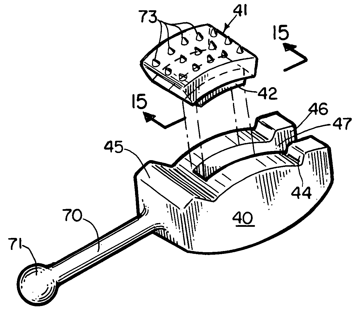
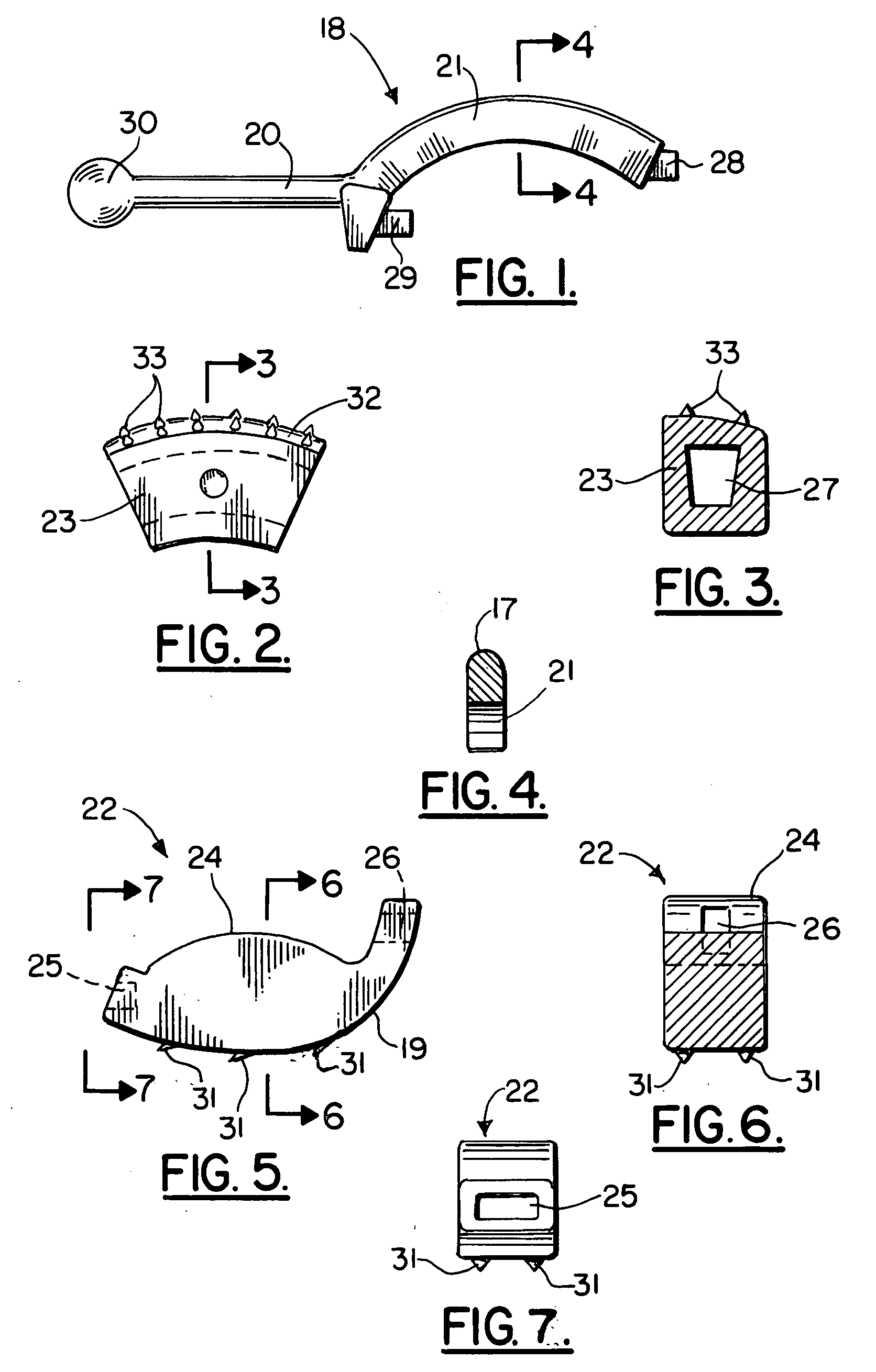
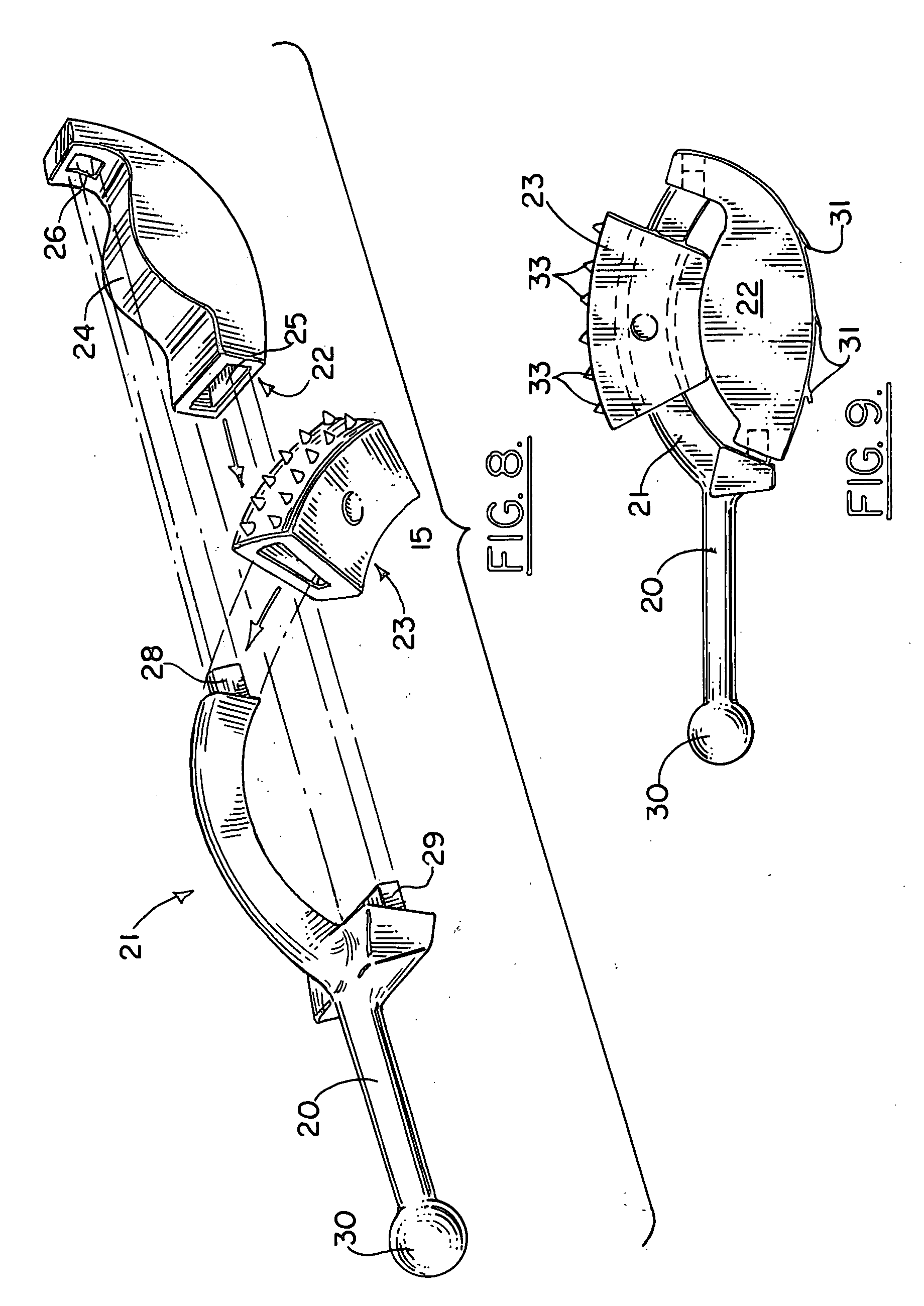