Methods and systems for treating fatty tissue sites using electroporation

Inactive Publication Date: 2006-12-28
ANGIODYNAMICS INC
View PDF98 Cites 162 Cited by
  • Summary
  • Abstract
  • Description
  • Claims
  • Application Information

AI Technical Summary

Benefits of technology

[0032] These and other objects of the present invention are achieved in, a system for treating fatty tissue sites of a patient. At least first and second mono-polar electrodes are configured to be introduced at or near the fatty tissue site of the patient. A voltage pulse generator is coupled to the first and second mono-polar electrodes. The voltage pulse generator is configured to apply sufficient electrical pulses between the first and second mono-polar electrodes to induce electroporation of cells in the fatty tissue site, to create necrosis of cells of the fatty tissue site, but insufficient to create a thermal damagin

Problems solved by technology

However, when used deep in the body, as opposed to the outer surface or in the vicinity of the outer surface of the body, it has a drawbac

Method used


Image

  • Methods and systems for treating fatty tissue sites using electroporation
  • Methods and systems for treating fatty tissue sites using electroporation
  • Methods and systems for treating fatty tissue sites using electroporation

Examples


example 1

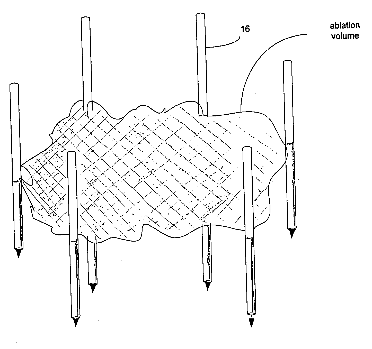
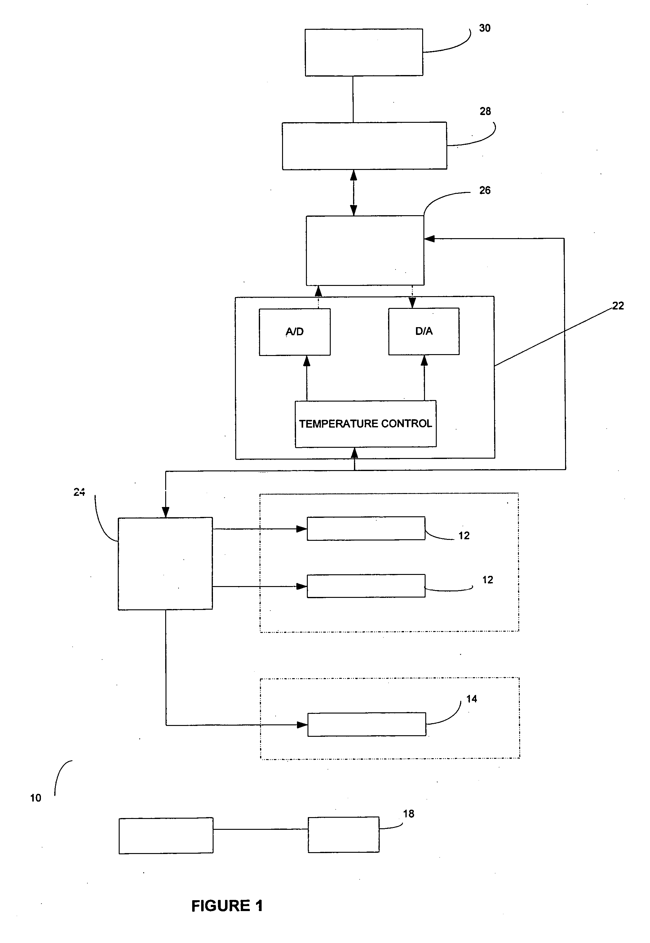
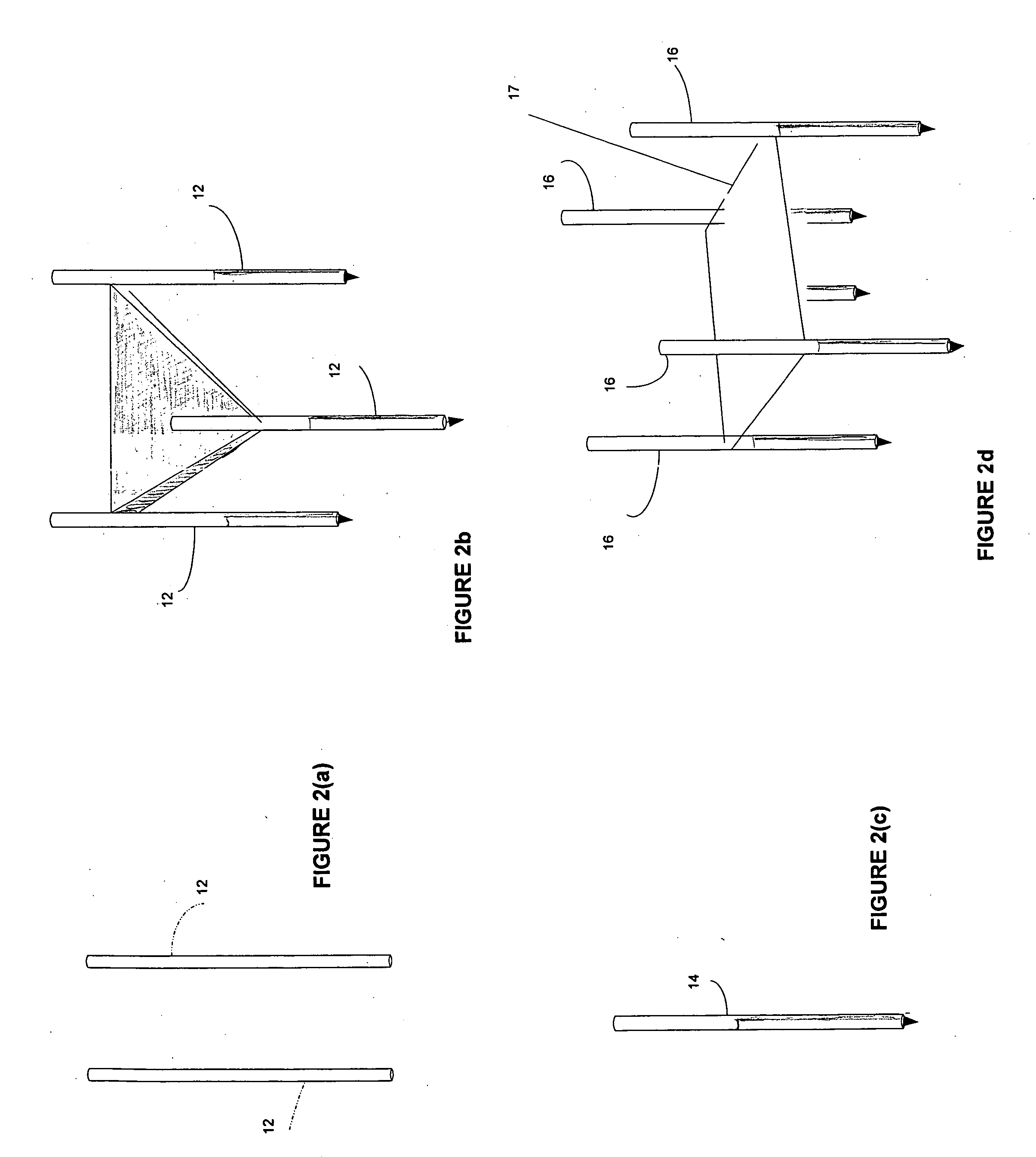
[0082] An area of the fatty tissue site is imaged. Two mono-polar electrodes 12 are introduced to the fatty tissue site of the patient. The area of the fatty tissue site to be ablated is positioned between the two mono-polar electrodes 12. Imaging is used to confirm that the mono-polar electrodes are properly placed. The two mono-polar electrodes 12 are separated by a distance of 5 mm to 10 cm at various locations of the fatty tissue site. A tumescent fluid is introduced. Pulses are applied with a duration of 5 microseconds to about 62 seconds each. Monitoring is preformed using ultrasound. The fatty tissue site is monitored. In response to the monitoring, pulses are adjusted to maintain a temperature of no more than 100 degrees C. A voltage gradient at the fatty tissue site in a range of from about 50 volt / cm to about 1000 volt / cm is created. A liposuction probe, coupled to a vacuum source, is provided and removes fatty tissue simultaneously during at least a portion of the electro...

example 2

[0083] An area of the fatty tissue site is imaged. Two mono-polar electrodes 12 are introduced to the fatty tissue site. The area of the fatty tissue site to be ablated is positioned between the two mono-polar electrodes 12. Imaging is used to confirm that the mono-polar electrodes 12 are properly placed. The two mono-polar electrodes are separated by a distance of 5 mm to 10 cm at various locations of the fatty tissue site. A tumescent fluid is introduced. Pulses are applied with a duration of about 90 to 110 microseconds each. Monitoring is performed using a CT scan. The fatty tissue site is monitored. In response to the monitoring, pulses are adjusted to maintain a temperature of no more than 75 degrees C. A voltage gradient at the fatty tissue site in a range of from about 50 volt / cm to about 5000 volt / cm is created. A liposuction probe, coupled to a vacuum source, is provided and removes fatty tissue after the electroporation. A volume of the fatty tissue site undergoes cell ne...

example 3

[0084] An area of the fatty tissue site is imaged. Two mono-polar electrodes 12 are introduced to the fatty tissue site of the patient. The area of the fatty tissue site to be ablated is positioned between the two mono-polar electrodes 12. Imaging is used to confirm that the mono-polar electrodes 12 are properly placed. The two mono-polar electrodes 12 are separated by a distance of 5 mm to 10 cm at various locations of the fatty tissue site. Pulses are applied with a duration of about 100 microseconds each. A monitoring electrode 18 is utilized. Prior to the full electroporation pulse being delivered a test pulse is delivered that is about 10% of the proposed full electroporation pulse. The test pulse does not cause irreversible electroporation. The fatty tissue site is monitored. In response to the monitoring, pulses are adjusted to maintain a temperature of no more than 60 degrees C. A voltage gradient at the fatty tissue site in a range of from about 50 volt / cm to about 8000 vol...


PUM


Abstract

A system is provided for treating fatty tissue sites of a patient. At least first and second mono-polar electrodes are configured to be introduced at or near the fatty tissue site of the patient. A voltage pulse generator is coupled to the first and second mono-polar electrodes. The voltage pulse generator is configured to apply sufficient electrical pulses between the first and second mono-polar electrodes to induce electroporation of cells in the fatty tissue site, to create necrosis of cells of the fatty tissue site, but insufficient to create a thermal damaging effect to a majority of the fatty tissue site. The system can be incorporated into standard liposuction devices or used simultaneously to treat and remove the tissue.

Description

CROSS-REFERENCE TO RELATED APPLICATIONS [0001] This application is related to U.S. Ser. Nos. ______ (Atty Docket 42218-0002), ______ (Atty Docket 42218-0003), and ______ (Atty Docket 42218-0005), filed on the same date as the instant application, all of which applications are fully incorporated herein by reference.BACKGROUND [0002] 1. Field of the Invention [0003] This invention relates generally to electroporation, and more particularly to systems and methods for treating fatty tissue sites of a patient using electroporation. [0004] 2. Description of the Related Art [0005] Electroporation is defined as the phenomenon that makes cell membranes permeable by exposing them to certain electric pulses (Weaver, J. C. and Y. A. Chizmadzhev, Theory of electroporation: a review. Bioelectrochem. Bioenerg., 1996. 41: p. 135-60). The permeabilization of the membrane can be reversible or irreversible as a function of the electrical parameters used. In reversible electroporation the cell membrane...

Claims


Application Information

Patent Timeline
IPC IPC(8): A61N1/00
CPCA61N1/0412A61N1/327A61N1/0476A61B2018/00613
Inventor RUBINSKY, BORISONIK, GARYMIKUS, PAUL
Owner ANGIODYNAMICS INC
Why Eureka
Try Eureka