Methods and apparatus for controlling the internal circumference of an anatomic orifice or lumen

a technology of anatomic orifices and lumens, applied in the field of implantable devices and associated delivery systems, can solve the problems of affecting the effect of the lumen, affecting the normal physiologic flow of blood, and the exact amount of narrowing required for the desired effect,

Inactive Publication Date: 2007-01-18
ST JUDE MEDICAL CARDILOGY DIV INC
View PDF99 Cites 366 Cited by
  • Summary
  • Abstract
  • Description
  • Claims
  • Application Information

AI Technical Summary

Problems solved by technology

In many physiologic settings, dysfunction may result from a structural lumen, which is either too large or too small.
Often, such surgical procedures require interruption in the normal physiologic flow of blood, other physiologic fluids, or other structural contents through the orifice or structure.
The exact amount of the narrowing required for the desired effect often cannot be fully appreciated until physiologic flow through the orifice or structure is resumed.
However, the ideal prosthetic valve has yet to be designed, which attests to the elegant form and function of the native heart valve.
However, the repairs of rheumatic insufficient valves have not met with good results due to the underlying valve pathology and the progression of disease.
Chordae rupture is a common cause of mitral insufficiency, resulting in a focal area of regurgitation.
The problem encountered in mitral valve repair is the surgeon's inability to fully assess the effectiveness of the repair until the heart has been fully closed, and the patient is weaned off cardiopulmonary bypass.
This increases overall operative, anesthesia, and bypass times, and therefore increases the overall operative risks.
If the prosthesis used to reduce the annulus is larger than the ideal size, mitral insufficiency may persist.
If the prosthesis is too small, mitral stenosis may result.
Significant clinical and logistical problems attend both of these existing technologies.
In the case of the coronary sinus procedures, percutaneous access to the coronary sinus is technically difficult and time consuming to achieve, with procedures which may require several hours to properly access the coronary sinus.
Moreover, these procedures employ incomplete annular rings, which compromise their physiologic effect.
Such procedures are typically not effective for improving mitral regurgitation by more than one clinical grade.
Finally, coronary sinus procedures carry the potentially disastrous risks of either fatal tears or catastrophic thrombosis of the coronary sinus.
Similarly, percutaneous procedures which employ sutures, clips, or other devices to affix the anterior mitral leaflets to the posterior mitral leaflets also have limited reparative capabilities.
Such procedures are also typically ineffective in providing a complete repair of mitral regurgitation.
Furthermore, surgical experience indicates that such methods are not durable, with likely separation of the affixed valve leaflets.
These procedures also fail to address the pathophysiology of the dilated mitral annulus in ischemic heart disease.
As a result of the residual anatomic pathology, no ventricular remodeling or improved ventricular function is likely with these procedures.
Additionally, “gas bloat” may cause the inability to belch, a common complication of over-narrowing of the GE junction.

Method used


Image

  • Methods and apparatus for controlling the internal circumference of an anatomic orifice or lumen
  • Methods and apparatus for controlling the internal circumference of an anatomic orifice or lumen
  • Methods and apparatus for controlling the internal circumference of an anatomic orifice or lumen

Examples


Embodiment Construction

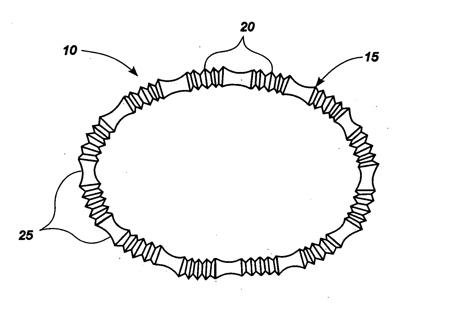
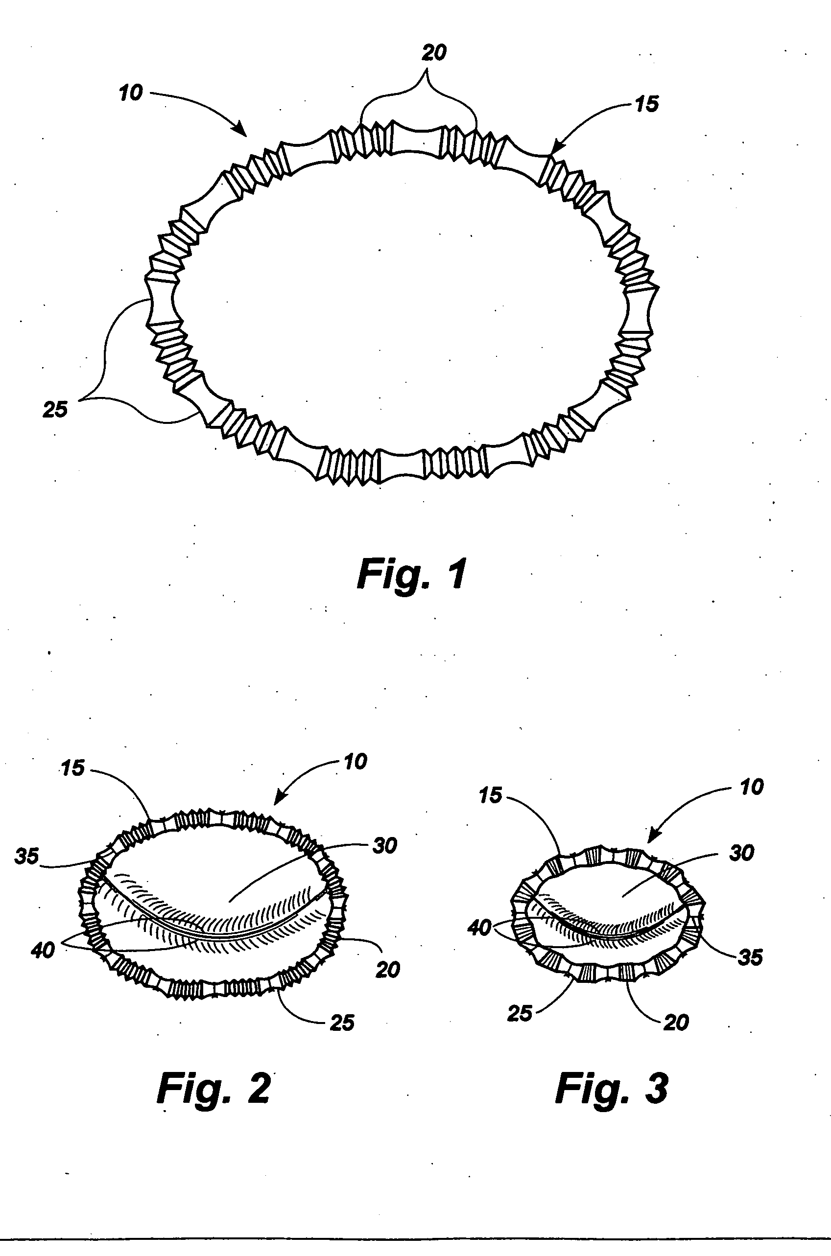
[0143] Referring now to the drawings, in which like numerals indicate like elements throughout the several views, an exemplary implant 10 comprising an implant body 15 is shown in FIG. 1. The implant body 10 may be provided in a shape and size determined by the anatomic needs of an intended native recipient anatomic site within—a mammalian patient. Such a native recipient anatomic site may be, by way of illustration and not by way of limitation, a heart valve, the esophagus near the gastro-esophageal junction, the anus, or other anatomic sites within a mammalian body that are creating dysfunction that might be relieved by an implant capable of changing the size and shape of that site and maintaining a desired size and shape after surgery. In various embodiments, the implant can be used for positioning an aortic valve, a triple A device positioning, aortic stent grafting applications, aortic endograph applications, aortic triple A stent graphs, ascending aortic aneurysm repair, for s...


PUM


Abstract

An implantable device is provided for controlling shape and/or size of an anatomical structure or lumen. The implantable device has an adjustable member configured to adjust the dimensions of the implantable device. The implantable device is housed in a catheter and insertable from a minimally invasive surgical entry. An adjustment tool actuates the adjustable member and provide for adjustment before, during or after the anatomical structure or lumen resumes near normal to normal physiologic function.

Description

CROSS-REFERENCE TO RELATED APPLICATION [0001] This application claims the benefit of U.S. Ser. No. 60 / 688,202, filed Jun. 7, 2005 and U.S. Ser. No. 11 / 390,984 filed May 27, 2006, which applications are fully incorporated herein by reference.BACKGROUND [0002] 1. Field of the Invention [0003] The present invention relates generally to implantable devices and associated delivery systems, and more particularly to an implantable device and an associated delivery system that controls shape and / or size of an anatomical structure or lumen. [0004] 2. Description of the Related Art [0005] Many anatomic structures in the mammalian body are hollow passages in which walls of tissue define a central lumen, which serves as a conduit for blood, other physiologic fluids, nutrient matter, or waste matter passing within the structure. In many physiologic settings, dysfunction may result from a structural lumen, which is either too large or too small. In most such cases, dysfunction can be relieved by ...

Claims


Application Information

Patent Timeline
Patent Type & Authority Applications(United States)
IPC IPC(8): A61F2/24A61F2/02
CPCA61F2/2448A61F2/2445A61F2250/0004A61F2/2466
Inventor CARTLEDGE, RICHARD G.LEE, LEONARD Y.FANN, JAMES I.
Owner ST JUDE MEDICAL CARDILOGY DIV INC
Why Eureka
Try Eureka