Oct-based imaging method

a catheter and imaging method technology, applied in the field of catheter-based imaging, can solve the problems of large distorted images, possible artifacts, and currently difficult for a doctor looking for the most minimal pathological changes, and achieve the effect of further improving the functional molecular process

Inactive Publication Date: 2007-02-15
SIEMENS AG
View PDF5 Cites 99 Cited by
  • Summary
  • Abstract
  • Description
  • Claims
  • Application Information

AI Technical Summary

Benefits of technology

[0008] The object of the present invention is to provide a method which enables catheter-based OCT im...

Problems solved by technology

However, the blood-filled vessels to be examined must first be rinsed out in order for imaging to be possible, since blood is not transparent with the light wavelength used.
The images (similar to those of the ultrasound imaging) are generally...

Method used


Image

  • Oct-based imaging method
  • Oct-based imaging method
  • Oct-based imaging method

Examples


Embodiment Construction

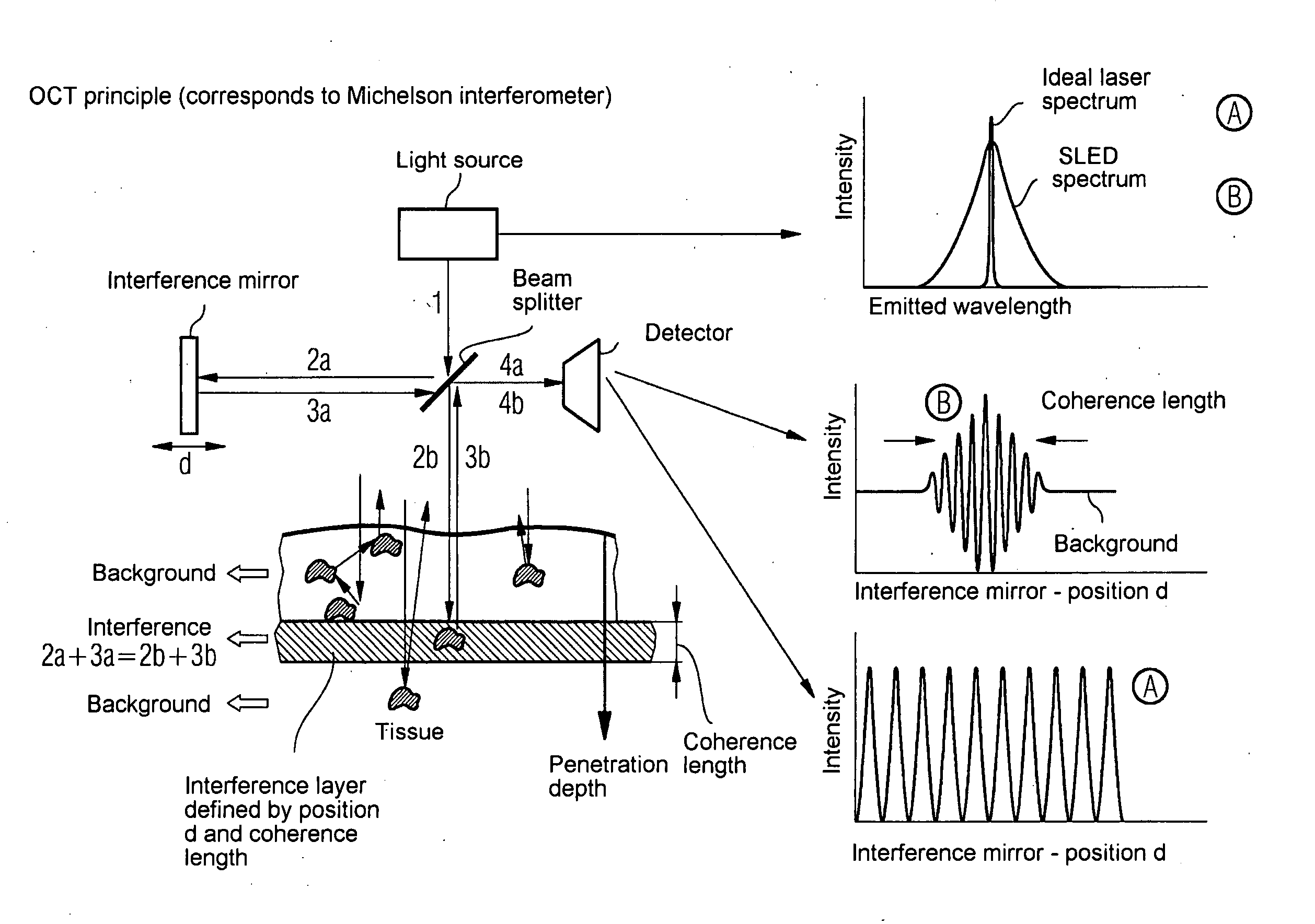
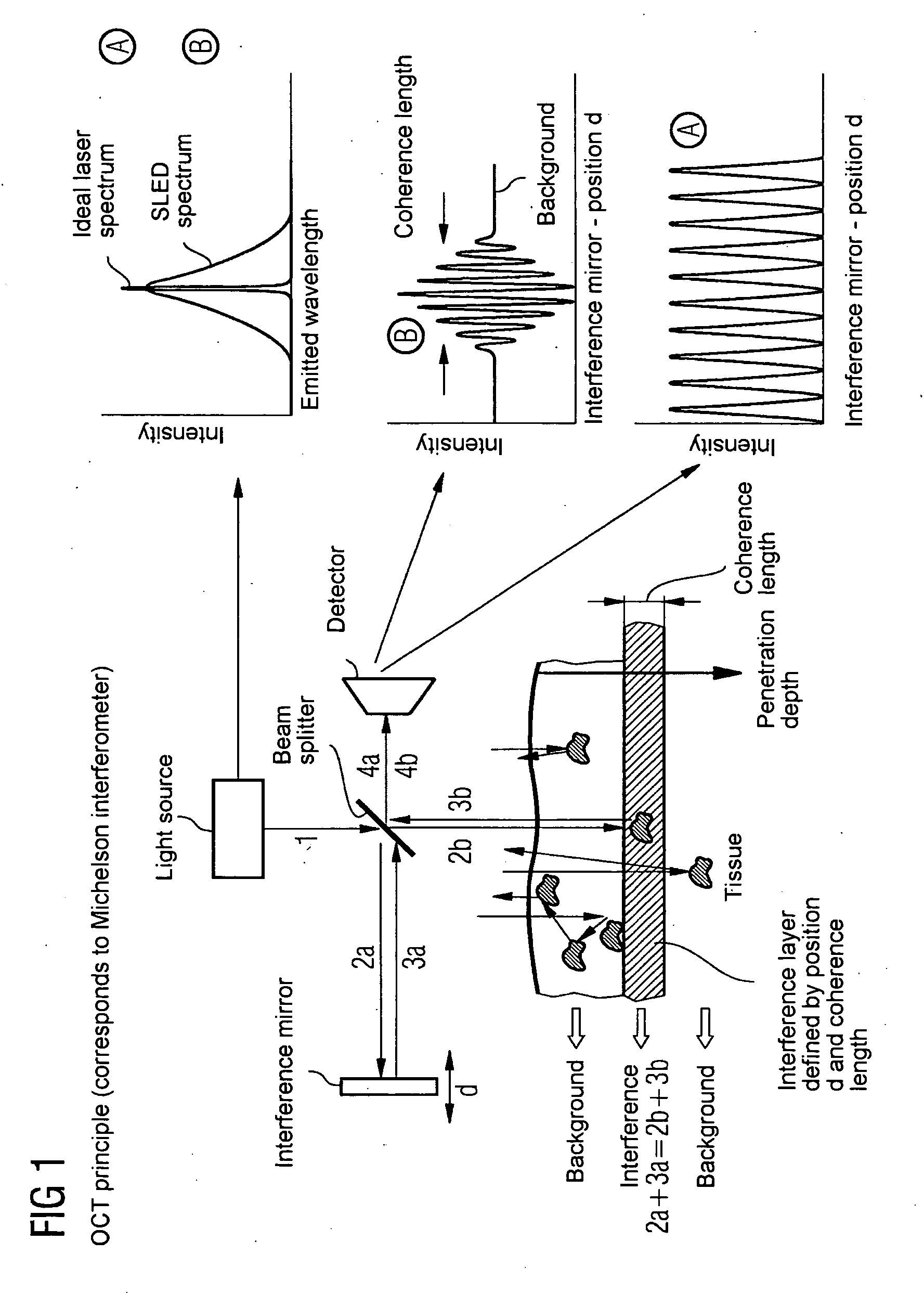
[0018] The principle of optical coherence tomography is to be explained below with reference to FIG. 1. The principle corresponds to the mode of operation of a Michelson interferometer. A light beam (e.g. laser beam) 1 emitted from a more or less coherent light source is divided by a beam splitter in the form of a semi-transparent mirror into two sub-beams 2a, 2b. The sub-beam 2a is directed onto an interference mirror, such that it strikes the beam splitter again in the form of a reflected beam 3a, penetrates said beam splitter and strikes a detector as beam 4a. In contrast the sub-beam 2b transmits the beam splitter immediately and is directed onto the tissue to be examined which comprises reflection and scatter centers, at which it strikes the beam splitter again in the form of a reflected beam 3b. This time however it is reflected by this and strikes the detector similarly as beam 4b. With the interference condition 2a+3a=2b+3b, the beam 4b coming from the interference layer int...


PUM


Abstract

The present invention relates to an imaging method with an OCT catheter for visualizing molecular functional processes in vulnerable plaques of a blood vessel of the blood vessel system of a patient, with OCT images of the contrast medium-marked vulnerable plaque being generated during continuously controlled movement of the light-emitting and light-absorbing OCT catheter head along the vulnerable plaque after the intravascular injection of a contrast medium into the blood vessel system and after the intravascular insertion of an imaging OCT catheter into the blood vessel comprising the vulnerable plaque.

Description

CROSS REFERENCE TO RELATED APPLICATIONS [0001] This application claims priority of German application No. 10 2005 029 897.4 filed Jun. 27, 2005, which is incorporated by reference herein in its entirety. FIELD OF THE INVENTION [0002] The present invention generally relates to catheter-based imaging, as used in medicine for the intravascular examination of patients. In this way, the present invention particularly relates to an OCT-based method, which enables and / or improves the visualization of functional molecular processes. BACKGROUND OF THE INVENTION [0003] Optical Coherence Tomography (OCT) is an imaging method for displaying up to 2 mm thick tissue structures with a resolution of approximately 15 μm. Similar to ultrasound diagnostics, in which acoustic structure transitions are reconstructed into images, in OCT optical structure transitions are displayed in two-dimensional cross-sectional depth images. The transitions are characterized by the tissue-specific refraction indices i...

Claims


Application Information

Patent Timeline
IPC IPC(8): A61B6/00
CPCA61B5/0066A61B5/6852A61K49/1818B82Y5/00H01L2924/0002G01B9/02091H01L2924/00
Inventor KLEEN, MARTINPFISTER, MARCUSREDEL, THOMAS
Owner SIEMENS AG
Why Eureka
Try Eureka