Devices and methods for tissue analysis

a tissue analysis and device technology, applied in the field of tissue analysis, can solve the problems of large equipment bulky and expensive to install, complicated techniques, and high cost, and achieve the effects of reducing the risk of infection, and improving the quality of tissue analysis

Inactive Publication Date: 2007-03-08
ISONEA LTD
View PDF71 Cites 98 Cited by
  • Summary
  • Abstract
  • Description
  • Claims
  • Application Information

AI Technical Summary

Problems solved by technology

These are generally expensive and involve some degree of risk which is usually associated with the use of x-rays, radioactive materials or gamma-ray emission.
Furthermore, these techniques are generally complicated and require equipment which is bulky and expensive to install and, in most cases, cannot be taken to the bedside to assess biological tissues in patients whose illness prevents them being moved.
However, the process requires sophisticated and sometimes expensive technology and cannot be used in tissues in which there is a substantial quantity of gas, such as the lung.
COPD places enormous economic burden on society.
Medical expenses for COPD patients are extremely high because of frequent visits to the emergency room, extended hospital stays and expensive medications.
The over-distension also markedly reduces the mechanical efficiency of the diaphragm.
Exercise is terminated early because of rapidly rising and unsustainable work of breathing.
The loss of alveolar tissue results in a loss of gas exchange surface area and decreases the number of capillaries available for gas exchange.
It also reduces the elastic recoil of the lung and leads to the collapse of the bronchioles and chronic airflow obstruction.
Thus, lung function is gradually lost through a reduction in gas-exchange area and in the amount of air that reaches the alveoli.
These methods are however somewhat complex and expensive, and are not suitable for intraoperative use, and are limited in resolution.
The techniques of bronchoscopy and endobronchical ultrasound can provide information about the interior surface of the lung and tissue in a specific site, but are limited to providing information on material at most a small distance below the surface.
However, that technique does not provide a means for learning about tissue in a specific location associated with an interventional device such as a bronchoscope or catheter.
Nor does it allow very well defined spatial localization of the information obtained.

Method used


Image

  • Devices and methods for tissue analysis
  • Devices and methods for tissue analysis
  • Devices and methods for tissue analysis

Examples


Embodiment Construction

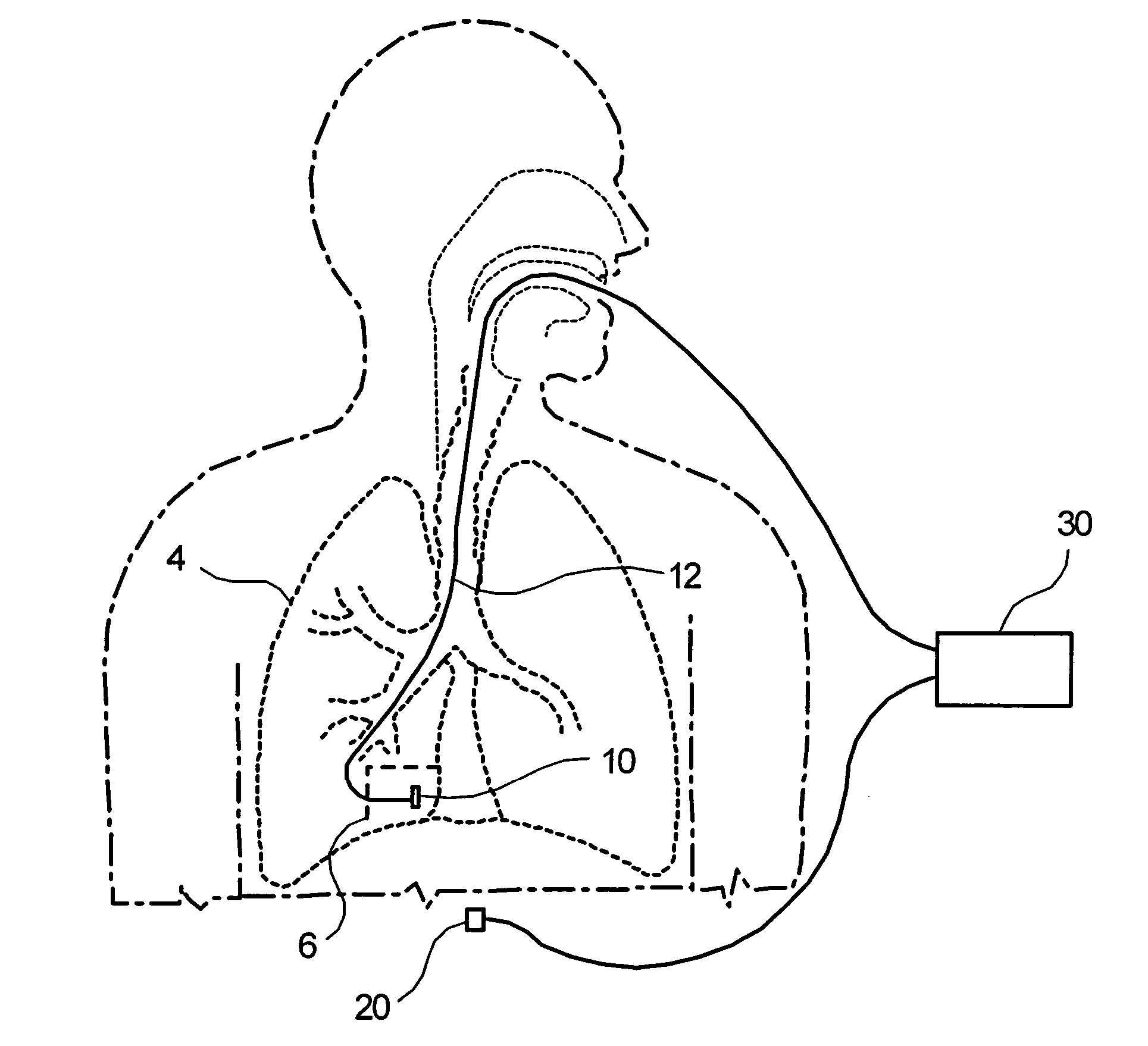
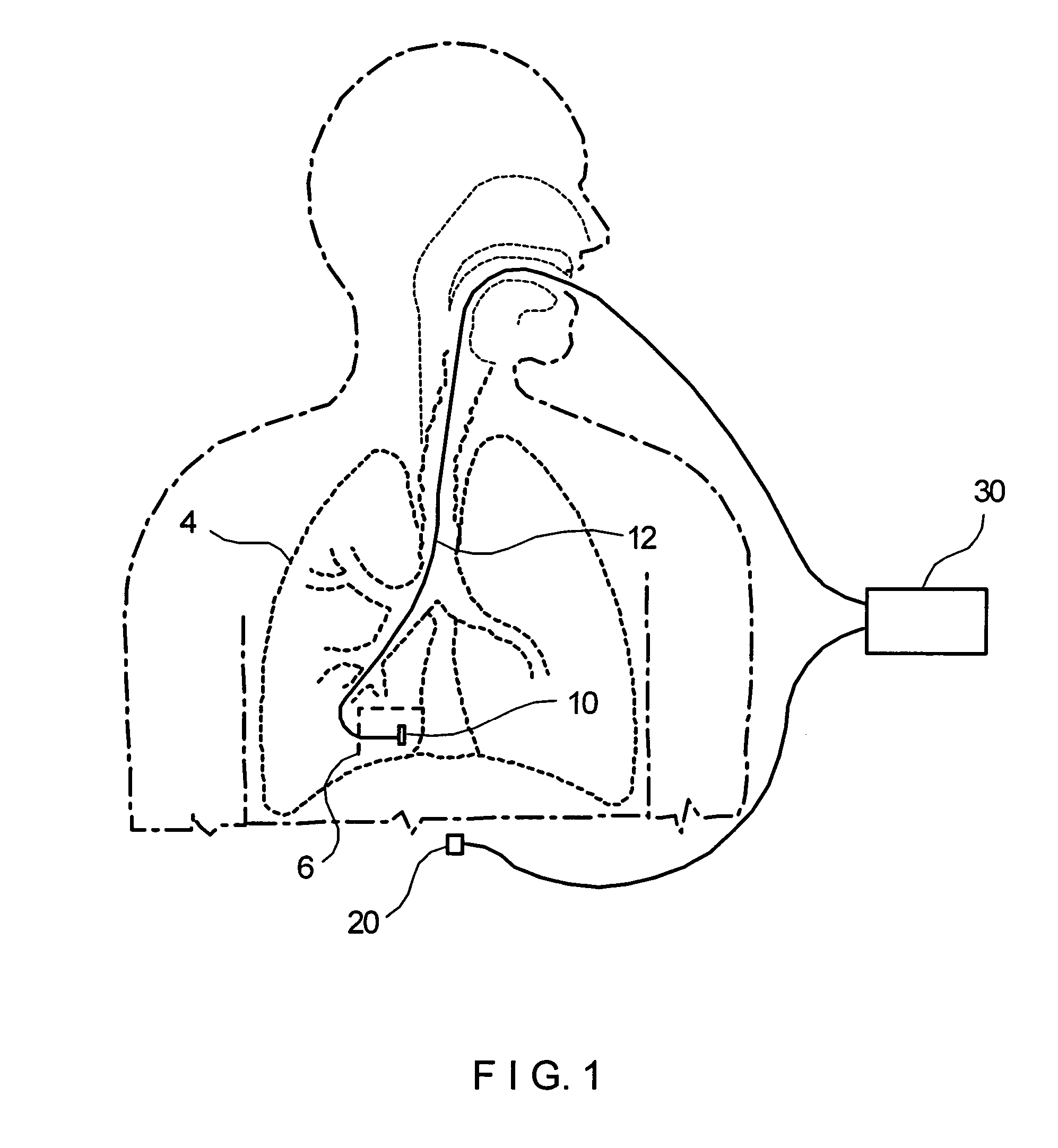
[0081] Methods and devices according to preferred embodiments of the present invention perform a localized measurement of the properties of tissue, such as lung tissue, with a view to determining whether or not the tissue is diseased, such as emphysematous or affected by cystic fibrosis, and the degree to which the disease has progressed. Additional results of this measurement would be a diagnosis of the disease stage of that portion of an organ, such as a lung.

[0082] The present description will primarily address preferred embodiments of methods and devices according to the invention in which lung tissue is assessed for the presence and stage of emphysema, or cystic fibrosis or chronic bronchitis. But as described in the previous paragraph, other methods and devices according the invention can assess the presence and stage of other diseases in other tissue or in other organs or portions of organs.

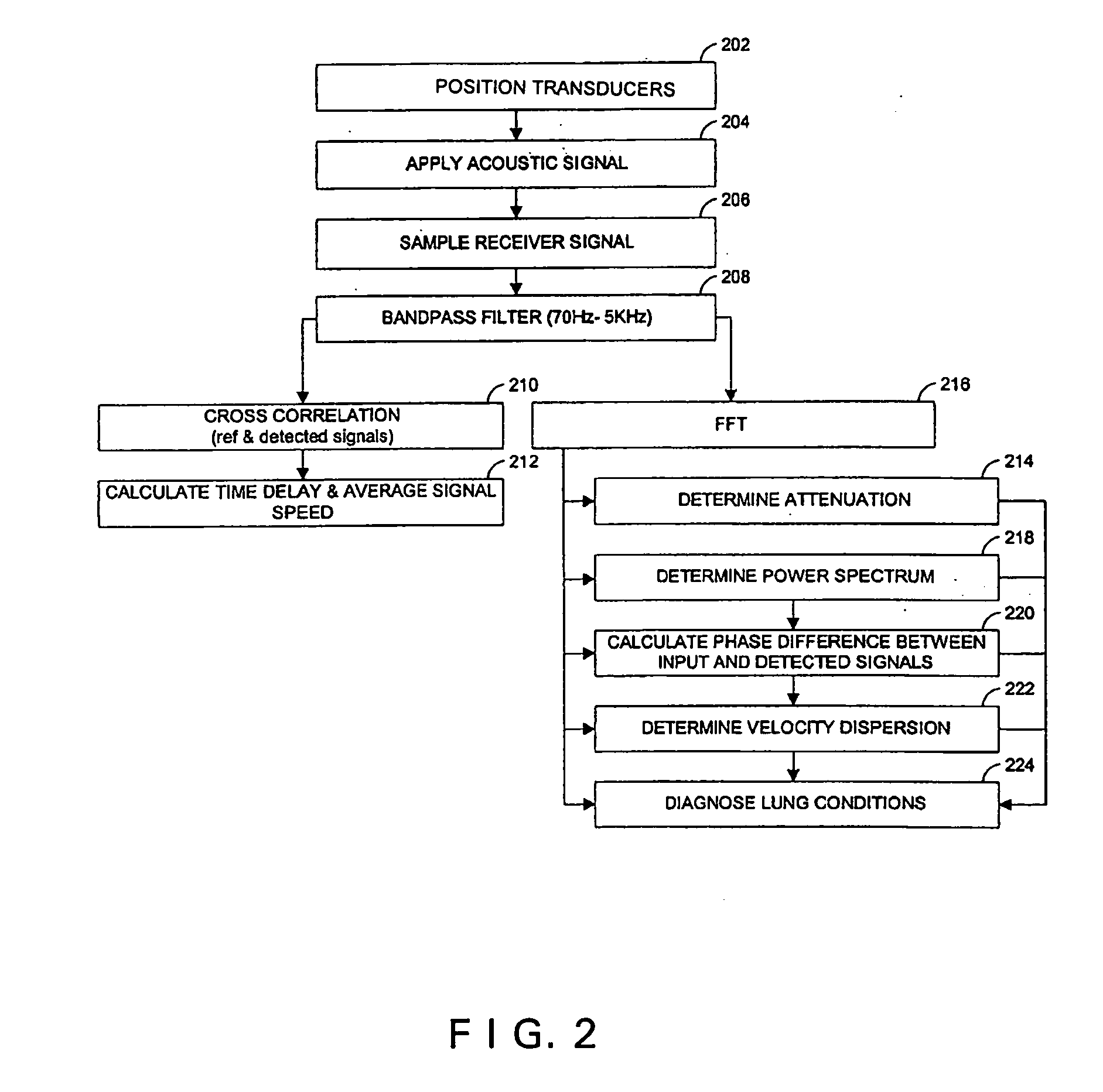
[0083] Characteristics of biological tissues can be determined by measuring the velo...


PUM


Abstract

Devices and methods are provided for assessing the condition of tissue, in particular to diagnose the location and stage of diseases such as emphysema. In one device or method according to the invention, sound is introduced into lung tissue. A portion of the sound is detected after it has passed through a portion of the lung. A sound-propagation parameter is determined by comparing a property of the introduced sound and a property of the detected sound. A region of examination of the tissue is identified, based in part on either a property of the component that introduces the sound or a property of the component that detects the sound. The condition of the tissue is assessed in the region of examination, based in part on the sound-propagation parameter.

Description

BACKGROUND OF THE INVENTION [0001] 1. Field of the Invention [0002] This invention relates assessing the condition of tissue, and more particularly to assessing whether and to what extent specific regions of the lung are affected by disease. [0003] 2. Background Information [0004] Determination of the condition of biological tissues without biopsy is useful in many circumstances: for example, when the region of tissue to be examined is inaccessible, or when the process of biopsy can cause pain or create medical complications, or be otherwise undesirable. [0005] Techniques presently used in determining the characteristics of biological tissues include x-rays, magnetic resonance imaging (MRI) and radio-isotopic imaging. These are generally expensive and involve some degree of risk which is usually associated with the use of x-rays, radioactive materials or gamma-ray emission. Furthermore, these techniques are generally complicated and require equipment which is bulky and expensive to ...

Claims


Application Information

Patent Timeline
Patent Type & Authority Applications(United States)
IPC IPC(8): A61B5/103A61B5/117
CPCA61B8/12A61B5/085A61B7/003
Inventor CARO, RICHARD G.
Owner ISONEA LTD
Why Eureka
Try Eureka