Method and apparatus for treating pelvic organ prolapse

a pelvic organ and prolapse technology, applied in the field of urogenital conditions, can solve the problems of increasing abdominal pressure, accelerating prolapse, and failure of the muscles to keep the pelvis floor supported, and achieve the effect of improving suture retention and facilitating and more secure suture attachmen

Active Publication Date: 2007-03-29
BOSTON SCI SCIMED INC
View PDF4 Cites 43 Cited by
  • Summary
  • Abstract
  • Description
  • Claims
  • Application Information

AI Technical Summary

Benefits of technology

[0034] In another embodiment, the apparatus is substantially one tape, in which the support portion is a wider center section, relative to the two end portions, in which the support and the end portions are substantially one tape. Such an embodiment would allow for easier and more secure suture attachment.
[0035] In another embodiment, the support portion is of a different material in order to provide for better suture retention.

Problems solved by technology

Increases in abdominal pressure, failure of the muscles to keep the pelvic floor supported, and damage to the ligaments and fascia all contribute to the development of prolapse.
In addition, if a woman has a hysterectomy, the vaginal angle may be altered and ligament support reduced, causing increased pressure at a more acute angle, accelerating the prolapse.
The loss of connective tissue strength may be associated with damage at childbirth, deterioration with age, poor collagen repair mechanisms, and poor nutrition.
Anterior vaginal wall prolapse causes the vaginal wall to fail to hold the bladder in place.
Mid-vaginal and high rectoceles may result from loss of lateral supports or defects in the rectovaginal septum.
Vaginal prolapse and the concomitant anterior cystocele can lead to discomfort, urinary incontinence, and incomplete emptying of the bladder.
Posterior vaginal prolapse may additionally cause defecatory problems, such as tenesmus and constipation.
However there can be complications due to vaginal wall ulceration.
The surgery may be performed through an abdominal incision or laparoscopically, however certain undesirable complications may occur.
Sacral colpopexy can also be a tedious, challenging surgical procedure, with an average procedure length of 247 minutes reported in Winters et al., Abdominal Sacral Colpopexy and Abdominal Enterocele Repair in the Management of Vaginal Vault Prolapse, Urology 56 (Suppl 6A) (2000): 55-63.
In addition, it is often required to correct multiple pelvic floor abnormalities simultaneously, which further increases the duration of the surgery.
This procedure involves attaching the vaginal vault to the sacrospinous ligament, which requires specialized skills and has the disadvantage of tending to place the vagina in an artificial anatomical position.
Although serious complications associated with sling procedures are infrequent, they do occur.
However, the smooth surface of the needles, which facilitates insertion through the tissues, prevents secure attachment of the forceps onto the needles, causing slippage or detachment of the forceps during the withdrawal procedure.
Improper placement of certain meshs is also particularly troublesome.
If the mesh is too loosely associated with its intended physiological environment, the mesh may be ineffective in supporting the urethra and treating incontinence.
Surgeons may exacerbate certain problems by improperly attempting to adjust the tension of a sling.
If insufficient adjustment force is applied, the sling will simply exhibit a memory property and return to its original, unacceptable position.
As a result, surgeons are tempted to use a great deal of force in order to loosen a sling that is perceived to be too tightly associated with its intended physiological environment.
Excessive deformation may also result in a lack of efficacy.
This tube passes through two separate regions of the patient's body with the attendant risk of cross-contamination.
The outer diameter is also relatively large (about 0.25 inches) with the attendant risk of tissue damage due to such large diameter.
The polypropylene tape supplied with the IVS Tunneller is of a thin, rectangular shape and is not believed to be optimally sized and shaped to afford concomitant procedures such as enterocele, cystocele, and / or rectocele repairs.

Method used


Image

  • Method and apparatus for treating pelvic organ prolapse
  • Method and apparatus for treating pelvic organ prolapse
  • Method and apparatus for treating pelvic organ prolapse

Examples


Embodiment Construction

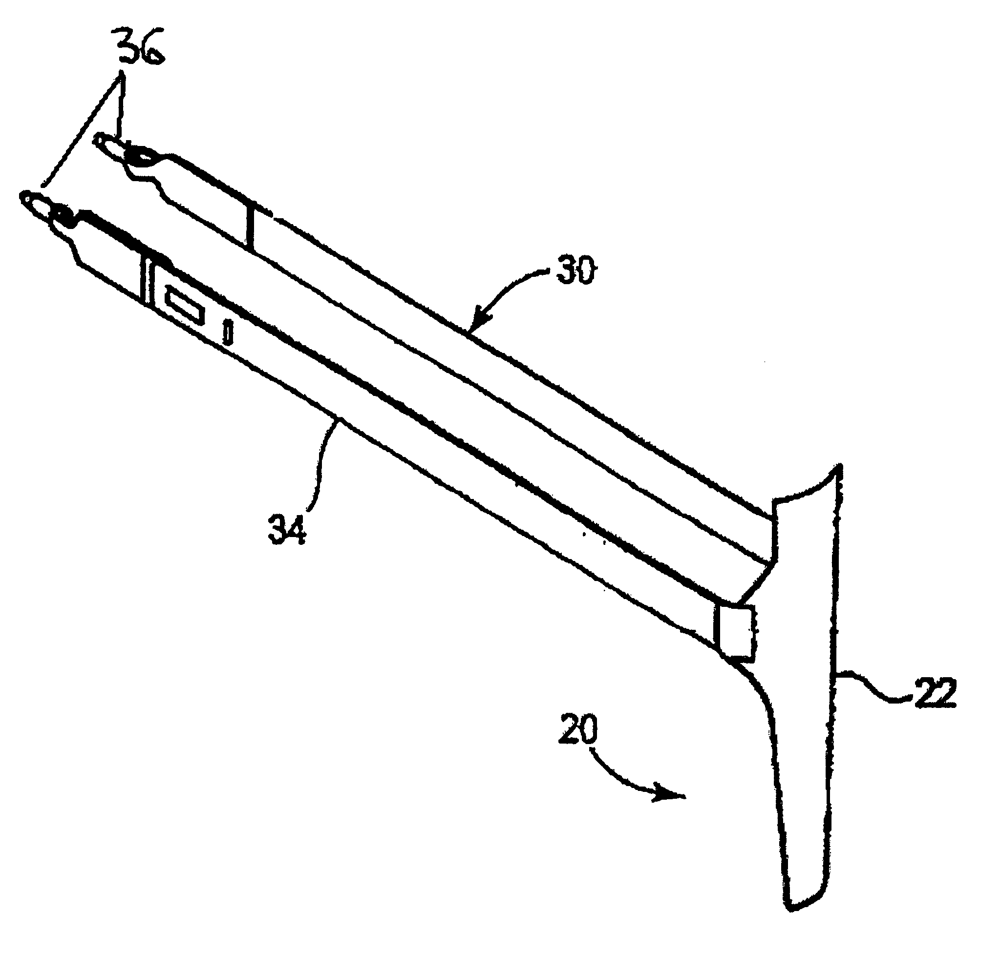
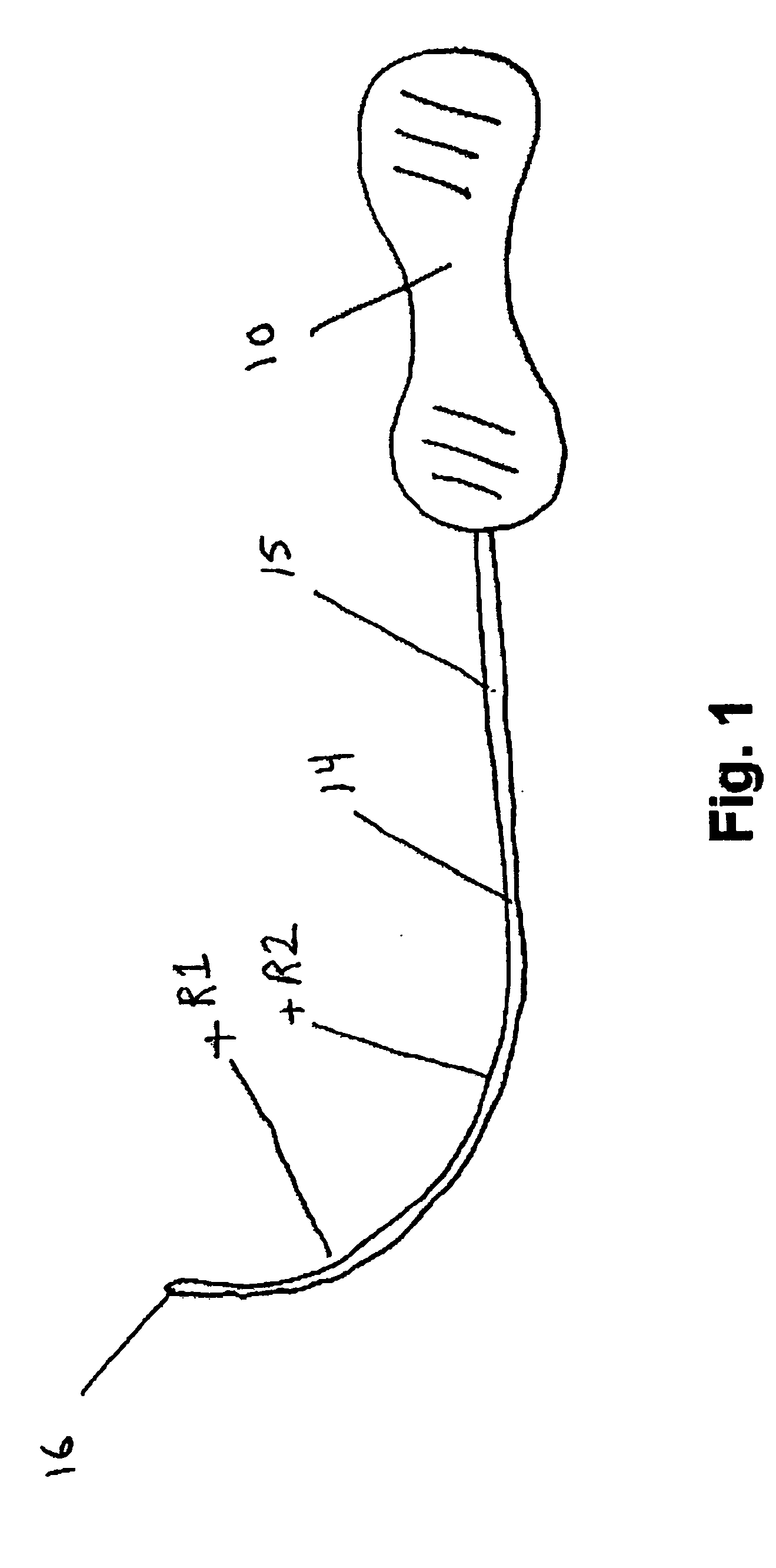
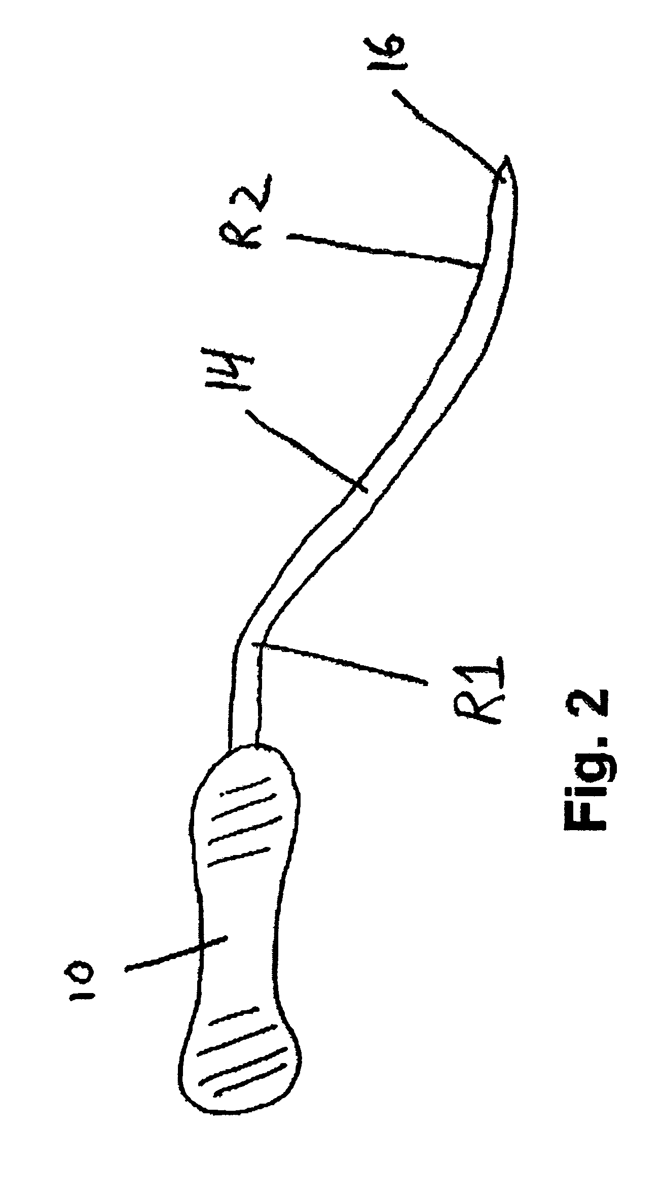
[0064] Referring now to the drawings, wherein like reference numerals designate identical or corresponding parts throughout the several views, FIG. 1 shows a needle 14 and handle 10 suitable for use in the present invention. Needle 14 terminates in a tip 16. Needle 14 comprises a generally straight section 15 near handle 10. In one embodiment, the straight section 15 shown in FIG. 1 is between about 4 inches and about 8 inches, preferably between about 5 inches and about 7 inches, more preferably between about 5.5 inches and about 6.5 inches.

[0065] The portion of needle 14 between straight section 15 and tip 16 includes a multi radii bend defined by a first radius R1 and a second radius R2, distinct from the first radius. The first radius R1 is generally between about 2 inches and about 4 inches, preferably between about 2.5 inches and about 3.5 inches. The second radius R2 is generally larger than R1. In one embodiment, R2 is between about 4 inches and about 6 inches, preferably b...


PUM


Abstract

An apparatus for treating pelvic organ prolapse in a patient is provided. The apparatus includes a support portion having first and second ends. A first elongated end portion is connected to said first end of said support portion. The first elongated end portion includes a first dilator configured to attach securely with a tip of a needle. A second elongated end portion is connected to said second end of said support portion. The second elongated end portion includes a second dilator configured to attach securely with a tip of a needle. The first and second needles include a straight portion, a tip, a first radius, and a second radius distinct from the first radius. The first radius and the second radius are disposed between the straight portion and the tip. A method and kit for said treatment is further provided.

Description

RELATED APPLICATIONS [0001] This application is a utility application claiming priority to U.S. Provisional Application 60 / 716,110, filed Sep. 12, 2005, the entire contents of which is incorporated by reference herein. This application is also a continuation in part of U.S. patent application Ser. No. 10 / 834,943 filed Apr. 30, 2004, and U.S. patent application Ser. No. 10 / 840,646 filed May 7, 2004 which are incorporated by reference herein.BACKGROUND OF THE INVENTION [0002] 1. Field of the Invention [0003] The invention relates generally to the treatment of urogenital conditions. More particularly, the invention relates to devices and surgical techniques for use in treating female pelvic organ prolapse. [0004] 2. Description of the Related Art [0005] When intra-abdominal pressure pushes the vagina outside the body, vaginal prolapse can develop. In normal circumstances, the levator ani muscles close the pelvic floor, supporting it from below while fascia a ligaments support. Increase...

Claims


Application Information

Patent Timeline
Patent Type & Authority Applications(United States)
IPC IPC(8): A61B19/00
CPCA61B17/06004A61F2250/0089A61B17/06109A61B2017/0046A61B2017/00805A61B2017/0496A61B2017/06009A61B2017/06076A61F2/0045A61F2002/3071A61F2002/30713A61F2002/30714A61F2250/0085A61F2250/0087A61B17/06066
Inventor ANDERSON, KIMBERLY A.COX, JAMES E.
Owner BOSTON SCI SCIMED INC
Why Eureka
Try Eureka