Vascular closure device

a technology of vascular closure and perforation, which is applied in the field of vascular closure devices, can solve the problems of ineffective utilization of practitioners, impaired patient comfort, and increased risk of hematoma, and achieve the effects of effective sealing of blood vessels, convenient use, and effective sealing of perforations

Inactive Publication Date: 2007-04-12
INNOVASIVE
View PDF20 Cites 132 Cited by
  • Summary
  • Abstract
  • Description
  • Claims
  • Application Information

AI Technical Summary

Benefits of technology

[0011] The present invention relates a closure device for effectively sealing a blood vessel or other body opening, and the structure and method of its introduction, application, and extraction. Embodiments of the present invention require little manual dexterity, are easy to use, and effectively seal a perforation by using three complementary methods: using grasping tines to appose the edges of the perforation together, folding the edges tightly together rather than simply pulling them together, and plugging the perforation site with a collar. Because embodiments of the present invention use three mechanisms to effectively seal a perforation, they provide an excellent seal and enable faster healing. Also, because they are particularly inexpensive to manufacture, they are especially well-suited for one-time use, making post-surgical sterilization unnecessary, thus cutting costs and increasing convenience.
[0012] It is an object of the present invention to fulfill one or more of the needs referred to above. In accordance with the principles of the present invention, this objective is obtained by providing a device and method for closing and sealing a puncture in a luminal wall. Embodiments of the present invention, in one aspect, provide a reliable and easily used device for promoting and achieving percutaneous vascular hemostasis at a perforation or puncture site in a subcutaneous bodily lumen, especially a blood vessel or a heart chamber, using a combination of sealing mechanisms to promote hemostasis in the most effective manner. In another aspect, the present invention relates to the method of using this hemostasis device to promote hemostasis at such a site.
[0014] In another embodiment, the present invention provides for a device for closing a vascular opening or other body cavity opening comprising: at least two tines including a distal end and a proximal end, wherein the tines are joined at the proximal end, and wherein the tines each have at least one barb adapted for catching tissue, and wherein the tines are deflected outward from the longitudinal axis of the tines; and a collar with an opening receiving the proximal end of the tines; wherein the collar can be moved from the proximal end of the tines towards the distal end of the two resilient tines to radially contract the tines, thereby decreasing the deflection.

Problems solved by technology

With externally-applied manual pressure, not only is patient comfort impaired, but practitioners are not being utilized efficiently.
Additionally, a risk of hematoma exists, since bleeding from the vessel may continue until sufficient clotting effects hemostasis.
Not only is direct pressure inefficient from both a medical and personnel perspective, the procedure may result in substantial reduction, if not complete arrest, of the flow of blood through the vessel.
Since thrombosis is one of the major calamities that can occur in the post-operative period, any reduction in blood flow is undesirable.
Also, external pressure application devices may be unsuitable for patients with substantial amounts of subcutaneous adipose tissue, since the skin surface may be a considerable distance from the vascular puncture site, thereby rendering skin compression inaccurate and thus less effective.
This method may entail some risk of disadvantageously introducing some of the adhesive or clotting agent into the bloodstream.
Unless pressure is applied, however, bleeding may occur around the plug into the subcutaneous tissue.
In fact, many of these techniques require a great deal of experience and manual dexterity to use successfully.
Thus, even experienced surgeons can have difficulty in using these techniques and devices.
While such devices, if properly employed, are capable of very effectively stemming blood flow, they may require a relatively high degree of dexterity to be operated properly.
Indeed, the vessel opening is often accessible through only small catheters making sutures even more difficult to tie.
Furthermore, the devices tend to be somewhat complex and expensive to manufacture, and thus are not practically employed as single use, disposable products.
Consequently, sterilization is required between uses to reduce the risk of infection, thereby increasing their cost and inconvenience.
Current devices for performing these functions suffer many of the same limitations as existing vascular closure devices.
All of these devices can potentially come loose, which would likely result in death, and there is a significant risk of thrombosis due to the implantation of foreign material.
Accordingly, even experienced surgeons may have difficulty using the devices increasing the risk of surgical complications.
In addition, existing devices are relatively expensive.

Method used


Image

  • Vascular closure device
  • Vascular closure device
  • Vascular closure device

Examples


Embodiment Construction

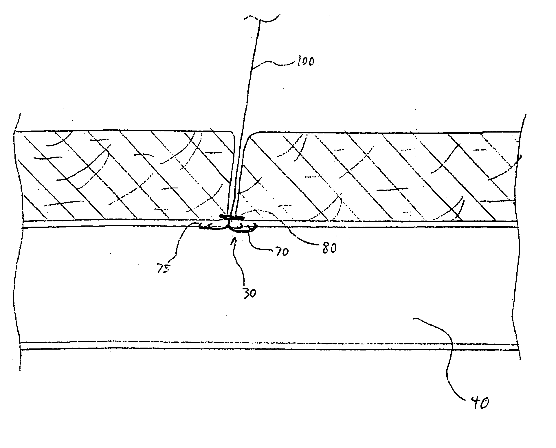
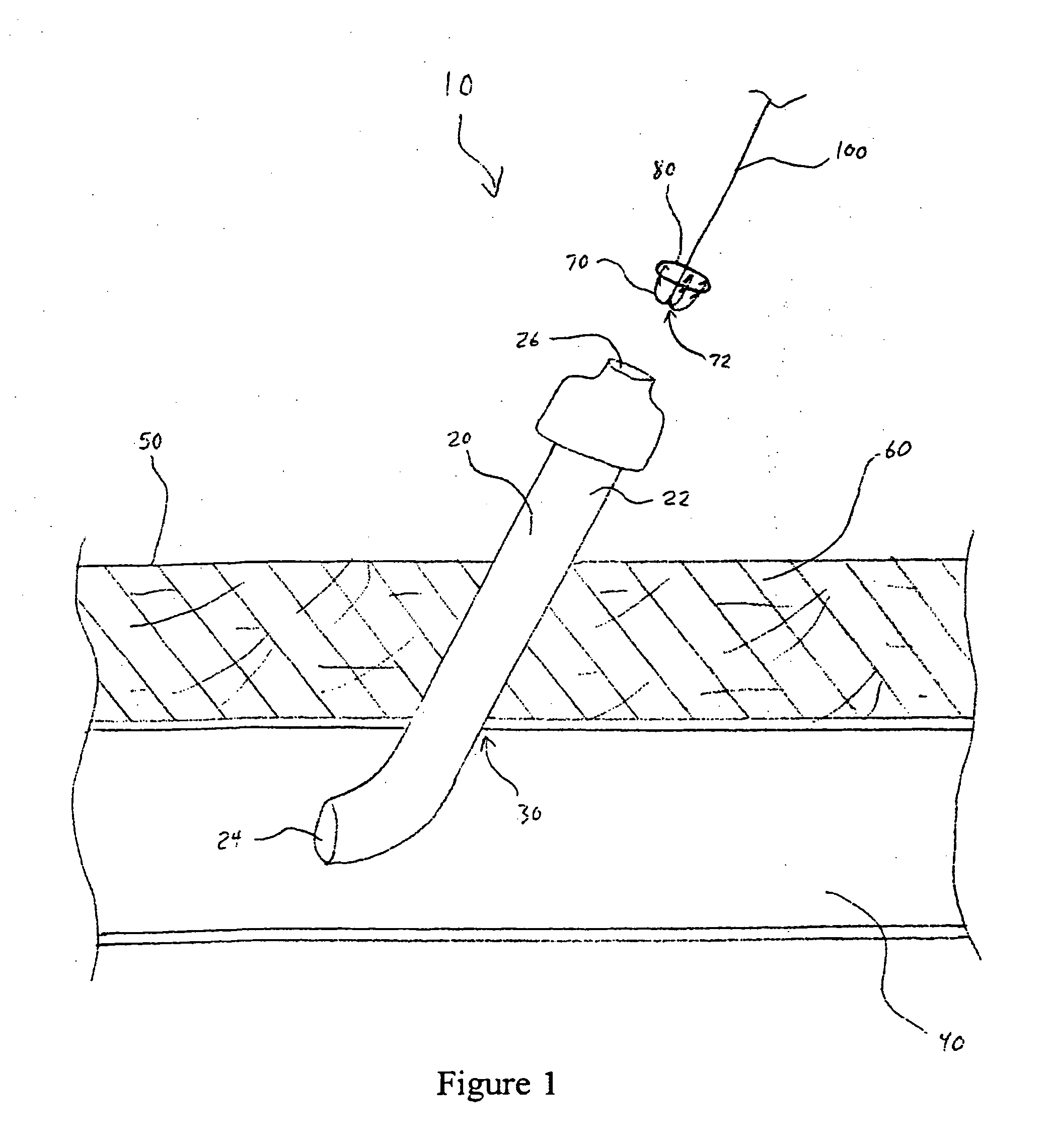
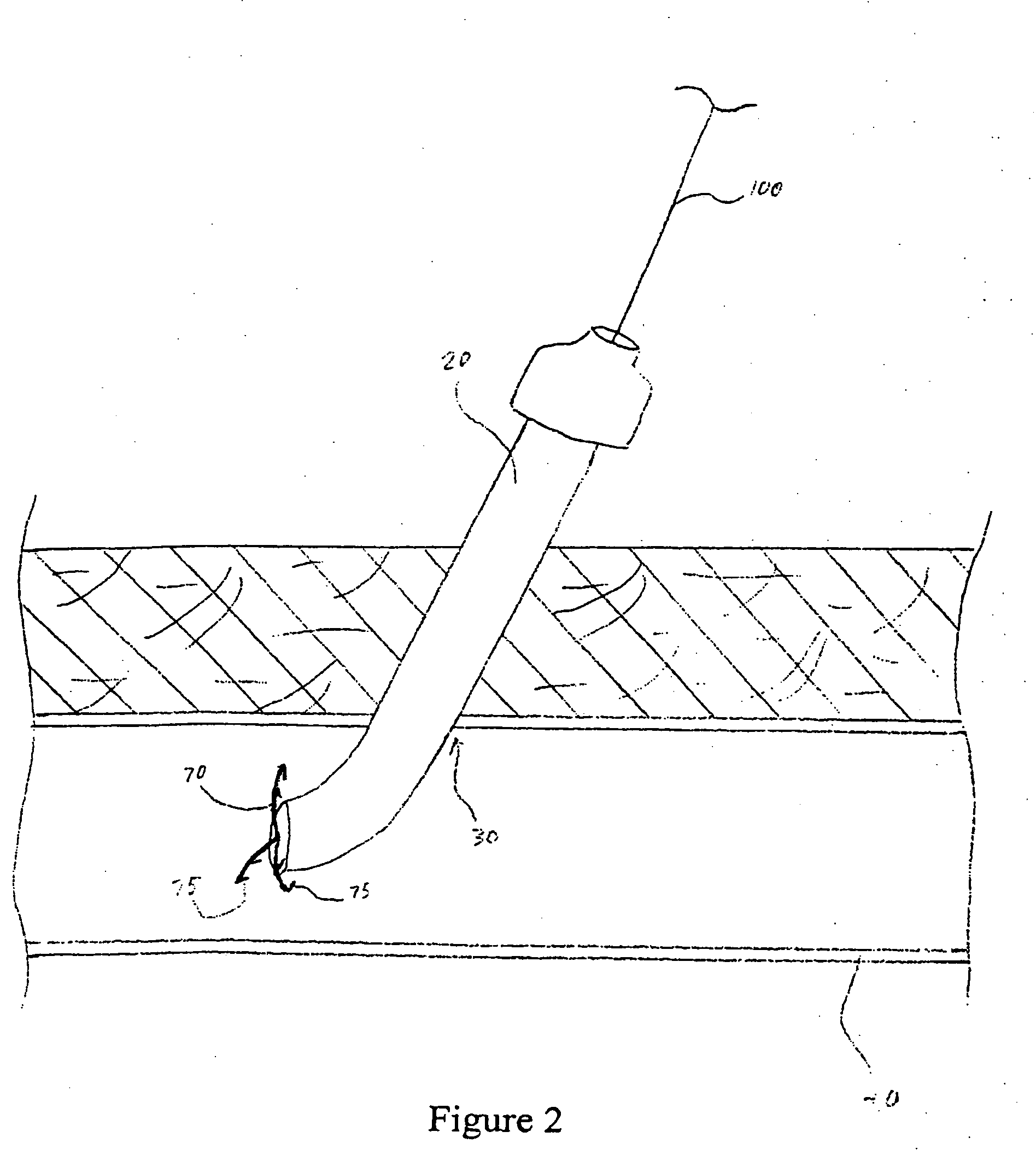
[0035] The present invention provides a reliable and easily-used vascular closure device for closing and effectively sealing an opening in a luminal wall, such as made during the course of a percutaneous surgical procedure, using three complementary sealing methods. The device comprises at least two resilient tines and a collar. The practitioner contracts the resilient tines from an open state to a closed state to grasp the interior edges of the opening together and folds and apposes the edges tightly together. The collar is then used to hold the tines in a closed position. In addition, the collar acts to plug the opening thereby acting as another means of sealing the opening. Because the present invention uses three mechanisms to effectively seal a perforation, it provides a better seal, enables faster healing, and better promotes and achieves percutaneous vascular hemostasis allowing earlier ambulation and patient discharge in the most effective manner. In addition, the use of the...


PUM


Abstract

The present invention provides a device for closing an opening to a body cavity and methods of closing an opening to a body cavity. The device and methods can be used to easily and effectively close a vascular puncture site resulting from a surgical procedure, an atrial or ventricular septal defect, a malfunctioning heart valve, or the left atrium appendage.

Description

[0001] This application is a continuation-in-part of application Ser. No. 11 / 245,437 filed on Oct. 7, 2005. The present invention relates to methods and devices used for closing openings in a body lumen, such as puncture sites in blood vessels, septal defects in heart chambers, heart valves that do not seal, and the mouth of the left atrial appendage.BACKGROUND OF THE INVENTION [0002] A growing number of therapeutic and diagnostic medical procedures involve the percutaneous introduction of instrumentation into a vein or artery. For example, in the treatment of vascular disease, such as atherosclerosis, it is a common practice to insert an instrument, such as a balloon, into an artery to carry out the procedure within the artery. Although a physician may elect to use a balloon to stretch out a vessel, he may alternatively use a laser to burn through any plaque present and open up the artery. Also, the physician may inject clot dissolving chemicals directly into the blocked artery or ...

Claims


Application Information

Patent Timeline
Patent Type & Authority Applications(United States)
IPC IPC(8): A61B17/08
CPCA61B17/0057A61B17/0643A61B17/068A61B2017/00637A61B2017/00668A61B2017/0641A61B17/12122A61B2017/00243A61B2017/00575
Inventor LEE, BENJAMIN
Owner INNOVASIVE
Why Eureka
Try Eureka