Method and apparatus for elasticity imaging

a technology of elasticity imaging and method, applied in the field of computation efficient algorithm for tissue compression analysis, can solve the problems of affecting the accuracy of tissue compression analysis,

Inactive Publication Date: 2007-04-26
ALOKA CO LTD
View PDF3 Cites 50 Cited by
  • Summary
  • Abstract
  • Description
  • Claims
  • Application Information

AI Technical Summary

Benefits of technology

[0022] In accordance with an aspect of the present invention, a process for performing elasticity imaging on a biological tissue broadly comprises selecting automatically based upon at least one criterion at least one frame pair comprising a pre-compression frame and a post-compression frame; analyzing the at least one frame pair; calculating an elasticity image; and displaying the elasticity image. The automatic selection step broadly comprises using a compression feedback algorithm. The at least one criterion broadly comprises an amount of tissue displacement and at least one tissue correlation result. The automatic selection step further broadly comprises predicting an elasticity image quality prior to calculating an elasticity image. The automatic selection step further broadly comprises providing to an operator at least one of the following: a visual feedback or an audible feedback or both visual feedback and audible feedback. The providing step further broadly comprises providing the visual feedback and the audible feedback to the operator upon achieving any one of the following: a compression motion, a decompression motion, an acceptable compression motion, an acceptable decompression motion, an unacceptable compression motion, an unacceptable decompression motion, a satisfactory compression motion, a satisfactory decompression motion, an unsatisfactory compression motion, or an unsatisfactory decompression motion. The process also broadly comprises confirming off-line the quality of a plurality of data used in the calculation of the elasticity image. The confirmation step broadly comprises displaying visually and projecting audibly at least one of the following: at least one quantitative data, at least one qualitative data, or both at least one quantitative data and at least one qualitative data. The confirmation step also broadly comprises displaying visually or projecting audibly at least one of the following: at least one quantitative data, at least one qualitative data, or both at least one quantitative data and at least one qualitative data.

Problems solved by technology

It has been proved that pathological conditions often produce changes in biological tissue stiffness.
However, a lesion may or may not possess echogenic properties that would make it detectable with conventional diagnostic ultrasound imaging systems.
As the boundary conditions and the modeling of theory of elasticity are highly dependent on the structure of the biological tissue, the implementation of the last step is rather cumbersome and typically not performed.
However, real-time elasticity imaging is indeed needed to acquire and process the ultrasonic echo data in such a way that patient-scanning time is relatively low and diagnostically relevant elasticity images are produced immediately during the scan.
Furthermore, the real-time processing of the ultrasonic echo data allows for freehand compression and scanning of the biological tissue rather than utilizing bulky and slow motorized compression fixtures.
Its disadvantage, however, consists of exhaustive operator training, as the sonographer constantly needs to adjust the compression technique to obtain strain images of good quality.
In short, due to the extremely complex nature of the tissue compression, obtaining elasticity images of consistent quality using free-hand strain imaging is neither trivial nor as expeditious as obtaining good quality B-mode images, thus real-time compression feedback is necessary to ensure proper operator training.
However, those implementations disclosed by the '768 patent and the Zhu et al. and Shiina et al. articles do not account completely for all the limitations mentioned above.
More particularly, neither the articles by Zhu et al. and Shiina et al. nor the teachings of the '768 patent provide a quantitative indication of the compression quality being achieved by the operator.
Moreover, the operator does not receive guidance in order to improve the compression quality when s / he is only provided strain images that may contain artifacts and poor SNR.
One of several drawbacks being that possible artifacts present in the strain image cannot be qualitatively linked to poor compression quality.
Therefore the computational burden placed upon the imaging system is extremely high while only select sets of strain images faithfully indicate the mechanical properties of the imaged tissue and are artifact-free.

Method used


Image

  • Method and apparatus for elasticity imaging
  • Method and apparatus for elasticity imaging
  • Method and apparatus for elasticity imaging

Examples


Embodiment Construction

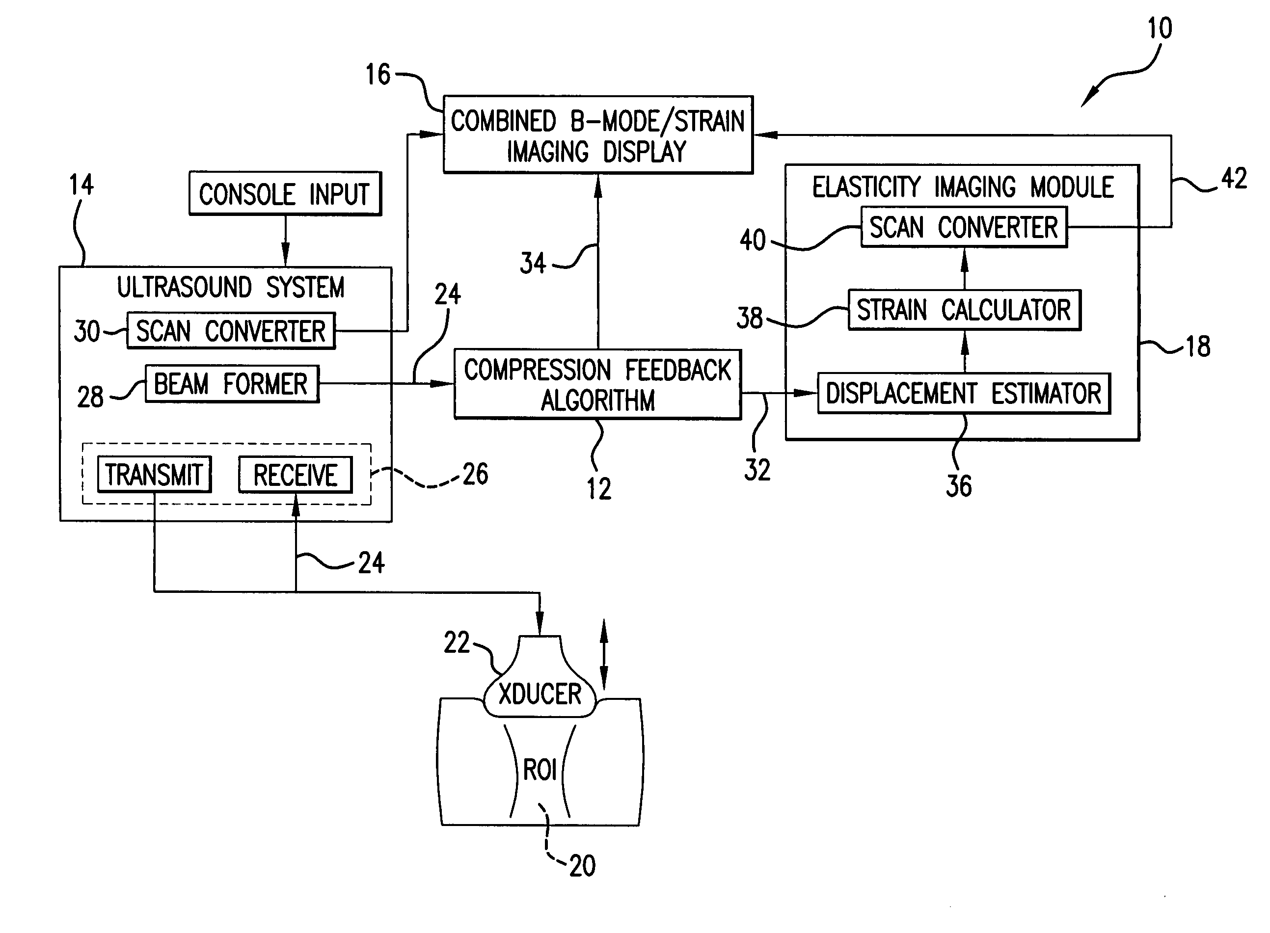
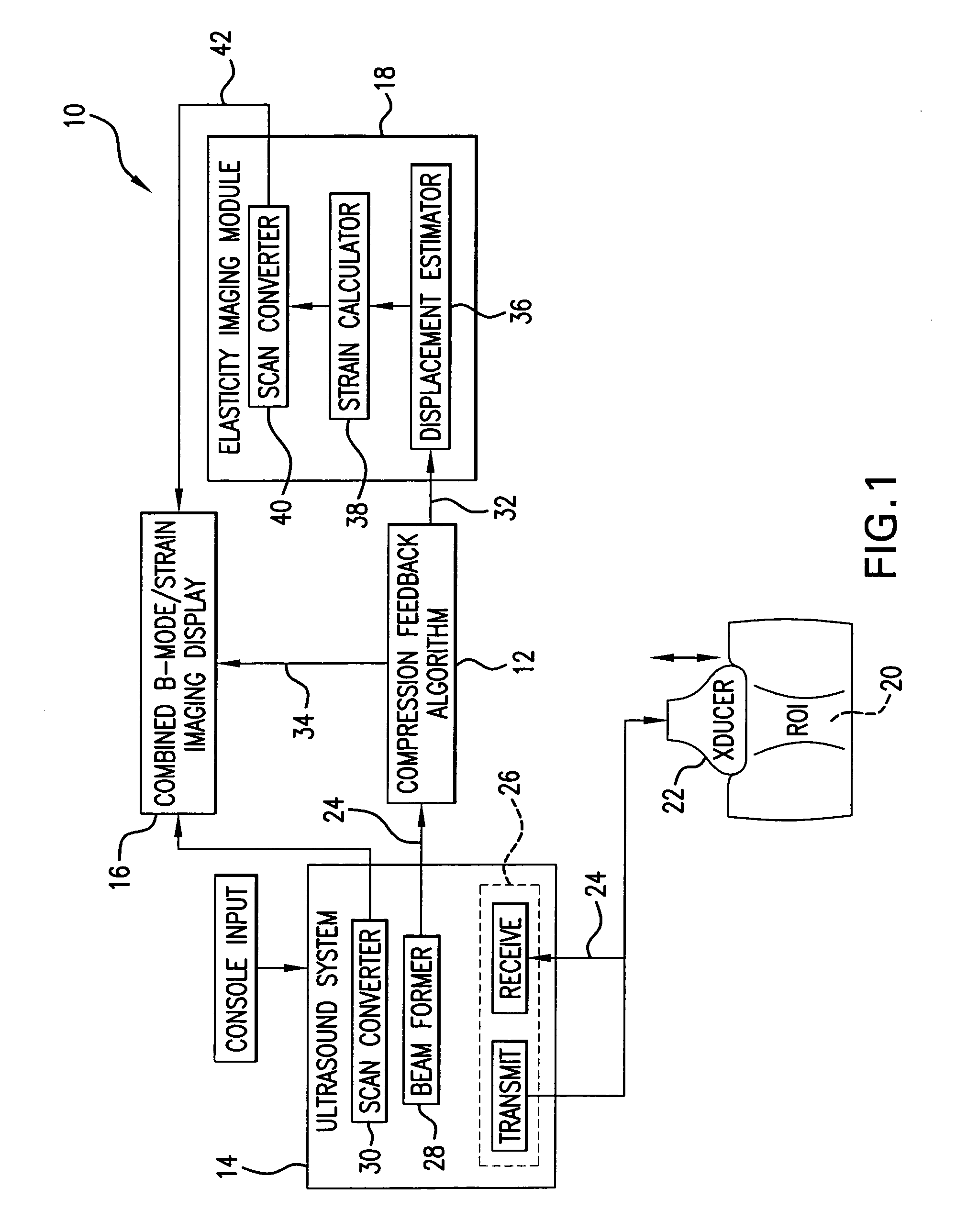
[0036] An elasticity imaging system, and method for using same, employs a tissue compression analysis algorithm for free-hand static elasticity imaging utilizing medical diagnostic ultrasound imaging equipment. The compression feedback algorithm's application offers tissue compression quality and provides quantity feedback to the operator. The compression feedback algorithm analyzes the pre- and post-compression frame pairs and provides an elasticity image quality prediction before an elasticity imaging module computes the elasticity image. The algorithm includes a criterion for the automatic selection of the most advantageous pre- and post-compression frame pairs for delivering elasticity images of optimal dynamic ranges and signal-to-noise ratios. The use of the algorithm in real time eases operator training and reduces significantly the amount of artifact in the elasticity images while also lowering the computational burden. In addition, operator training and confirmation of the ...


PUM


Abstract

A computational efficient algorithm for compression analysis of free-hand static elasticity imaging performed using medical diagnostic ultrasound imaging equipment offers tissue compression quality and quantity feedback to the operator. The algorithm includes a criterion for automatic selection of the most advantageous pre- and post-compression frame pairs delivering elasticity images of optimal dynamic ranges (DR) and signal-to-noise ratios (SNR). The use of the algorithm in real time eases operator training and reduces significantly the amount of artifact in the elasticity images while lowering the computational burden.

Description

CROSS-REFERENCE TO RELATED APPLICATION [0001] Benefit is claimed of U.S. patent application Ser. No. 60 / 730,709, filed on Oct. 26, 2005, and entitled “Method and Apparatus for Elasticity Imaging”, the disclosure of which is incorporated by reference herein as if set forth at length.BACKGROUND OF THE INVENTION [0002] 1. Field of the Invention [0003] The present invention relates to a computational efficient algorithm for tissue compression analysis for free-hand static elasticity imaging. More specifically, this invention relates to an elasticity imaging system that employs medical diagnostic ultrasound imaging equipment to produce strain images. [0004] 2. Description of Related Art [0005] It has been proved that pathological conditions often produce changes in biological tissue stiffness. Tumor tissues, for example, are known to exhibit mechanical properties different from the surrounding tissue, as indicated by the use of palpation as a diagnostic tool. Breast and prostate tumors a...

Claims


Application Information

Patent Timeline
Patent Type & Authority Applications(United States)
IPC IPC(8): A61B8/00
CPCA61B5/0048A61B8/08A61B8/14A61B8/485G01S7/52026G01S7/52034G01S7/52042G01S7/5206A61B5/0051A61B5/0053
Inventor RADULESCU, EMIL G.
Owner ALOKA CO LTD
Why Eureka
Try Eureka