

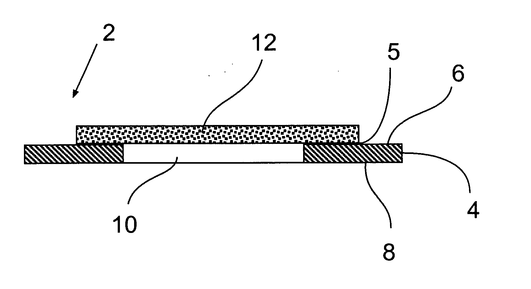
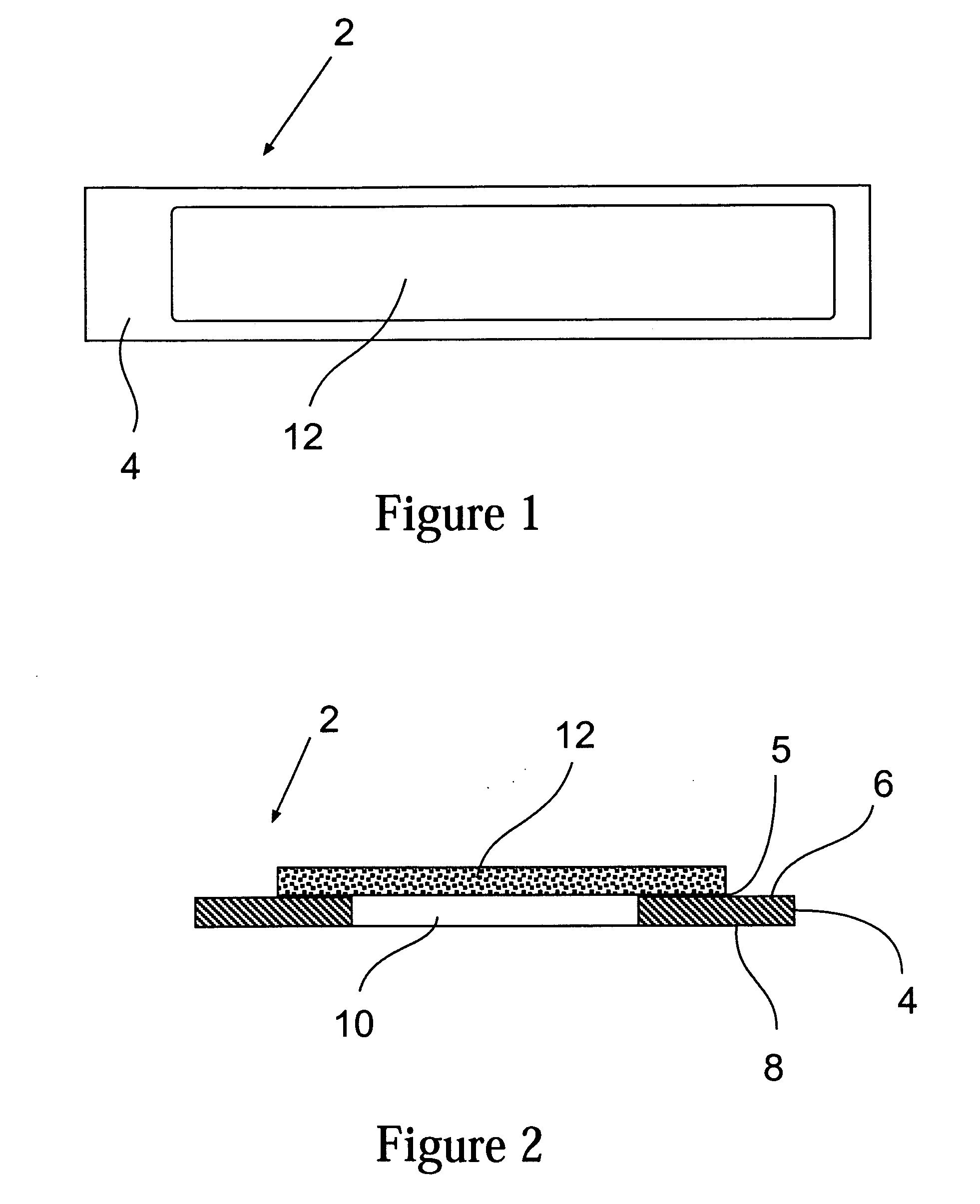
Protein microarray slide
a technology of protein microarrays and slides, applied in the field of slides, can solve the problems of limiting the ability to detect fluorescent signals, limiting the ability to distinguish fluorescent signals, and difficult to distinguish fluoresced protein molecules from substrates, etc., and achieves the effects of reducing the amount of fluorescent light, reducing and eliminating background fluorescence generation, and improving background fluorescence performan
- Summary
- Abstract
- Description
- Claims
- Application Information
AI Technical Summary
Benefits of technology
Problems solved by technology
Method used
Image

Examples
example 1
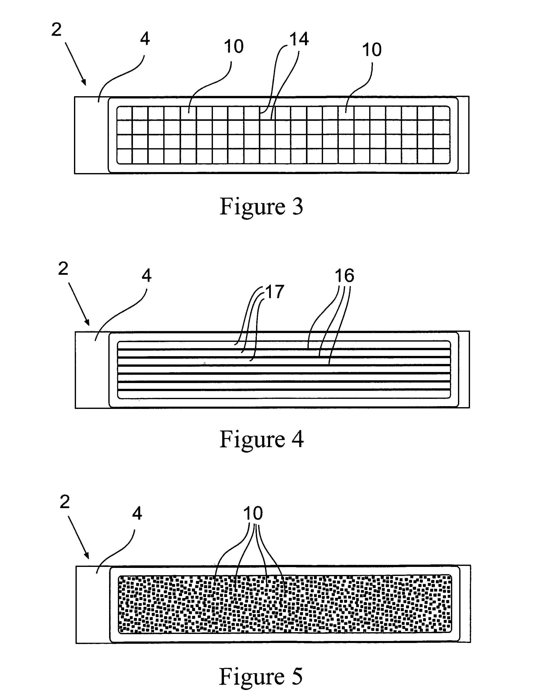
[0100] A carbon filled polystyrene slide support was formed in the same configuration as a conventional glass microscope slide. One opening centrally located in the support was formed of a size having a length of 46 (mm) and a width of 18 (mm). A PVDF asymmetric membrane, Immobilon™ FL membrane available from Millipore Corporation of Billerica, Mass. was solvent bonded to the slide with acetone. The acetone was applied to the edges of the smaller pored side of the membrane and allowed to wick into the membrane for 1 second. The solvent was allowed to evaporate for 3 seconds and then the smaller pored side of the membrane was pressed against the desired major surface of the slide over the opening using a one eighth inch thick silicone elastomer sheet to distribute the clamping load as evenly as possible across the sheet. FIG. 22 A-C show a series of SEMs of the example.
PUM
| Property | Measurement | Unit |
|---|---|---|
| angle of incidence | aaaaa | aaaaa |
| angle of incidence | aaaaa | aaaaa |
| size | aaaaa | aaaaa |
Abstract
Description
Claims
Application Information