Remote Virtual Medical Diagnostic Imaging Viewer

a remote medical and imaging viewer technology, applied in the field of remote virtual medical diagnostic imaging viewer, can solve the problems of unacceptably slow system, unfavorable patient safety, and inability to monitor the patient's medical condition, and achieve the effect of efficient method of remote medical image viewing

Inactive Publication Date: 2007-06-07
TAILSTREAM TECH +1
View PDF9 Cites 3 Cited by
  • Summary
  • Abstract
  • Description
  • Claims
  • Application Information

AI Technical Summary

Benefits of technology

[0010] In the existing embodiment of the invention a computer or other capture device, captures a lossy image file then compresses and transmits the compressed image file which is then downloaded onto a computer hard drive and decompressed by the remote receiver for viewing. In contrast, the preferred embodiment of the invention does not require transmitting the actual medical image files to a receiver and is therefore a more efficient method of remote medical image viewing.
[0012] a) to lower the cost and provide high security or regulatory compliance with the need for remote viewing of medical images and data for medical diagnostics and any other field requiring secure remote viewing of image files and data.
[0013] b) to provide a faster way to view digital images remotely
[0015] d) To be able to use a digital imaging viewer without having to buy and install medical imaging or any other viewing or compression / decompression software.
[0016] e) To be able to see large numbers of images in sequence animated in rapid succession and manipulate them remotely faster than existing methods.

Problems solved by technology

Present methods for this all rely on transmitting a medical image file to the viewer which is prohibitively slow and uses too much transmission bandwidth.
A present embodiment of the invention for remote image viewing is U.S. Pat. No. 5,432,714 to Novik; (1995) which discloses a system of compressing and transmitting data to be decompressed and viewed by an experienced or trained viewer, however, for medical diagnostic imaging, this system is unacceptably slow and costly compared to our inventive method and system.
However, the prior art methods of image data transmission and remote image viewing, particularly when applied to medical diagnostic imaging, do not make use of our novel method of using an image storing application provider server to enable a remote secure executable environment, independent of the operating system of a viewing computer to temporarily reconstruct medical image files, rather than compressing, transmitting and then and uncompressing the actual medical image files interactively.

Method used


Image

  • Remote Virtual Medical Diagnostic Imaging Viewer
  • Remote Virtual Medical Diagnostic Imaging Viewer
  • Remote Virtual Medical Diagnostic Imaging Viewer

Examples


Embodiment Construction

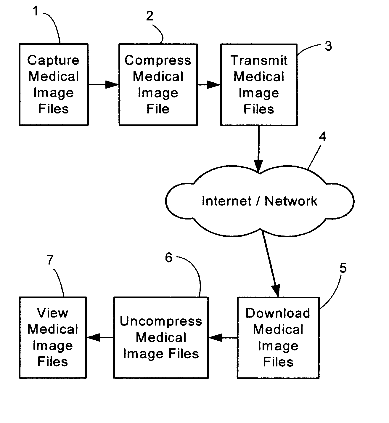
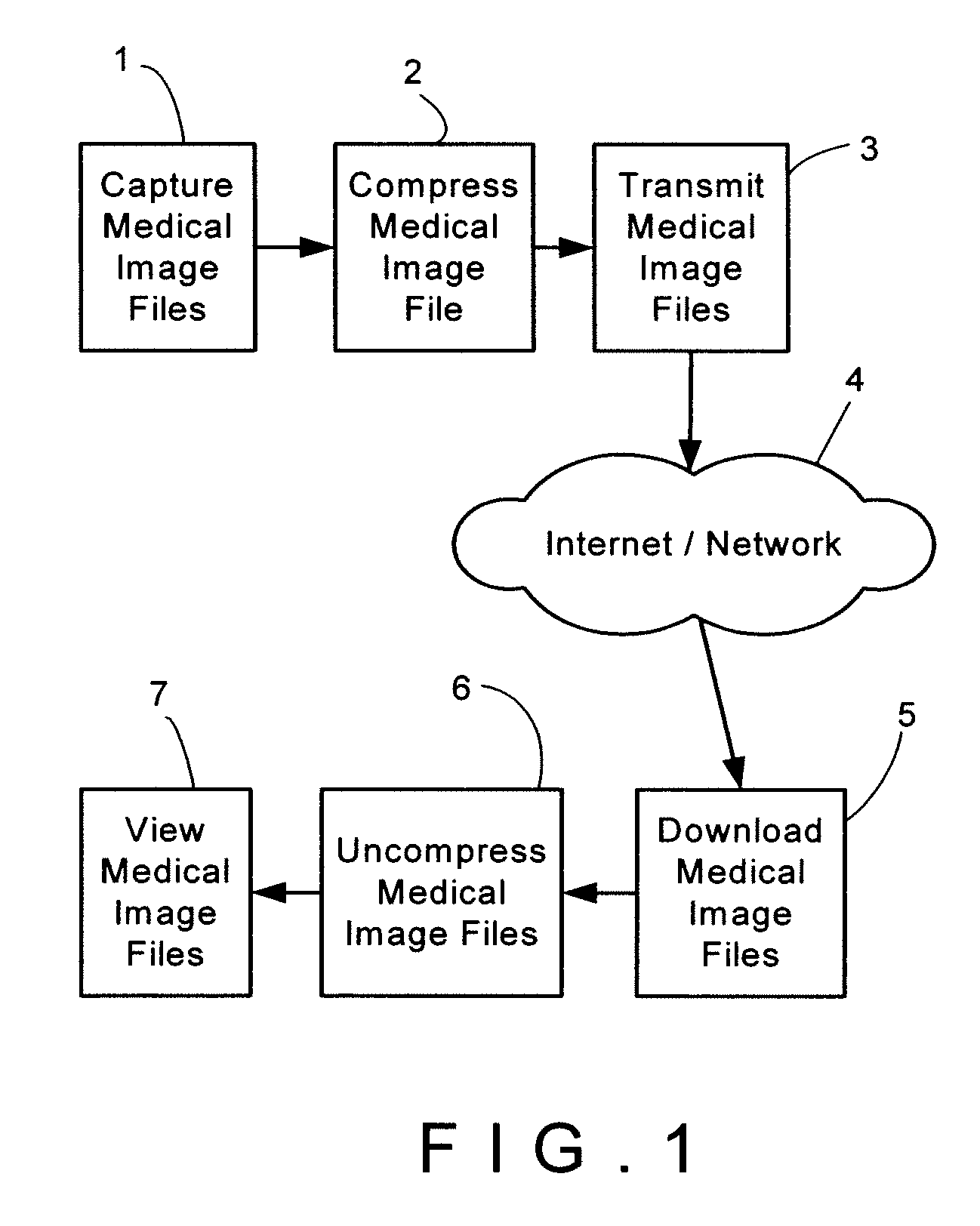
[0034] The present invention describes an apparatus for capturing and transmitting the image file for remote viewing interactively. The invention will be described in FIG.1, which is a block diagram schematic of one preferred embodiment of the present invention. Medical image files are captured (1) and then compressed (2) transmitted (3) over a network or the Internet (4) which are downloaded on a users computer (5) and uncompressed (6) and viewed remotely (7).

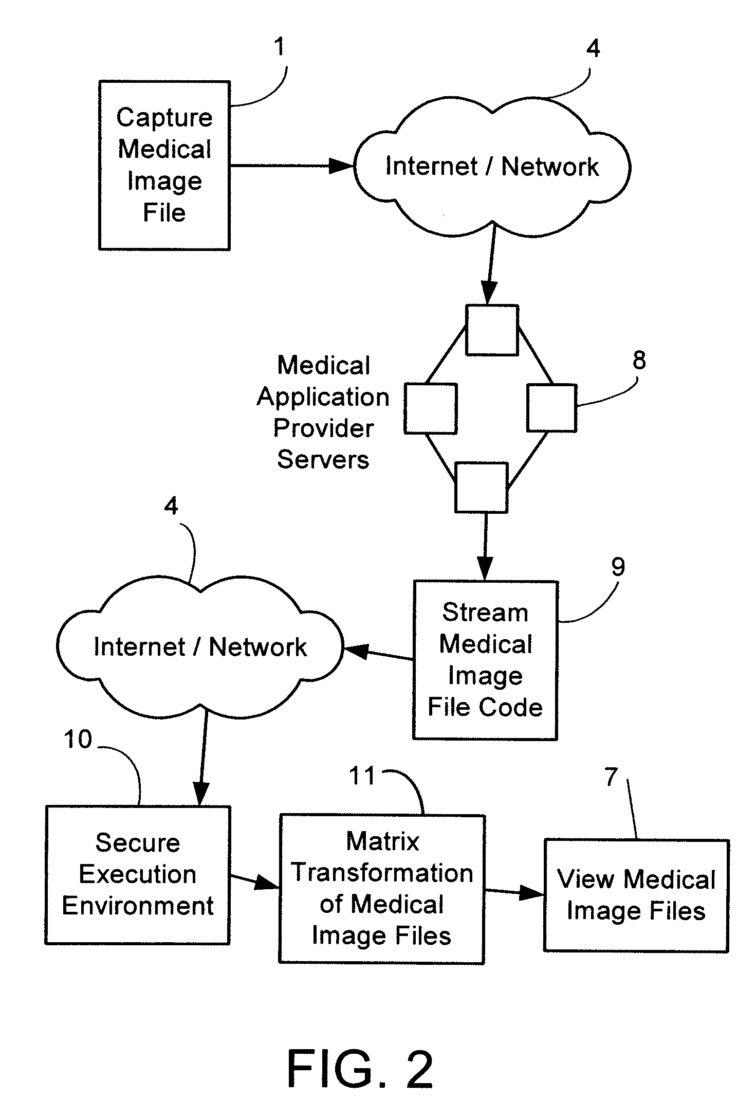
[0035]FIG. 2 is a flowchart of the preferred operation of the present invention, and will be explained with reference to the apparatus of FIG. 1, although other appropriate apparatus may be substituted in performing the inventive method. In this method for viewing a file remotely, the medical image files are first captured (1) transmitted (3) over a network or the Internet (4) to one or more servers running application service provider software (8) which are stored with meta data including access control information, origin o...


PUM


Abstract

A medical image and data application service provider system provides a way of remotely viewing and manipulating medical images and data for diagnostic and visualization purposes by users unconstrained by geography. Medical images and data are stored on one or more servers running application service provider software along with meta-data such as access control information, origin of information and references to related data. A set of medical data consisting related information is sent as an encrypted stream to a viewing station running client software in a secure execution environment that is logically independent of the viewing station's operating system.

Description

CROSS-REFERENCE TO RELATED APPLICATIONS [0001] This invention relates to an improved method for interactive remote viewing of medical image data over prior invention: U.S. Pat. No. 5,432,714—Novick—Jul. 11, 1995.STATEMENT REGARDING FEDERALLY SPONSORED RESEARCH OR DEVELOPMENT [0002]“Not Applicable”REFERENCE TO A MICROFICHE APPENDIX [0003]“Not Applicable”BACKGROUND OF THE INVENTION [0004] It is very desirable to obtain medical imaging data with CT, MRI, PET, or other diagnostic imaging systems or any type of image capture system and then to permit persons to view remotely all the medical images without having to transmit the actual image files or allow the image files to actually download into the receiving viewing computer. Physicians for example, or other imaging users have a need to quickly access and analyze large numbers of image files from remote image capture systems securely, without downloading or storing the image files onto their computer. Present methods for this all rely ...

Claims


Application Information

Patent Timeline
Patent Type & Authority Applications(United States)
IPC IPC(8): H04L9/00G16H10/60G16H30/20G16H40/67H04N1/00
CPCG06F19/321G06F21/6245G06F2221/2149G06Q50/24H04N1/00209H04N1/00244H04N2201/0079G16H40/67G16H30/20G06F21/53G06F2221/032G06F21/606G06T11/00G16H10/60
Inventor WISE, KELLYSHAEBANYAN, MEREDITH
Owner TAILSTREAM TECH
Why Eureka
Try Eureka