
Method and system for radiographic imaging with organ-based radiation profile prescription

Inactive Publication Date: 2007-06-28
GENERAL ELECTRIC CO
View PDF42 Cites 48 Cited by
  • Summary
  • Abstract
  • Description
  • Claims
  • Application Information

AI Technical Summary

Benefits of technology

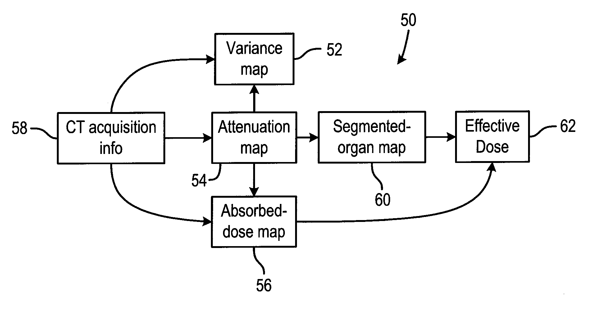
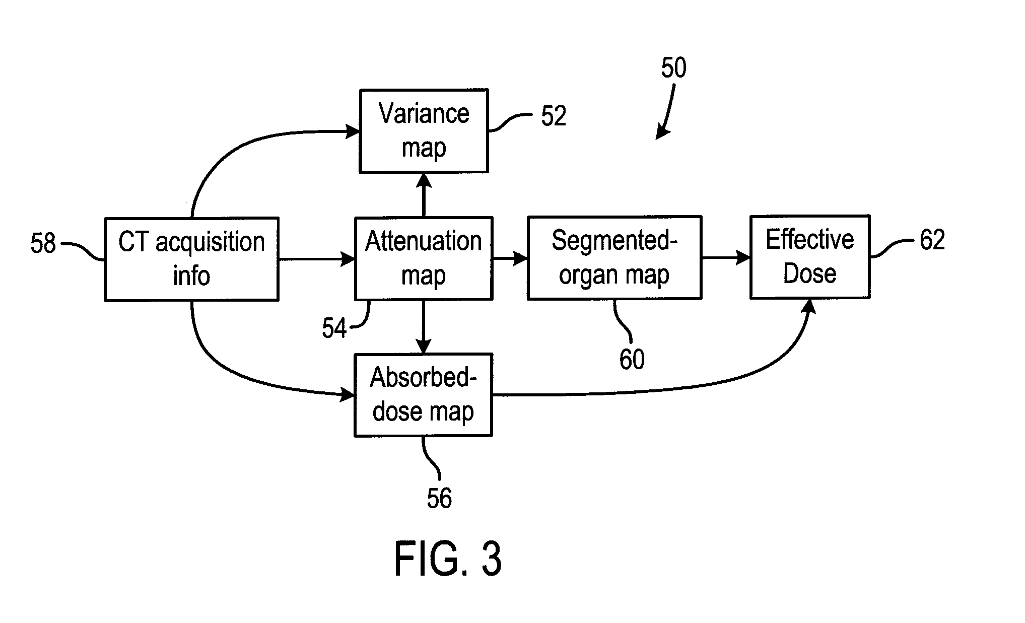
[0008] In accordance with another aspect, a radiographic imaging system is presented and includes an x-ray source configured to project x-rays towards a detector according to a certain radiation profile, which establishes number of x-rays projected and energy level of the x-rays projected as a function of time and location, and possibly a finite time interval during which x-rays are produced for each view. The detector is configured to output electrical signals in response to a reception of x-rays. The system further has a computer programmed to acquire an organ map for a subject to be imaged and determine a parameterized dose absorption map for the subject to be imaged and determine a parameterized noise variance map for the subject to be imaged. The computer further determines an irradiation profile that minimizes effective dose for each organ of the organ map and maximizes image quality for an image of the subject.

Problems solved by technology

Thus, if the radiation dose is set to the maximum permitted for the sensitive anatomical structures, the entire image will have poor spatial and contrast resolution.
In other words, in conventional CT scans, the radiation profile used to define the scan considers the total radiation, but does not consider the effective dose for the patient.

Method used


Image

  • Method and system for radiographic imaging with organ-based radiation profile prescription
  • Method and system for radiographic imaging with organ-based radiation profile prescription
  • Method and system for radiographic imaging with organ-based radiation profile prescription

Examples


Embodiment Construction

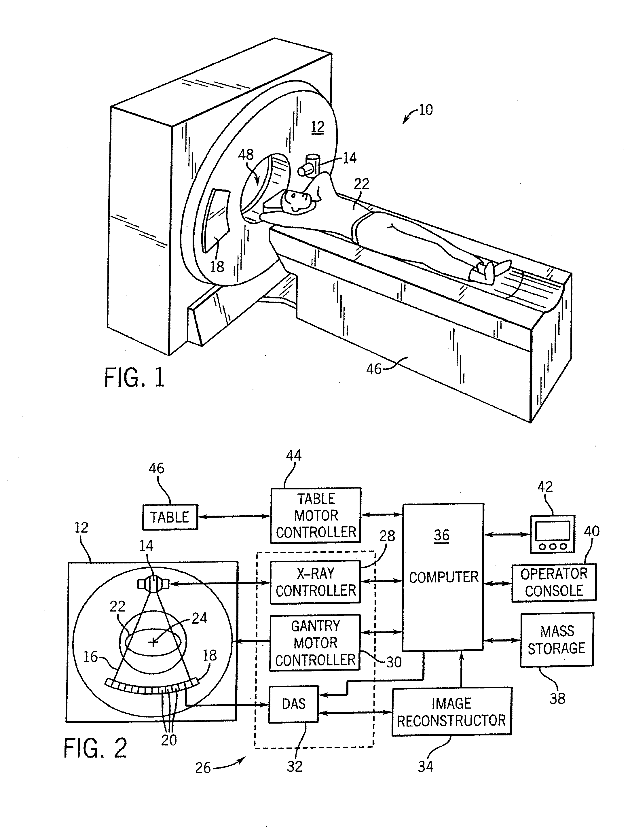
[0021] The operating environment of the present invention is described with respect to a four-slice computed tomography (CT) system for imaging of a multi-component object, such as a medical patient. However, it will be appreciated by those skilled in the art that the present invention is equally applicable for use with single-slice or other multi-slice configurations. Moreover, the present invention will be described with respect to the detection and conversion of x-rays. However, one skilled in the art will further appreciate that the present invention is equally applicable for the detection and conversion of other types of radiation. The present invention will be described with respect to a “third generation” CT scanner, but is equally applicable with other CT systems. For example, the invention is also applicable with systems having multiple source spots for increased flexibility in determining an optimal radiation profile by individually steering the different sources.

[0022] R...


PUM


Abstract

A method and system is disclosed that minimizes the effective dose to an object by determining a segmented component map for the object, parametrizing tube current / energy level / x-ray filtration / x-ray pulse width as a function of time, determining a corresponding absorbed dose map and variance map, and determining an energy level / tube current profile or curve that results in a desirable effective dose to the object and a desirable noise variance throughout the image.

Description

BACKGROUND OF THE INVENTION [0001] The present invention relates generally to diagnostic imaging and, more particularly, to a method and apparatus for maximizing image quality for an image of a multi-component object while minimizing the absorbed dose by the object on a per-component basis. [0002] Generally, four key properties define the performance of a computed tomography (CT) scan: spatial resolution, temporal resolution, image noise, and radiation dose. Spatial resolution defines the degree of small object detail in an image and is generally affected by a number of factors including detector aperture, number of acquisition views, focal spot size, object magnification, slice thickness, slice sensitivity profile, helical pitch, reconstruction algorithm, pixel matrix, patient motion, and field-of-view. Temporal resolution defines the length of the temporal interval over which the scan data is acquired for a given slice. Generally, it is desirable to increase temporal resolution (i...

Claims


Application Information

Patent Timeline
IPC IPC(8): H05G1/60A61B6/00G01N23/00G21K1/12
CPCA61B6/032A61B6/503A61B6/542
Inventor DE MAN, BRUNO K. B.SENZIG, ROBERT FRANKLIN
Owner GENERAL ELECTRIC CO
Learn More