Diagnostic imaging device for the analysis of circulating rare cells

a rare cell and diagnostic imaging technology, applied in the field of automatic sample imaging, can solve the problems of limiting the fraction of total acquistion that is time spent on motion, and so as to reduce the total acquistion time, reduce the time period required, and reduce the bleaching of samples, especially with pe

Active Publication Date: 2007-07-05
MENARINI SILICON BIOSYSTEMS SPA
View PDF16 Cites 1 Cited by
  • Summary
  • Abstract
  • Description
  • Claims
  • Application Information

AI Technical Summary

Benefits of technology

[0013] The present invention provides apparatus and methods to an imaging system for automated operation in a clinical laboratory, improving upon diagnostic imaging devices for rare cell detection used in clinical laboratories. The device of the present invention is a compact and self-contained unit, designed to fit on a laboratory bench top with minimum space in a simple, exterior box-like design. The illumination and collection optics are structured to provide a clinical laboratory technician easy access and use in the analysis of large numbers of patient samples.
[0014] Further, stepper motor technology, used in changing the filter cubes and stage movement, has been replaced with nanomotion motors in a closed feedback loop. These movements are critical if an image is to be developed from multiple fluorescent detectors or dyes whereby more than one fliter cube is required. More specifically the motion time for each cube is reduced from 2 to 0.2 seconds for two consecutive filter cubes. The time spent moving between two consecutive positions is reduced from 0.5 to 0.1 seconds. Because the area needed for image acquistion is larger than the system field of view, the acquisition of an entire image requires acquisition of images at several different positions. Nanomotion motors reduce the time period required for these functions and limits the fraction of the total acquistion that is time spent on motion. The majority of time will then be spent on acquition of the image itself, instead of time spent moving the sample and / or cubes. Further with the total acquistion time drastically reduced, the bleaching of the sample, especially with PE, is reduced. The substantial reduction in acquisition time allows for a position-by-position scan of the entire image. With a position-by-position scan, images are acquired for all filter cubes before moving on to the next position in a boustrophedonic motion. Any possible movement of the image during a color-by-color scan is eliminated.
[0015] The automated fluorescent optical imaging device of the present invention further provides a prealigned bulb and method for installation / use in the enumeration of fluorescently labeled circulating tumor cells (CTC) that are immuno-magnetically selected and aligned. This invention provides for the standardization of the lamp housing stage through the establishment of a master lamp / stage unit. This master lamp is seated within an adjustment collar as shown in the drawings. The collar has machined gaps which are used to microalign the bulb filament. Thus the collar is used to ensure exact alignment (for X-Y-Z orientation) of the electrodes with respect to any instrument unit. Once aligned, the collar-bulb unit is inserted into a V-block structure for final support. A further improvement to any subsequent shifting in the alignment is the use of a braded wire as a flexible connecting lead. These combined improvements allow for a prealigned bulb to be used in any corresponding imaging unit. This reduces the costs of aligning individual units at their respective locations and provides for efficient installation.
[0016] Thus, this automated rare cell imaging device improves upon the speed of sample analysis. The system's compact design in a clinical diagnositic laboratory enables enumeration and identification of target cells for targeting the clinical status of diseases such as metastatic cancer. The pre-aligned buld structure provides rapid, convienent maintenance. Together, these concepts aid in the detection and quantification of target cells in a sample specimen, such as but not limited to, screening and detection in early stage cancer, monitoring for disease remission in response to therapy and selection of more effective dose regimens or alternative therapies in case of relapse.

Problems solved by technology

Nanomotion motors reduce the time period required for these functions and limits the fraction of the total acquistion that is time spent on motion.

Method used


Image

  • Diagnostic imaging device for the analysis of circulating rare cells
  • Diagnostic imaging device for the analysis of circulating rare cells
  • Diagnostic imaging device for the analysis of circulating rare cells

Examples


Embodiment Construction

[0028] The apparatus of this invention improves upon current CellSpotter® technology to provide a more robust application with less user intervention in a device that is more condusive to a clinical laboratory environment.

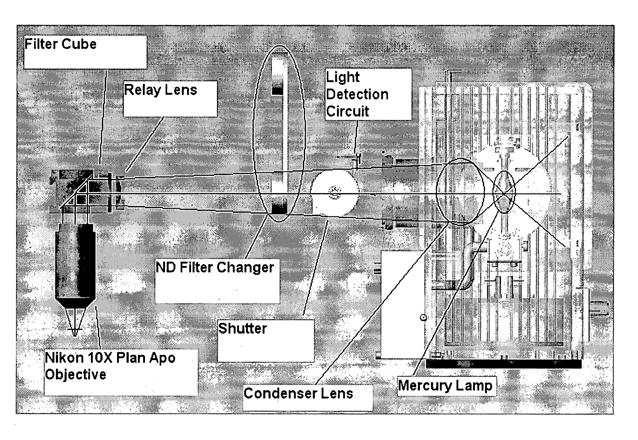
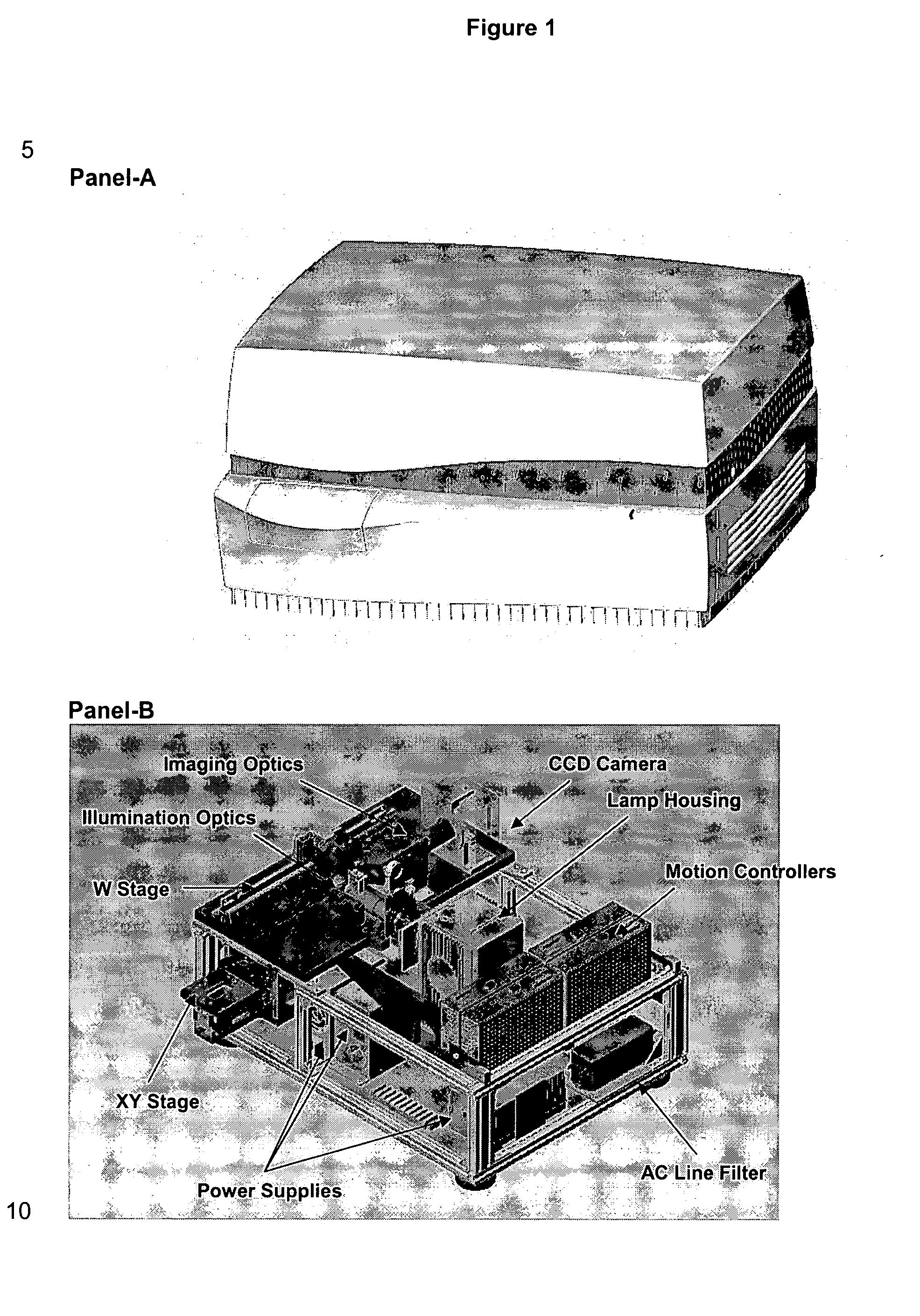
[0029] First, the overall layout is a non-microscope type design to provide a compact benchtop profile for efficient space management in the laboratory (see FIG. 1). The system is encased in a simple, streamline boxed unit to avoid connecting-cable clutter and to discourage access by laboratory personnel (FIG. 1A). The internal components are designed to provide efficient illumination and collection optics in a confined space (FIG. 1B)

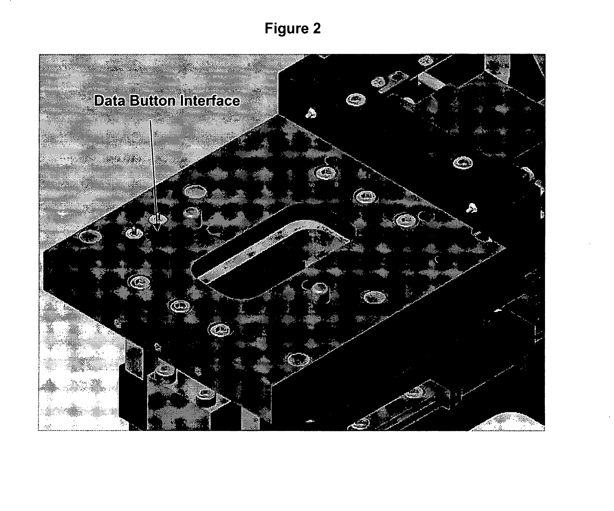
[0030] Second, improvements to individual components provide more versatility. The unit can scan an individual sample for multiple analyses. Further, the unit is expandable to include multiple analyte specific reagents (ASR) and the associated software algorithms. MagNest™, the magnetic device, communicates to the stage through a da...


PUM

PropertyMeasurementUnit
fluorescence excitation wavelengthsaaaaaaaaaa
motion timeaaaaaaaaaa
timeaaaaaaaaaa

Abstract

The present invention provides a system for imaging circulating tumor cells from blood after enrichment. The system is designed to provide optimum use in a clinical laboratory setting with minimum laboratory bench top space. Operator intervention is minimized compared to other analytical methodologies. The system is useful in the enumeration and identification of target cells in a sample specimen for screening and detection of early stage pre-metastatic cancer, monitoring for disease remission in response to therapy and selection of more effective dose regimens or alternative therapies for individual patients.

Description

PRIORITY INFORMATION [0001] This application claims priority under 35 USC 119(e) to U.S. Provisional Application Nos. 60 / 602,504, filed 17 Aug. 2004; 60 / 645,683, filed 21 Jan. 2005; and 60 / 648,477, filed 31 Jan. 2005.FIELD OF THE INVENTION [0002] This invention relates generally to the field of automated sample imaging, and more particularly to image cytometry of isolated rare cells from body fluids by an automated device for analysis relating to the diagnosing, monitoring and managing of specific diseases, particularly cancer. BACKGROUND ART [0003] Tumor cells are often present in blood of carcinoma patients as rare cells at very low frequencies (<10 cells per ml). These levels may provide clinically useful information. However, the laborious procedures required to detect and quantify the presence of circulating tumor cells and the statistics of low numbers introduce a high level of variability in the results. The system of this invention improves upon imaging devices used in a ...

Claims


Application Information

Patent Timeline
Patent Type & Authority Applications(United States)
IPC IPC(8): G01N21/25
CPCG01N15/1475G01N21/6458G01N2021/6421G01N2015/1488G01N2015/1006
Inventor COUMANS, FRANK A.W.MARLIN, ARTHUR G.MODICA, FRANK P.VERRANT, JOHN V.
Owner MENARINI SILICON BIOSYSTEMS SPA
Why Eureka
Try Eureka