

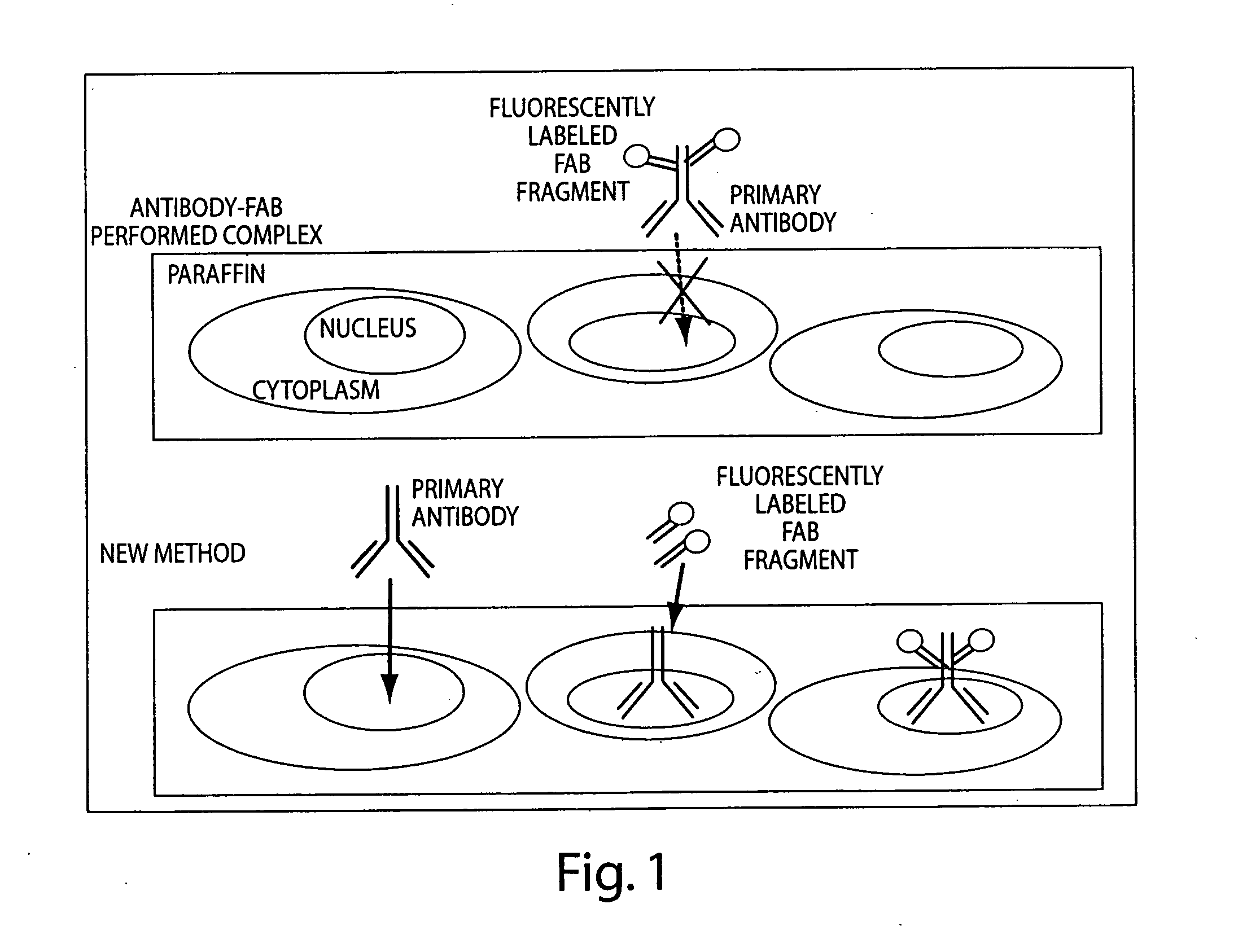
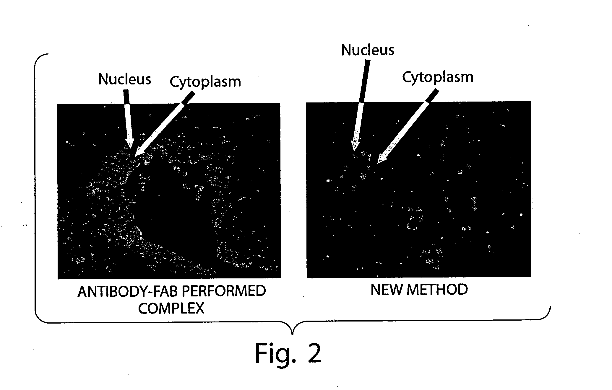
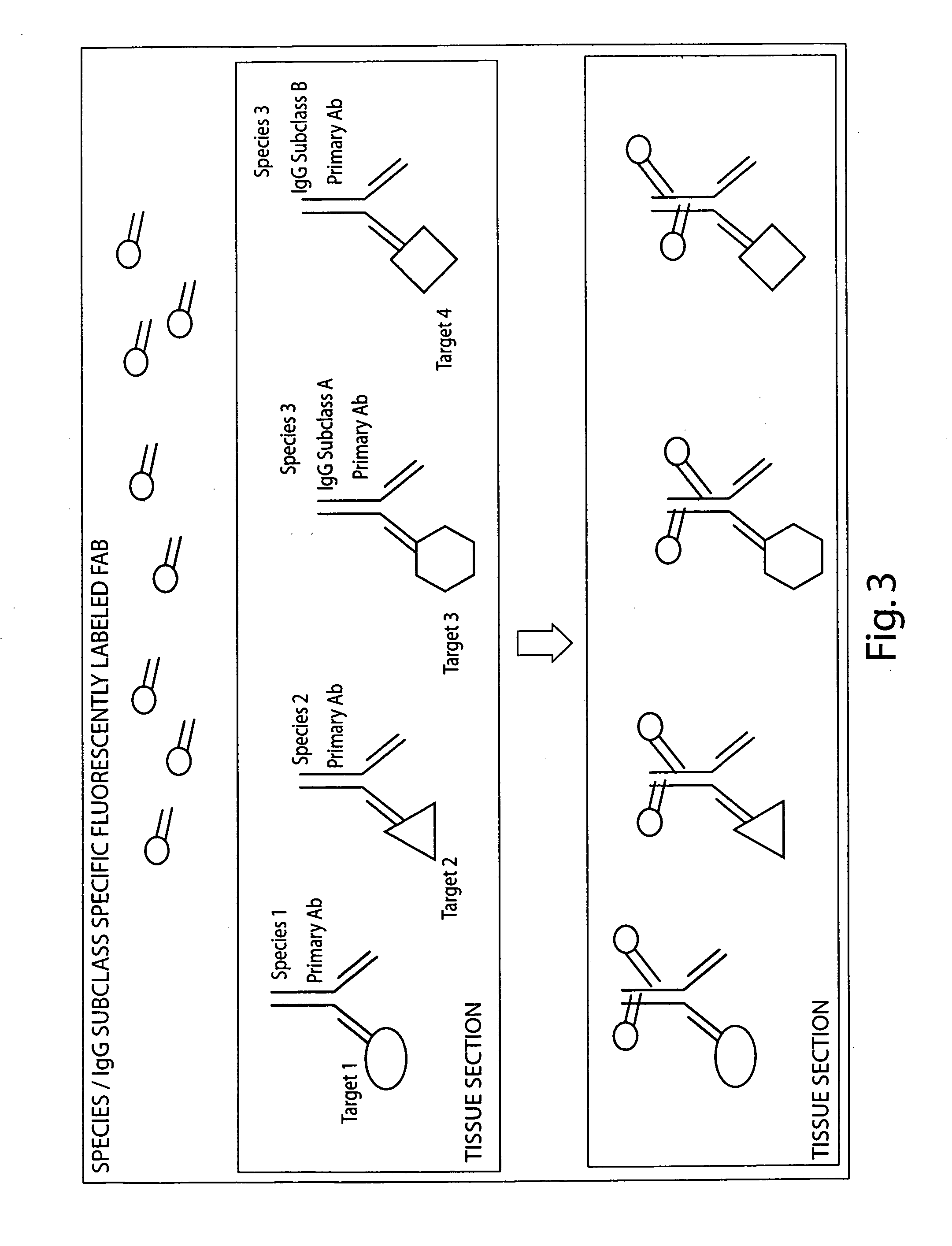
Multiplex in situ immunohistochemical analysis
a multi-in-situ immunohistochemical and immunohistochemical technology, applied in the field of immunofluorescence, can solve the problems of background problems, labor-intensive methods, and certain amount of antibodies
- Summary
- Abstract
- Description
- Claims
- Application Information
AI Technical Summary
Benefits of technology
Problems solved by technology
Method used
Image

Examples
example 1
General Sample Preparation Methods
[0124] The following methods are generally used to during the Multiplex detection methods according to the invention
Antizen Retrieval:
[0125] 1. De-paraffinize and re-hydrate the tissue samples as per the standard Leica 5020 SOP. [0126] 2. Pre-heat 250 ml of 1× Reveal antigen retrieval solution to boiling in water bath in microwave (heat solution for seven (7) minutes at power level seven (7)). [0127] 3. Place slides in container of boiling 1× Reveal solution. Allow to boil for 8.0 minutes as described above. [0128] 4. When completed, remove container from microwave water bath and allow to cool for 20 minutes. [0129] 5. Rinse slides in PBS briefly followed by 1×5 minutes at room temperature. [0130] 6. Place slides on Nemesis 7200 and begin auto-staining program.
Tissue Permeabilization:
[0131] Incubate slides in PBT (PBS with 0.2% Triton-X) for 30 minutes. PBT is made as follows:
Dilution / FinalReagentVendorCatalog #[Conc.]AmountVolumeDifco FA B...
example 2
Multiplex Detection of Androgen Receptor, Cytokeratin and AMACR
[0142] Androgen Receptor (AR), Cytokeratin 18 and AMACR have been found to be important biomarkers for the evaluation of prostate cancerous tissue. The qualitative and quantitative distribution of these markers in formalin fixed, paraffin embedded tissue sections or Tissue Microarrays were detected as described below. Samples up to 16 years old were used in this study.
[0143] 1.) Antigen Retrieval (in Reveal Solution, Citrate Buffer or Proteinase K)
[0144] For antigen retrieval, tissue sections or TMAs were heated in 1× Reveal Solution (BioCare Medical) in a decloaking chamber according to standard protocol and then allowed to cool for 15 minutes. Alternative methods of antigen retrieval include: 1) heating tissue sections or TMAs in 10 mM Citrate Buffer, pH6.0, for 15 minutes in a calibrated microwave followed by cooling for 15 minutes or 2) enzymatically digesting tissue sections or TMAs in a Proteinase K solution (co...
example 3
Multiplex Detection of EGFR, Phospho-EGFR and Cytokeratin 18
[0166] The Epidermal Growth Factor Receptor (EGFR) and downstream signaling members have recently been shown to be over-expressed in certain tumor types. As a result another group of biomarkers under analysis are EGFR, phospho-EGFR and Cytokeratin 18. In order to measure the qualitative and quantitative distribution of these biomarkers in formalin fixed, paraffin embedded tissue sections or Tissue Microarrays were detected as follows:
[0167] Deparaffinization and re-hydration of tissue samples performed on the Discovery XT Automated Slide Processing Machine (Ventana Medical, Tuscan Ariz.).
[0168] 1.) Antigen Retrieval (Proteinase K)
[0169] For antigen retrieval, tissue sections or TMAs were incubated in a Proteinase K solution (commercially available as a Ready-to-Use reagent for antigen retrieval) for 12-15 minutes. This was applied to slides in “pre-treatment 1” step of machine protocol using a user fillable dispenser (p...
PUM
 Abstract
Description
Claims
Application Information