Method for accurate in vivo delivery of a therapeutic agent to a target area of an organ

Inactive Publication Date: 2007-07-19
SIEMENS AG
View PDF3 Cites 13 Cited by
  • Summary
  • Abstract
  • Description
  • Claims
  • Application Information

AI Technical Summary

Problems solved by technology

The most significant difficulty involved in using interventional cardiology for this purpose is to precisely guide the catheter (sheath) to a location close to the site of damaged myocardial tissue, and to subsequently guide the injection needle precisely to the damaged myoca

Method used


Image

  • Method for accurate in vivo delivery of a therapeutic agent to a target area of an organ
  • Method for accurate in vivo delivery of a therapeutic agent to a target area of an organ
  • Method for accurate in vivo delivery of a therapeutic agent to a target area of an organ

Examples


Embodiment Construction

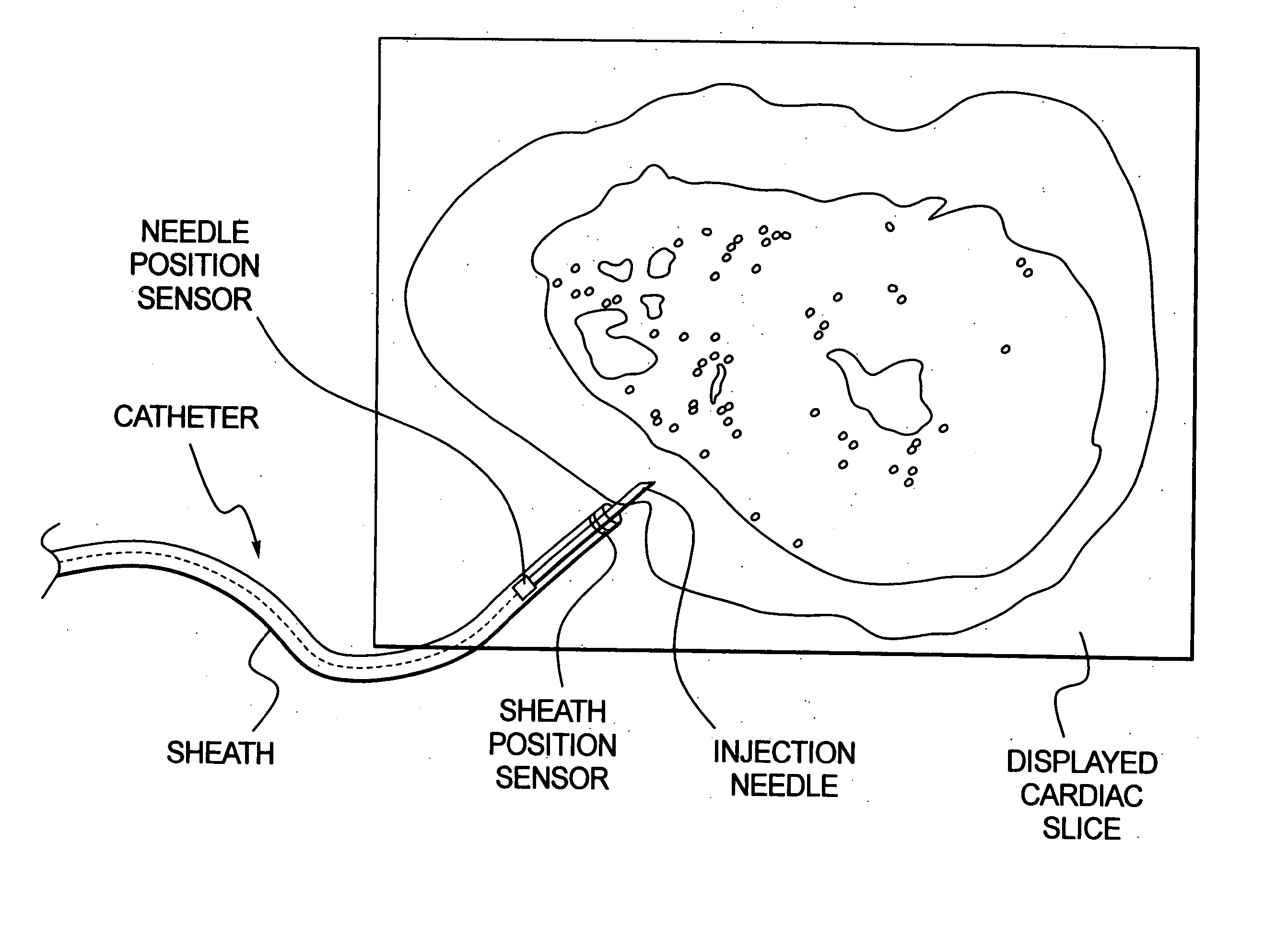
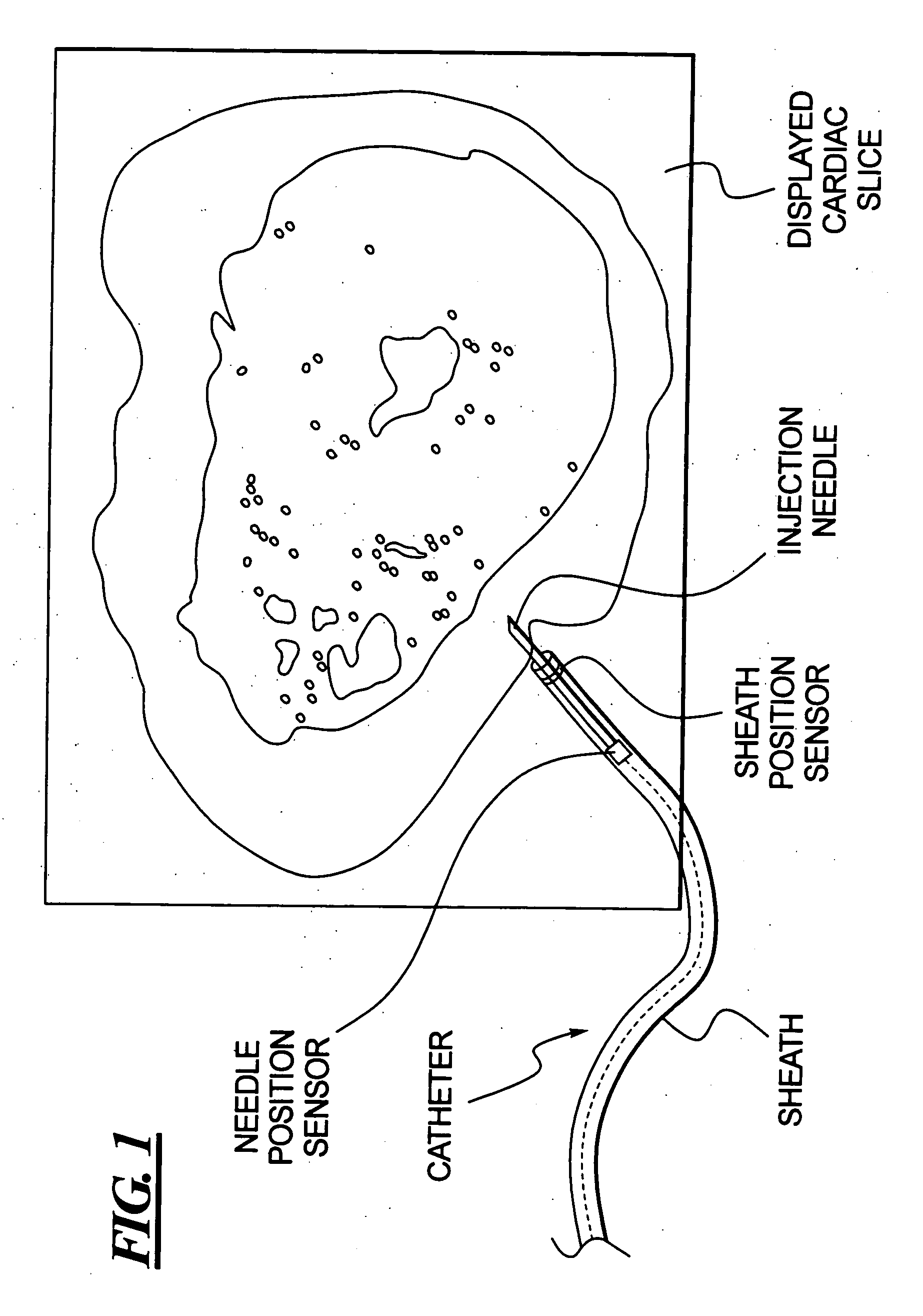
[0023]FIG. 1 schematically illustrates a displayed cardiac slice, in this case an axial slice, that is used in accordance with the invention to guide and monitor the administration of a therapeutic agent via a catheter. The catheter has a catheter sheath containing an injection needle via which a therapeutic agent, in liquid or emulsified form, can be delivered to a delivery site, in this case the myocardium. The injection needle and the sheath each have a position sensor that allows the respective positions of the sheath and the needle to be identified using a known navigation system.

[0024] In practice, the displayed cardiac slice is a 3D image that is obtained using a suitable imaging modality. The schematic representation of the displayed cardiac slice that is necessary for illustrative purposes in FIG. 1 will, in practice, be a conventional 3D medical image in which all of the features conventionally contained in, and identifiable in, such a 3D medical image will be present.

[0...


PUM


Abstract

In a method for accurately delivering a therapeutic agent to a target area of an organ of a living subject, such as for injecting stem cells into the myocardium of the heart, a 3D image, in which the target area and a delivery path thereto are visible, is obtained prior to delivery of the therapeutic agent. The 3D image is displayed, and a catheter is introduced into the subject and a real time positional indication of the catheter in the subject is obtained and incorporated into the displayed image, providing visual support for guiding the catheter to the target area. When the catheter is at the target area, the therapeutic agent is injected into the target area via the catheter. The distribution of the injected therapeutic agent relative to the target area is then monitored in the displayed image.

Description

BACKGROUND OF THE INVENTION [0001] 1. Field of the Invention [0002] The present invention concerns a method for delivering a therapeutic agent to a target area of an organ in a living subject, and in particular to a method for accurately injecting stem cells into the myocardium of a heart. [0003] 2. Description of the Prior Art [0004] Treating damaged myocardial areas of a heart by the injection of stem cells is an area of current biomedical research that appears promising. An advantage of the renewal of damaged myocardial areas by means of stem cells of an adult is that stem cells from the body of the patient can be propagated in cultures, and then re-supplied to the patient, without concerns about rejection thereof by the patient's own immune system. [0005] Two possibilities currently exist for placement of the stem cells relative to the myocardium. One technique is to inject stem cells intra-arterially into coronary arteries that supply the damaged myocardium areas. Another known...

Claims


Application Information

Patent Timeline
IPC IPC(8): A61B5/05
CPCA61B17/3478A61B19/52A61B19/5244A61B2017/00247A61B2018/00392A61M2025/0091A61B2019/5238A61B2019/5251A61K35/12A61M25/0082A61M25/0084A61B2019/5236A61B34/20A61B90/36A61B2034/2051A61B2090/374A61B2090/376
Inventor RAHN, NORBERTBOESE, JANKLEEN, MARTIN
Owner SIEMENS AG
Why Eureka
Try Eureka