Ergonomic needle tissue harvesting instrument not requiring a stylet

a needle tissue and ergonomic technology, applied in the field of needle tissue harvesting instruments, can solve the problems of not providing sufficient data to establish important diagnoses, little functional improvement in design since, and non-invasive diagnostic techniques, and achieve the effect of effective and safe specimen harvesting

Inactive Publication Date: 2007-07-19
LSI SOLUTIONS
View PDF2 Cites 49 Cited by
  • Summary
  • Abstract
  • Description
  • Claims
  • Application Information

AI Technical Summary

Benefits of technology

[0014] Briefly stated and in accordance with certain presently preferred embodiments of the invention, a medical diagnostic instrument for the ergonomic, effective and safe harvesting of specimen at targeted

Problems solved by technology

Non-invasive diagnostics techniques (e.g., taking a patient's temperature, radiographic imaging, electrocardiographic monitoring, etc.) alone often do not provide sufficient data to establish important diagnoses; directly obtaining actual tissues samples for pathologic evaluation is routinely necessary to confirm or rule out critical diagnoses, such as the presence of cancerous cells.
While the first EUS-FNA devices proved reasonably effective, very little functional improvement in design has occurred since.
This is difficult, or at least time consuming.
To date, design improvements to these known products seem to be limited to alternative bevels on needle tips, a variety of stylet tips, more kink resistant stylet materials (e.g., nitinol instead of just stainless steel), needle size offerings (now typically 19, 22 and 28 gauge), tip surfaces alterations for improved echogenicity, more flexible and puncture resistant sheath styles, extendable sheath connectors to accommodate a variety of endoscope working channel lengths, etc.
Claims of improved ergonomics in these available products appear to be limited

Method used


Image

  • Ergonomic needle tissue harvesting instrument not requiring a stylet
  • Ergonomic needle tissue harvesting instrument not requiring a stylet
  • Ergonomic needle tissue harvesting instrument not requiring a stylet

Examples


Embodiment Construction

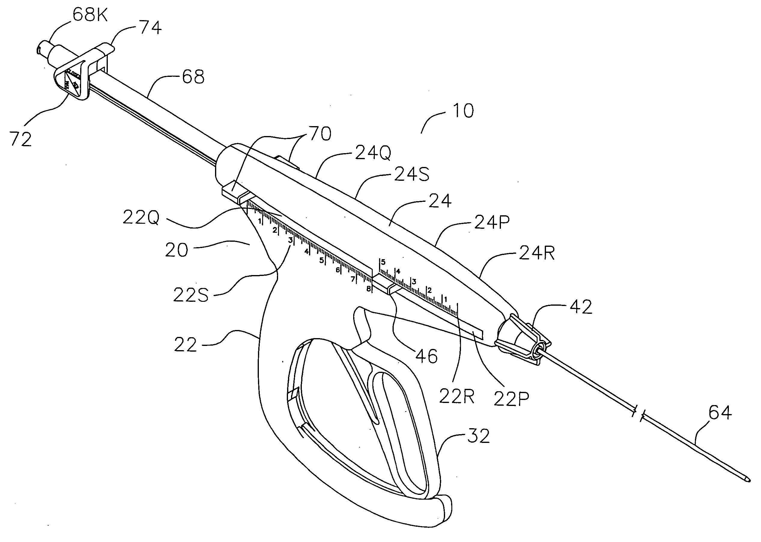
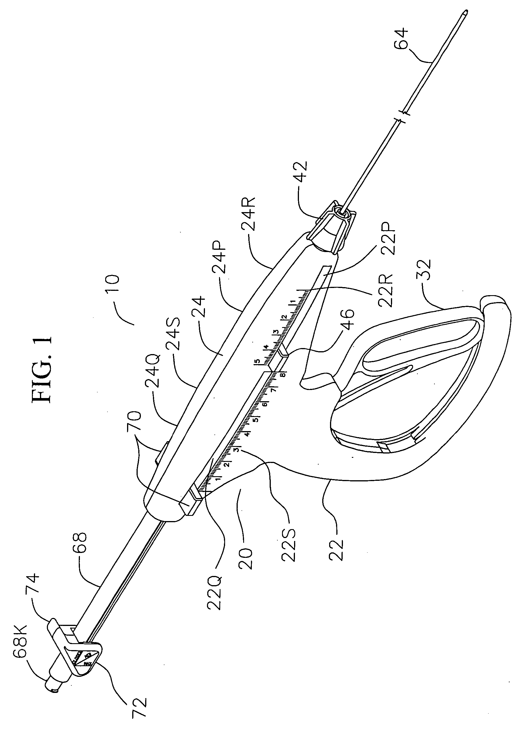
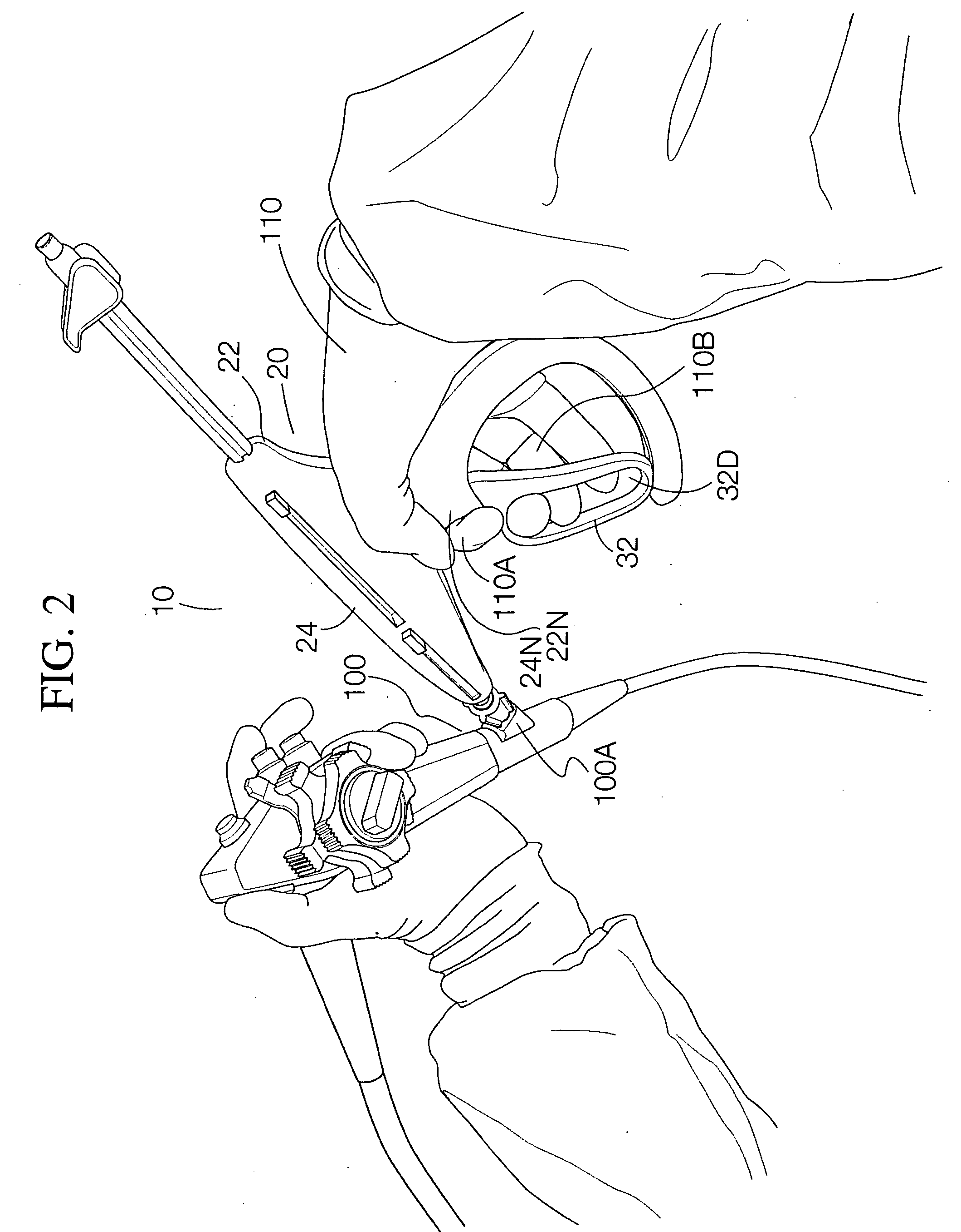
[0068] In accordance with a presently preferred embodiment of the invention, a medical diagnostic instrument is provided for the ergonomic, effective and safe harvesting of specimen at targeted remote tissue sites. The instrument includes a pistol grip style handle with a hand activated lever, customized adjustment features and a specialized elongated flexible double tube needle shaft inside of a protective sheath. For clarity, these novel design features will be presented here in the sequence that they are typically encountered in a routine EUS-FNA procedure. The instrument is attached to port in the proximal end of an echoendoscope with its sheathed flexible needle shaft placed within the scope's working channel. The length of needle shaft positioned inside the echoendoscope is adjusted using the button mediated needle shaft length adjustment feature. After imaging the location of the targeted lesion, the needle penetration depth is set by moving another set of buttons. With a squ...


PUM


Abstract

A medical diagnostic instrument for the ergonomic effective and safe harvesting of specimen at targeted remote tissue sites includes a pistol grip style handle with a hand activated lever and a specialized elongated flexible double tube needle shaft inside of a protective sheath for use, in a preferred embodiment, within an endoscope. The length of needle shaft is adjusted using locking buttons. Moving another set of buttons sets the needle penetration depth. With a squeeze of the lever, a novel thin band drive mechanism advances the needle shaft out of its sheath, through adjacent tissue and into the targeted site. The double tube needle shaft incorporates a pointed distal tip attached to an innermost “grater” needle, which has sharp edged tissue cutting holes or “grating” features that are exposed by retracting back an overlying cover tube. Sliding back the cover tube is achieved via a cam. The sharp cutting surfaces of the grater tube communicate directly with a vacuum source attached near the handle. Oscillation of the vacuum augmented grater tube back and forth within the targeted tissue yields small pieces of tissue for harvest. When adequate amounts of tissue are drawn into the grater tube, the cover tube is re-advanced over the distal grater tube, the needle shaft is retracted back into the sheath and the instrument along with the harvested specimen are removed from the patient.

Description

CROSS-REFERENCE TO RELATED APPLICATIONS [0001] Not applicable. STATEMENT REGARDING FEDERALLY SPONSORED RESEARCH OR DEVELOPMENT [0002] Not applicable. REFERENCE TO A “SEQUENCE LISTING”[0003] Not applicable. BACKGROUND OF THE INVENTION [0004] 1. Field of the Invention Field of the Invention [0005] This invention relates generally to a needle tissue harvesting instrument and more particularly to such a device including a needle having one or more tissue scraping apertures in a side wall of the needle and a cover tube coaxially disposed on the needle and slidably movable from an extended position covering the needle to a retracted position exposing one or more of the tissue scraping apertures. [0006] 2. Description of Related Art [0007] Diagnostic patient interventions are used to learn more about a patient's physical condition. Non-invasive diagnostics techniques (e.g., taking a patient's temperature, radiographic imaging, electrocardiographic monitoring, etc.) alone often do not provi...

Claims


Application Information

Patent Timeline
IPC IPC(8): A61B10/00
CPCA61B10/0233A61B2010/0208A61B10/04
Inventor SAUER, JUDE S.
Owner LSI SOLUTIONS
Why Eureka
Try Eureka