Actinic light colposcope and method to detect lesions in the lower female genital tract produced by human papilloma virus using an actinic light colposcope

a technology of human papilloma virus and colposcope, which is applied in the field of actinic light colposcope and method to detect lesions in the lower female genital tract, can solve the problems of high cost, large investment in equipment, and inability to diagnose hpv-induced lesions and cins

Inactive Publication Date: 2007-08-16
AVM LICENSING CORP
View PDF29 Cites 9 Cited by
  • Summary
  • Abstract
  • Description
  • Claims
  • Application Information

AI Technical Summary

Benefits of technology

[0029]The mechanical component further comprises a base or support, an arm, a bolster. The base or support comprises a rectangular steel plate, which provides overall stability to the equipment; four independent rotating wheels further comprising a brake mechanism which allows the equipment to be moved and set in place; a horizontal support further comprising a rotating articulation to maneuver and set the optical handle and bolster, and further holding in place the feeding source for the illumination equipment.
[0030]The arm comprises a counterbalanced mechanism which allows tri-axial movement, which in turn allows the user of the present invention to easily set the equipment in the precise location required by the gynecological procedure being conducted.
[0035]During the illumination step, the light emitted by the light bulb's filament (FIG. 1010) passes through the collector lens (FIG. 1020) which focuses the light beam which, in turn, is reflected at 90 degrees through a mirror placed for that purpose 45 degrees in the axis of the light's trajectory (FIG. 1,2030, 030). It cannot be overemphasized that the apparatus and method of the present invention results in an episcopic illumination. Episcopic illumination is based on the illumination norms which adhere to optical and microscopic principles, including Kohler's illumination. Kohler's illumination allows the use of luminescent energy with minimal waste and heat emission.
[0045]The following figure shows where, within the apparatus of this invention, the exciting filter (FIG. 1, 2040) is located. It is an easy-to-set-up and easy-to-use system. The light beam goes through the exciting filter (FIG. 1, 2040), and the ray goes through a first prism where it is sent to the frontal lens (FIG. 1, 2, 4070) of the object, which condenses the light to then illuminate the point of the tissue being observed within the patient's lower genital tract (FIG. 1, 4060).

Problems solved by technology

HPV induced lesions and CINs are not often easily diagnosed using the procedure outlined above.
Accordingly, many gynecologists have to use laboratory tests with wide margins of error, or use highly sophisticated and costly equipment, e.g., spectroscopy.
Such equipment usually requires a large investment of time and resources to train personnel, long waiting periods to obtain results and multiple visits by the patient.

Method used


Image

  • Actinic light colposcope and method to detect lesions in the lower female genital tract produced by human papilloma virus using an actinic light colposcope
  • Actinic light colposcope and method to detect lesions in the lower female genital tract produced by human papilloma virus using an actinic light colposcope
  • Actinic light colposcope and method to detect lesions in the lower female genital tract produced by human papilloma virus using an actinic light colposcope

Examples


Embodiment Construction

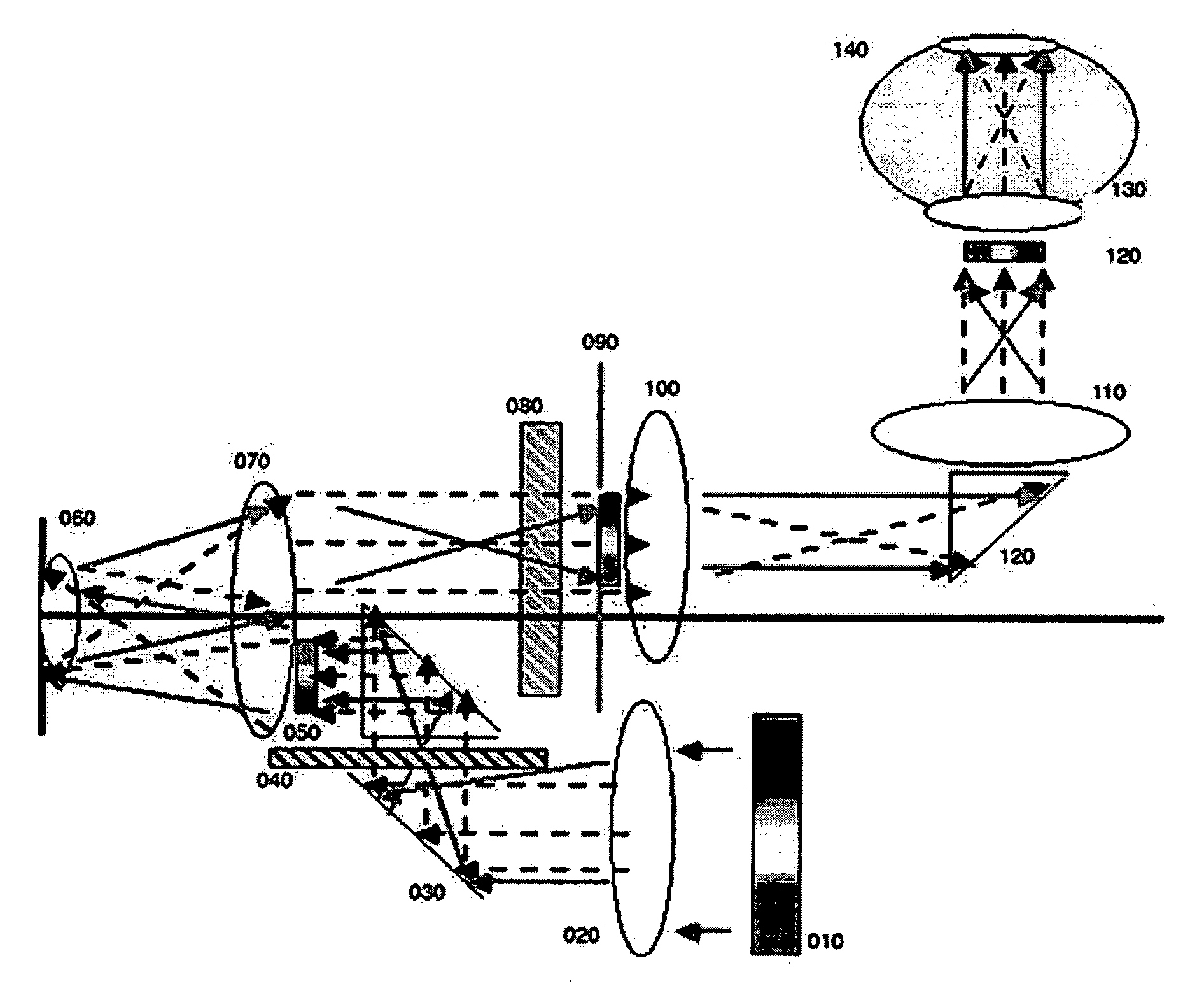
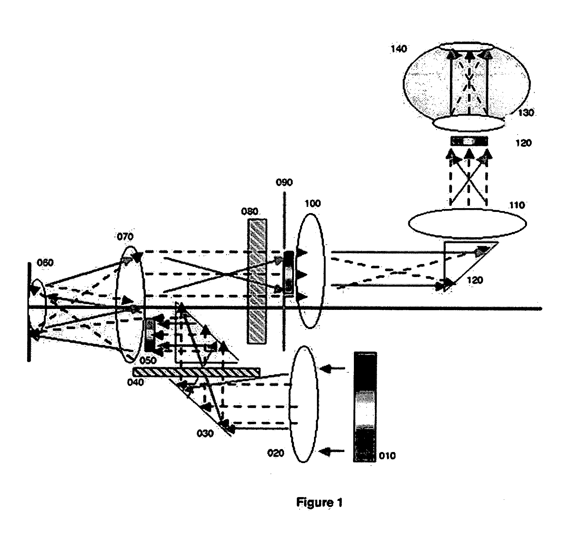
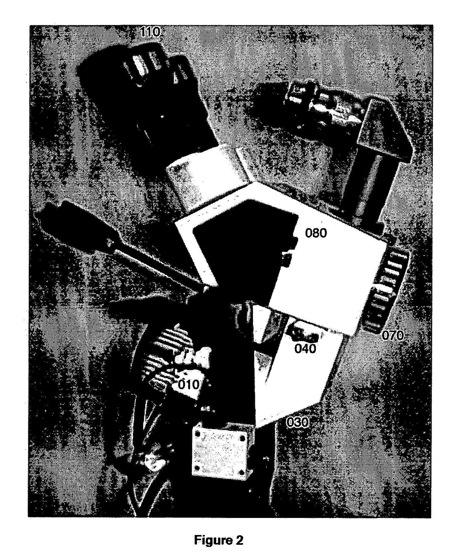
[0050]In its preferred embodiment, the present invention uses the fluorochrome Fluorescein Isothiocyanate for several important reasons. This system is commonly used by ophthalmologists to conduct retinal fluorangiography, and in a solution to see corneal lesions. Ophthalmologists perform those procedures using an actinic lamp which has a cobalt filter and a wide spectral emission. The actinic lamp used by ophthalmologists does not have suppressor filters. The method of this invention uses a fluorochrome because, among other reasons, it does not present toxic or adverse effects in humans. In the clinical context, the method of this invention comprises the steps of:[0051]setting the patient in the gynecological position;[0052]inserting the plastic vaginal speculum to avoid undesirable reflexes;[0053]visually locating the patient's cervix;[0054]taking a vaginal pH swab (frequently HPV infections present with other bacterial illnesses or parasitical infections are characterized by alka...


PUM


Abstract

The present invention involves an apparatus and method to detect lesions in the lower female genital tract produced by the Human Papilloma Virus using an actinic light colposcope. The apparatus of the present invention comprises mechanical, electrical and optical (also referred to herein as the actinic light) components. The method of the present invention comprises the general steps of: (1) illumination, (2) excitement, and (3) suppression. The method disclosed and claimed herein in the clinical context takes between 15 and 20 minutes and reliable results are obtained during the patient's office visit.

Description

RELATED APPLICATIONS [0001]Applicant claims the benefit of PCT Application No. PCT / MX 03 / 0088 filed with the PCT Receiving Office in Mexico City, Mexico on Oct. 27, 2003.ENTERING THE NATIONAL STAGE IN THE U.S. [0002]This application constitutes the entry of the National Stage in the U.S. of Application No. PCT / MX 03 / 0088 pursuant to 35 U.S.C. §371. Applicant is attaching hereto copy of PCT Application No. PCT / MX 03 / 0088 pursuant to C.F.R. §1.490(b) (1).TECHNICAL FIELD OF THE INVENTION [0003]Gynecological tools and methods, gynecological detection methods, tools and methods to detect lower female genital tract lesions, actinic light-based tools and methods to detect lower female genital tract lesions.BACKGROUND OF THE INVENTION[0004]The viral gender Papilloma is an extremely extensive DNA virus, which infects many animal species populations, including humans. There are over 100 types of known Papilloma viral sources, which when infecting humans are known as Human Papilloma Virus (HPV...

Claims


Application Information

Patent Timeline
Patent Type & Authority Applications(United States)
IPC IPC(8): A61B1/00
CPCA61B1/00188A61B1/00193A61B1/0646A61B1/00186A61B1/043A61B1/303
Inventor DIAZ SANCHEZ, JOEL GERARDO
Owner AVM LICENSING CORP
Why Eureka
Try Eureka