Method for detecting a functional signal in retinal images

a functional signal and image technology, applied in the field of retinal image functional signal detection, can solve the problems of difficult detection of functional signal and difficult monitoring of changes, and achieve the effect of removing noise and assessing the health status of the retina

Inactive Publication Date: 2007-09-13
KESTREL CORP
View PDF6 Cites 6 Cited by
  • Summary
  • Abstract
  • Description
  • Claims
  • Application Information

AI Technical Summary

Benefits of technology

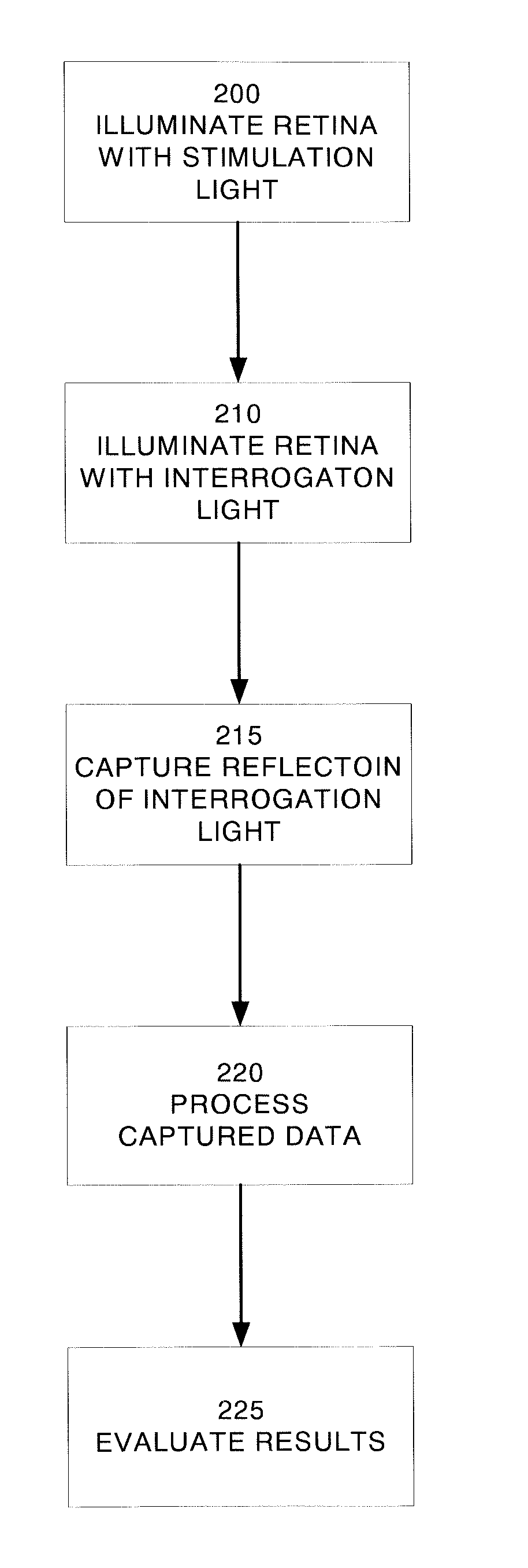
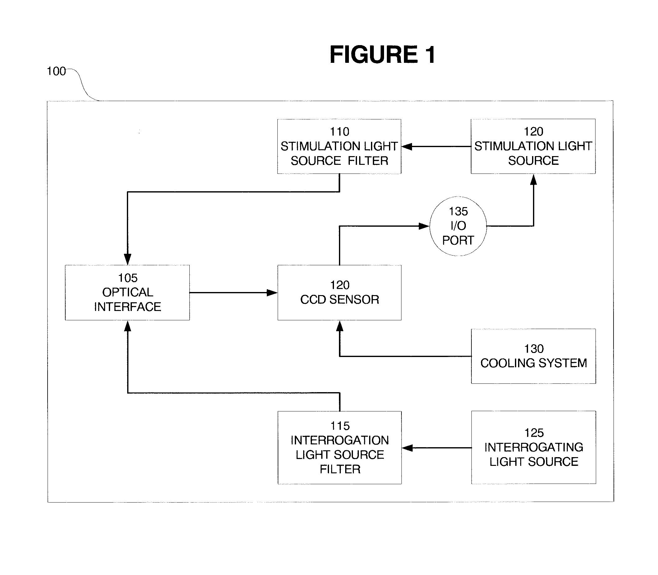
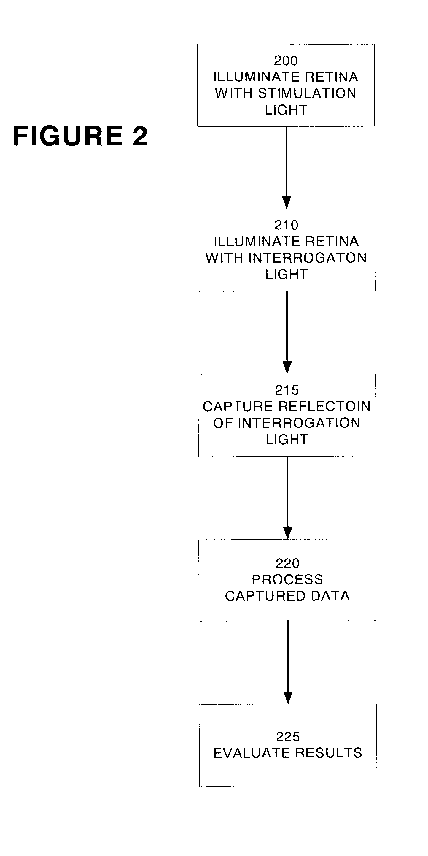
[0012] An embodiment of the present invention is a system and method for optical mapping of retinal function using retinal imaging. In this embodiment, the retina is stimulated in a selected spectral band (for example and without limitation 530 nm±5 nm) centered on the green maximum sensitivity of the retina under photopic conditions. The reflected intensity from the retina is measured at an interrogating spectral band that indicates the state of hemoglobin saturation before and after visual stimulation. To maximize the signal to noise ratio (SNR), an interrogation wavelength centered on, for example and without limitation, 700±20 nm was used, where retinal stimulation is minimal and the difference in absorption between oxyhemoglobin and reduced hemoglobin is greatest. The optical changes that result from retinal neuronal activity are mapped by registration of recorded CCD frames that have been corrected for noise effects (as more fully set forth below), with subsequent comparison of post-stimulation images from pre-stimulation images. In an alternative embodiment of the present invention, a hemifield of the retina is stimulated in the selected spectral band (530 nm±5 nm) centered on the green maximum sensitivity of the retina stimulated so that both stimulated regions of the retina and non-stimulated regions of the retina can be imaged simultaneously, thereby further reducing variability due to temporal recording of images. Variable stimulation patters may be used in either the hemifield stimulation or the full retinal stimulation to maximize the resultant reflectance recorded.
[0016] It is yet another aspect of the present invention to increase the sensitivity of current visual field testing methods.
[0017] It is yet another aspect of the present invention to improve diagnosis of eye disease with improved retinal images.
[0024] The optical changes that result from retinal neuronal activity are captured by the detector. The changes are mapped by registration of recorded image frames. The raw data is preprocessed to eliminate unwanted artifacts, such as blinking or excessive eye movement. Because the resulting signal from the retinal activation contains noise from other sources (for example, the non-stimulated retinal background and other unknown physiological changes), the data is further processed to remove noise. In an embodiment of the present invention, principal components analysis (PCA) is used to isolate the signal representing the state of hemoglobin saturation before and after visual stimulation. In another embodiment of the present invention, blind source separation (BSS) (using the extended spatial decorrelation (ESD) algorithm) and independent component analysis (ICA) (using the Fast-ICA algorithm) are used to extract the functional signal from the retinal videos. By comparison of post-stimulation images from pre-stimulation images, and applying the data analysis techniques of the present invention, measurements of changes in blood perfusion due to neural activity resulting from visual stimulation of the photoreceptors in the human retina can be made, and hence the health status of the retina can be assessed.

Problems solved by technology

However, perimetry remains a subjective test that requires the subject to make important judgments during the test that can be clouded by anxiety, fatigue, or lack of concentration.
A second problem with the current perimetry tests is that almost 40-50% of the optic nerve may be damaged before a significant perceptual change can be detected on the visual field test, making it relatively insensitive for detecting early damage when intervention may still save vision.
A third problem is that the visual field test is highly variable in areas of defects where damage has occurred, making it difficult to monitor changes.
However, the measured changes in reflectance in response to the visual stimulus are on the order of 0.1% to 1.0% of the total reflected intensity level that makes the functional signal difficult to detect by standard methods since it is masked by the other signals (noise) that are present.

Method used


Image

  • Method for detecting a functional signal in retinal images
  • Method for detecting a functional signal in retinal images
  • Method for detecting a functional signal in retinal images

Examples


Embodiment Construction

[0038] Independent component analysis (ICA) is a statistical and computational technique used to reveal hidden factors that underlie a set of random variables, in this case, measurements of reflectance from a retina. The goal is to recover independent sources given only the sensor observations that are unknown linear mixtures of the unobserved independent source signals. Thus ICA is use to analyze mulitvariate data stemming from the production of images of the retina. ICA is related to Principle Component Analysis (PCA) and factor analysis but is more capable of finding underlying sources or factors in a data set because it takes into account higher order statistical properties of the data. For example, PCA is a correlation based transformation of data. In contrast, ICA not only decorrelates the signals (i.e. 2nd order statistics) but also reduces the higher order statistical dependencies (i.e. 4th order cumulants) and attempts to make the signals detected as statistically independe...


PUM


Abstract

A system and method for detecting a functional signal in retinal images. An optical imaging device comprises a stimulation light source, an interrogating light source, and a detector. The retina is stimulated by the stimulation light source. The retina is then illuminated by an interrogation light, and the reflected intensity from the retina is measured at an interrogating spectral band that indicates the state of hemoglobin saturation before and after visual stimulation. The optical changes that result from retinal neuronal activity are captured by the detector. The signal representing the state of hemoglobin saturation before and after visual stimulation is isolated. In an embodiment of the present invention, this signal is isolated using principle components analysis (PCA). In another embodiment of the present invention, blind source separation (BSS) and independent component analysis (ICA) algorithms such as extended spatial decorrelation and fast-ICA are used to isolate the functional signal from the retinal videos.

Description

CROSS REFERENCE TO OTHER APPLICATIONS [0001] This application claims benefit under 35 U.S.C. § 119(e) of U.S. Provisional Application Ser. No. 60 / 401,224, entitled “Method for Determining Signal Detection in Retinal Videos,” filed Aug. 5, 2002, which is incorporated by reference herein, in its entirety, for all purposes. [0002] This application is a continuation of U.S. patent application Ser. No. 10 / 628,292, entitled “Method of Detecting A Functional Signal in Retinal Images,” filed Jul. 28, 2003.FIELD OF INVENTION [0003] The present invention relates generally to a process for detecting functional signal in retinal images. More particularly, the present invention relates to a process for extracting the functional signal from the background noise via advanced statistical techniques yielding a functional signal from retinal activation in the presence of noise from other sources. BACKGROUND OF THE INVENTION [0004] Visual field testing (perimetry) is the most widely used method for de...

Claims


Application Information

Patent Timeline
Patent Type & Authority Applications(United States)
IPC IPC(8): A61B3/14A61BA61B3/00A61B3/10A61B3/12A61B5/00A61B13/00
CPCA61B3/12A61B5/0059A61B5/14555
Inventor SOLIZ, PETERBARRIGA, EDUARDO
Owner KESTREL CORP
Why Eureka
Try Eureka