
Intracochlear Nanotechnology and Perfusion Hearing Aid Device

a hearing aid and nanotechnology technology, applied in the field of intracochlear hearing aid devices, can solve the problems of inability to convert mechanical energy into electrical energy, poor result of cochlear implants, and often destroyed anatomy within cochlear ducts

Inactive Publication Date: 2007-09-27
DOMESTIC ASSET LLP
View PDF36 Cites 113 Cited by
  • Summary
  • Abstract
  • Description
  • Claims
  • Application Information

AI Technical Summary

Benefits of technology

[0028]The present invention will be understood in more detail from the drawings and description presented be

Problems solved by technology

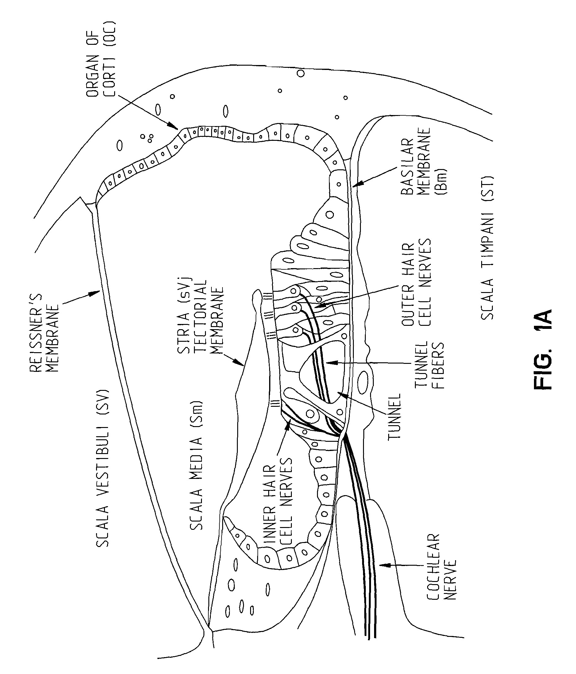
Even though the ear bones are vibrating and the inner ear fluids are vibrating, if the Organ of Corti is not functioning, then mechanical energy cannot be turned into electrical energy.
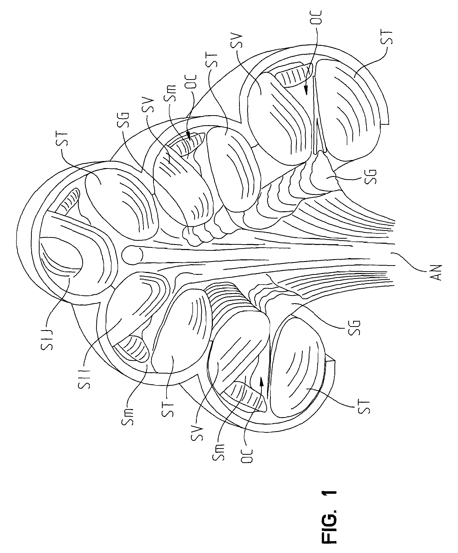
In a hearing impaired person, the anatomy within the cochlear duct is often destroyed.
Further, when medical care of hearing loss is rendered, specifically with cochlear implantation, if the anatomic structures, in addition to the Organ of Corti, are decreased in number or not present to transmit the stimulus provided by the implant, a poor result would be expected from the cochlear implant.
When cochlear implants were first being conceived and tested, it was not known how best to approach the cochlear nerve with the electrical impulse electrode studs.
One of the problems in dealing with intra-cochlear implants is that the small size of the inner ear structures such as the Organ of Corti, dendrites, spiral ganglion cells, and all the other anatomic structures within the cochlea severely limits the size of any intra-cochlea implant.
Such implant-induced damage deleteriously impacts the performance of the spiral ganglion SG and the cochlear nerve AN that are in the modiolus.
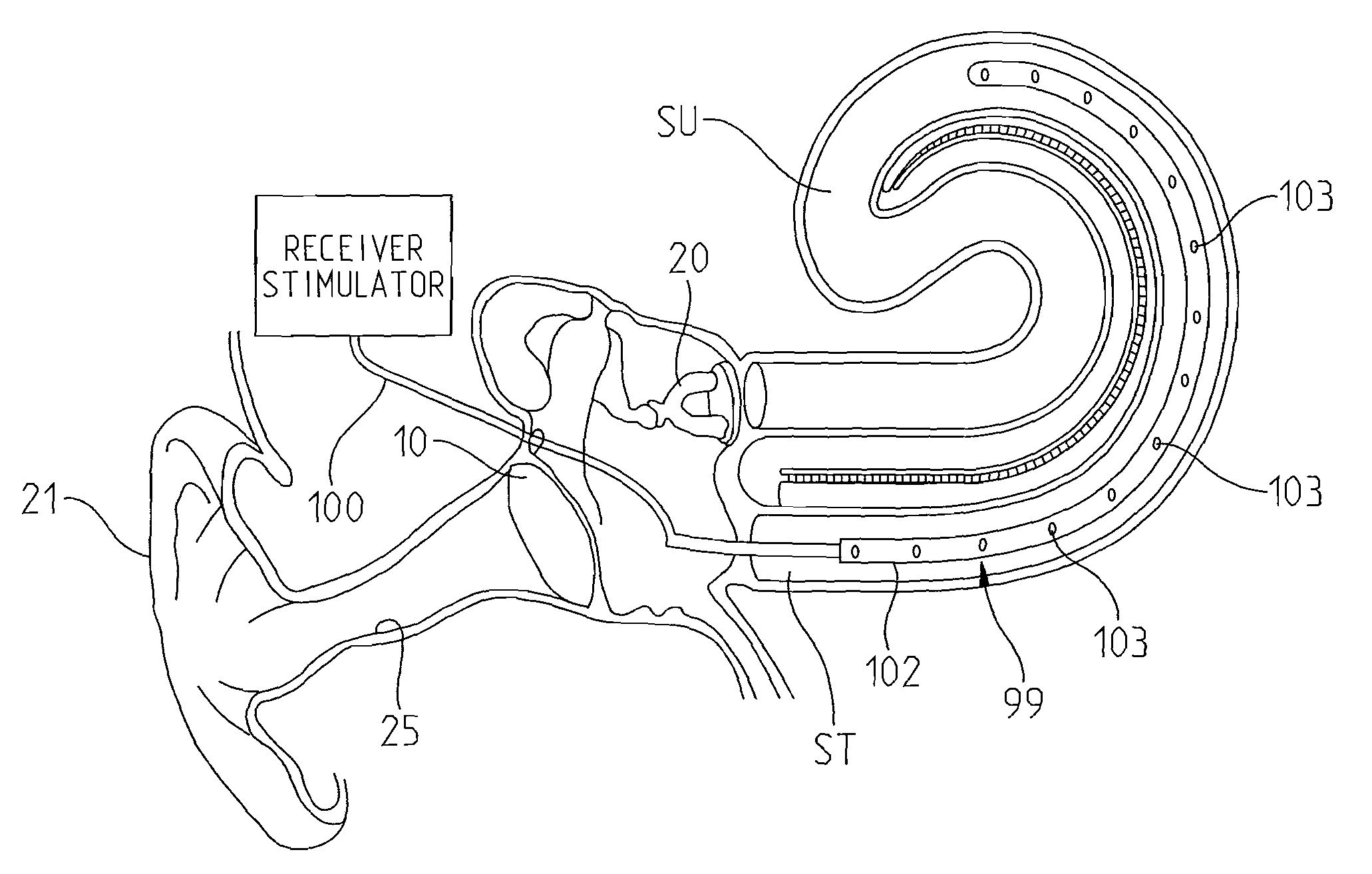
Thus, an intra-cochlear implantation technique can be very traumatic and reduce the ability of the implant to successfully stimulate residual neural elements in patients with hearing loss. FIG. 2 diagrammatically shows the prior art implant electrode wire 100 of an intra-cochlear implant attached to the electrode 102 with stimulating points 103 within the cochlea.
There may be lesser degrees of trauma with these newer implants although the damage they can potentially cause is still considerable.
Another major problem with present implants is that they have a relatively limited number of electrodes from which to stimulate the cochlea.
Thus, a major deficiency of present implants is that they do not adequately stimulate in numbers all along the cochlea.
The result is lack of resolution within the sound-perception spectrum.
Another major problem with presently available implants is that they do nothing to help regenerate any of the anatomic deficiencies just described.
With present implants there are no means of delivering chemicals, medications, nutrients, nucleotides, and cells into the cochlea and / or perfusion of these substances and cells into and out of the cochlea.
Another deficiency with presently available implants is that electrical stimulus transmission is not transmitted from an electrode stud to a nerve structure by directly touching or even being intimate to one.
Therefore, a major deficiency of the intra-cochlear implant prior art is the relatively low number of stimulating electrode points that are placeable on an implant.
As alluded to above, the primary drawbacks of current known implants are: (1) the internal destruction they cause to the cochlea; (2) the size limitations that they impose; (3) the lack of tonal and tonotopic specificity; (4) the lack of direct nerve touching electrical stimulation; and (5) the lack of a mechanism to achieve anatomic regeneration.
One of the vexing problems that face those designing implants is the limitations that exist on the potential size of implant.
Implant size limitations exist because there is only so much internal diameter to the cochlea to allow an electrode to pass.
Thus, current technology only allows for so many electrode stimulating studs inside the cochlea because the sum diameter of the many numbers of wires to the electrode studs is limited by the cochlear diameter.
The electrode sizes are destructively large and stiff.
Additionally, the space limitations do not allow for perfusion hardware and cell-nerve regeneration techniques within the cochlea.

Method used


Image

  • Intracochlear Nanotechnology and Perfusion Hearing Aid Device
  • Intracochlear Nanotechnology and Perfusion Hearing Aid Device
  • Intracochlear Nanotechnology and Perfusion Hearing Aid Device

Examples


Embodiment Construction

[0040]A schematic view of the intra-cochlear implant system 300 of the present invention is shown in FIG. 4, it being understood that the view in FIG. 4 is drawn highly schematically to acquaint the user with the primary components of the device.

[0041]The implant system 300 includes an intra-cochlear implant 302 that is designed for implantation preferably within the scala tympani ST of the cochlea, although it may also be placed with the scala vestibuli SV or scala media (also known as the cochlear duct) SM of the cochlea. In FIG. 4, the implant 302 is placed within the scala tympani ST. The implant 302 comprises a body portion 301 comprised of a bundle of electrodes that includes a large number of individual electrodes, as represented by electrode 304 for conducting a signal to an internal area of the scala tympani ST that is preferably adjacent to the hair-like nerve endings NE of the Cochlea, Organ of Corti OC, and spiral ganglion cells of the cochlea. The body 301 has a primary...


PUM


Abstract

An intra-cochlear implant is provided for aiding in the hearing of a patient. The implant includes a body portion implantable within an interior of a cochlea of a patient. The body portion has a proximal end, a distal end and a primary axis. A plurality of signal carrying electrodes extends along the body portion. The electrodes have proximal ends and distal ends, with the proximal ends being capable of receiving a signal from a signal generator, and the distal ends being capable of delivering the received signal to an anatomical receptor within a cochlea. At least several of the plurality of electrodes have a nanoelectrode-sized portion. The implant also may include a fluid delivery system of tubules, reservoirs, and pumps for the delivery of chemicals and cells to activate regeneration of neural elements lost during the hearing loss process.

Description

I. FIELD OF THE INVENTION [0001]The present invention relates to hearing aid devices, and more particularly to an implantable intra-cochlear hearing aid device using nano-technology.II. BACKGROUND OF INVENTION[0002]The genesis of the instant invention was in the multiple deficiencies that the Applicants found with existing intra-cochlear hearing aid implants of the ear. To understand the issues that give rise to the need for implantable hearing aids, it is helpful to first understand the anatomy and physiology of the ear, and in particular, the cochlea and its associated structures.[0003]Referring to FIG. 3, there is shown a sectional view of the anatomy of the human auditory system A. At a macro level, the human auditory system A consists of the outer ear 0, the middle ear M, and the inner ear I. The outer ear 0 includes the pinna or auricle 21 and the ear canal 25 that provides communication from the outer ear 0 to the middle ear M. The middle ear M includes the eardrum (tympanic ...

Claims


Application Information

Patent Timeline
IPC IPC(8): A61N1/36
CPCA61N1/36032A61N1/0541A61N1/36038
Inventor FRITSCH, MICHAEL H.FRITSCH, JOHN H.FRITSCH, JOSEPHINE
Owner DOMESTIC ASSET LLP
Learn More