

Kinetic metabolic assay for antifungal susceptibility testing
a susceptibility testing and metabolic assay technology, applied in the field of metabolic assays, can solve the problems of increasingunable to predict clinical outcomes, and unable to meet the needs of patients, and achieves the effect of reducing the cost of patient health care, and improving the quality of li
- Summary
- Abstract
- Description
- Claims
- Application Information
AI Technical Summary
Problems solved by technology
Method used
Image


Examples
example
Example 1
Materials and Methods
[0035]C. albicans strain and medium. C. albicans CA-1 isolate obtained from the culture collection of Diane Brawner (Microbiology Department, Montana State University)(14). The strain was stored at −80° C. Planktonic cells were cultured in 2% YEPD medium (2% glucose, 1% bacto yeast extract, and 2% bacto peptone). The solid agar medium for the CFU assay was was 1% glucose, 0.5% bacto yeast extract, 2% bacto agar, 0.1% ammonium sulfate dissolved in 20% tap water and 80% D.I. water.
[0036] Modified growth medium. In addition to the 2% YEPD medium, the modified growth medium contained optimal concentrations of ergosterol (Alfa Aesar; catalog no. 57-87-4), MgCl2(Sigma; catalog no. M4880), and KCl (Sigma; catalog no. P5405). The final concentrations of these reagents in the kinetic assays were 40 μM ergosterol, 88 μM MgCl2, and 42.5 μM KCl.
[0037] CFU assay. CFU were estimated for both AmB treated and untreated planktonic sample. 100 μl planktonic C. albica...
example 2
Time Course of Metabolic Reduction
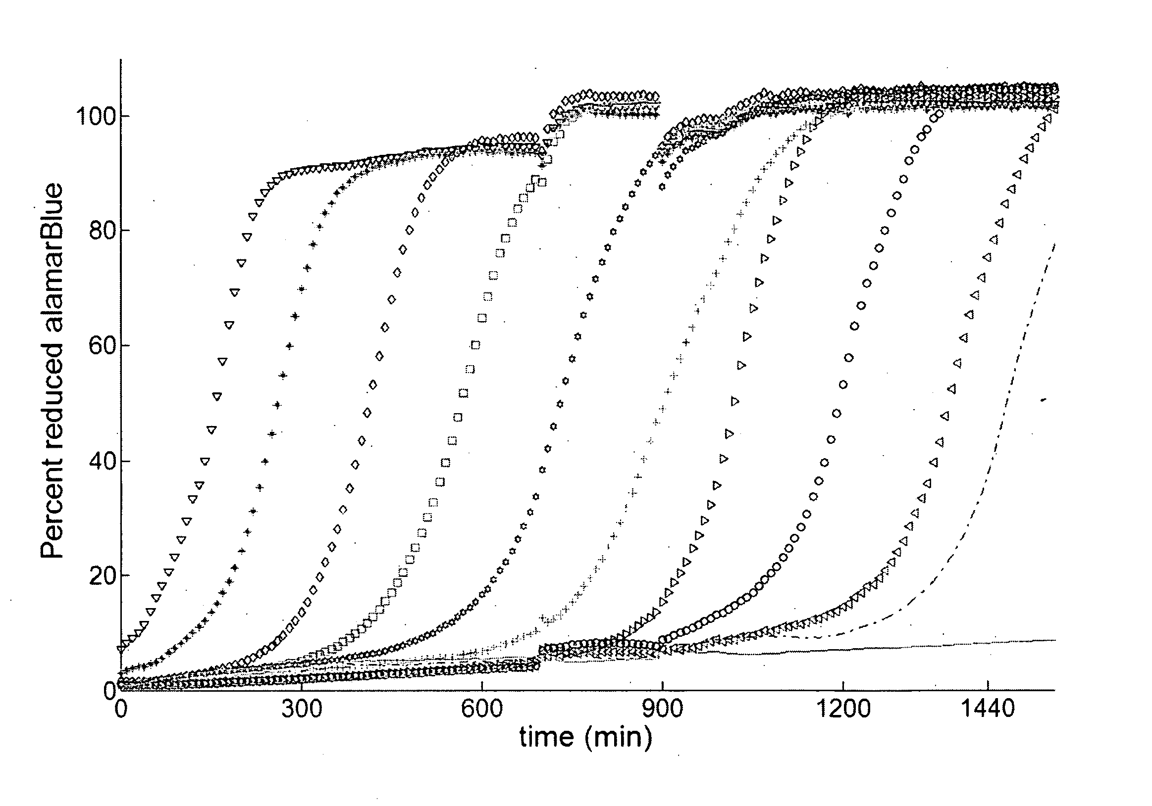
[0049] Metabolic assays are either performed during (10, 37, 48), or after treatment of the antifungal agent (1, 5, 45). In either case these assays measure the degree of metabolic reduction by endpoint analysis (5, 20, 39, 45). KMA presents an alternative approach in which, for example, the reduction of the metabolic indicator is followed in real-time. The KMA enhances the dynamic range up to six orders of magnitude by taking advantage of cell proliferation to amplify viable cells.
[0050] The kinetic curves of metabolic reduction of XTT and alamarBlue followed a sigmoidal shape (FIG. 1). The corresponding curves of turbidimetric changes had an overlapping profile implying that metabolic reduction was primarily occurring due to cell proliferation. This was expected because all cells were placed in a fresh growth medium after drug treatment by isolating them from the solution with drug. FIG. 1 shows the kinetic curves for the alamarBlue assay for a ...
example 3
Calibration and Dynamic Range of the KMA
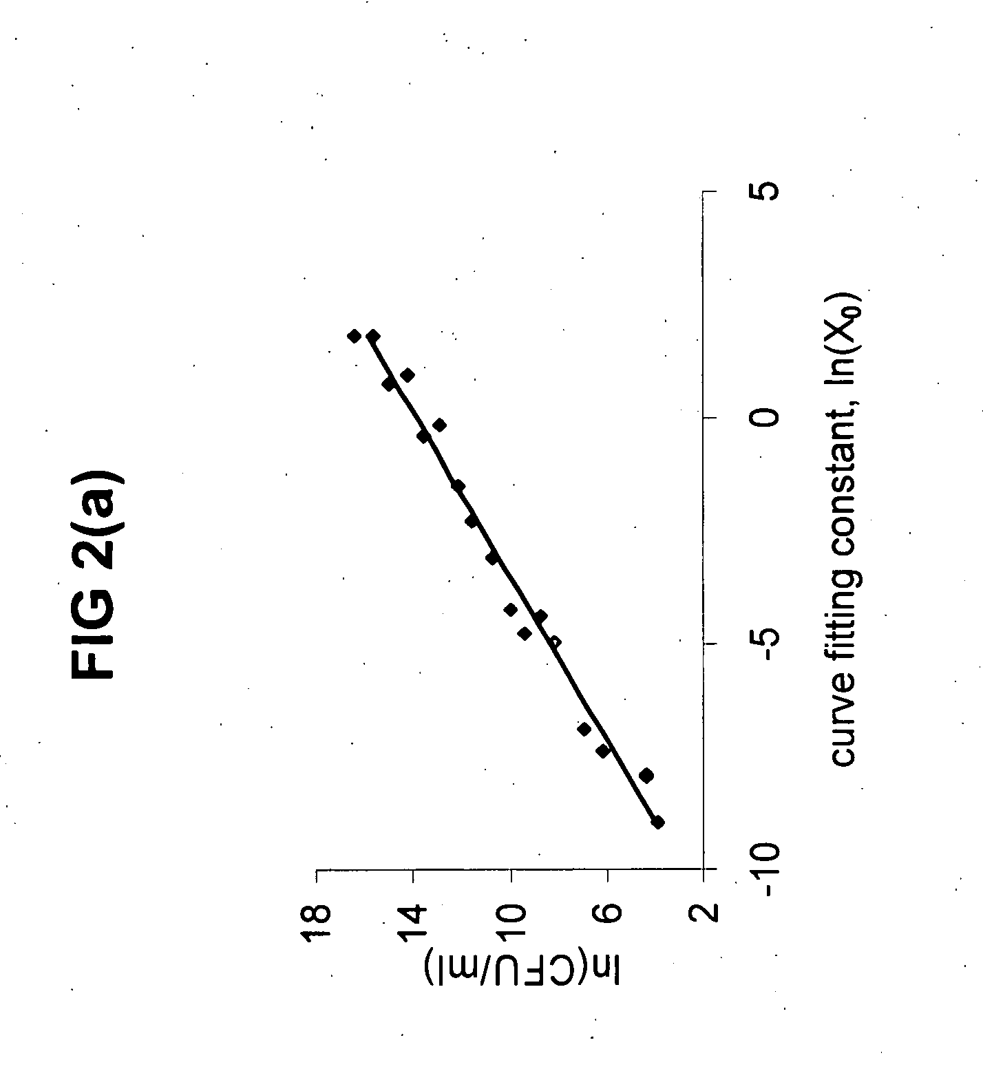
[0051] The KMA based on XTT or alamarBlue were calibrated with CFU. An additional assay based on the same kinetic principle was developed using turbidimetric changes as an indicator. As a result, four assays were simultaneously applied to every planktonic sample. Three assays constituted kinetic analysis and the fourth was a CFU assay. In addition, two data techniques curve fitting and t-threshold) were used to analyze data from each kinetic assay. The kinetic assays were performed in the presence of an unmodified- and also a modified 2% YEPD medium designed to quench the action of residual AmB after treatment. C. albicans chosen for all the calibration experiments were from two planktonic growth phases, exponential- and stationary-phase cells.
[0052] Calibrations were constructed for all combinations of variables (Table 2). FIG. 2a and 2b show an example of the calibrations constructed for the alamarBlue indicator using both techniques of da...
PUM
 Abstract
Description
Claims
Application Information