Simulated heart valve root for training and testing

Active Publication Date: 2007-11-01
EDWARDS LIFESCIENCES CORP
View PDF99 Cites 22 Cited by
  • Summary
  • Abstract
  • Description
  • Claims
  • Application Information

AI Technical Summary

Benefits of technology

[0008] The present invention provides a simulated heart valve root that more faithfully re-creates the anatomy of a diseased patient. The aortic root may be used for training purposes, or alternatively in a flow tester to examine the implanted valve in use for paravalvular leaks. The simulated heart valve root may be incorporated into a simulated heart for more realistic training purposes.
[0014] In a supplement to the training system, a means for vibrating the simulated heart is provided for more realism. Furthermore, a simulated radiopaque rib cage or spinal column may be provided around the simulated heart.
[0015] In a further aspect of the invention, a method of training a physician to implant a prosthetic heart valve utilizes a simulated heart valve root including a tubular body formed of a flexible material and defining an annular ledge. The heart valve root is mounted in a fixture having an access port, and physicians are instructed in delivering and implanting a prosthetic heart valve within the heart valve root. For more realism, the fixture may be subject to vibratory motion, or fluid may be directed through the heart valve root.

Problems solved by technology

Conventional heart valve surgery is an open-heart procedure that is highly invasive, resulting in significant risks include bleeding, infection, stroke, heart attack, arrhythmia, renal failure, adverse reactions to the anesthesia medications, as well as sudden death.
This is sometimes difficult given the highly calcified condition of the aortic annulus in particular.
Furthermore, due to the remote nature of expandable valve replacement procedures, the physician does not have the luxury of carefully positioning and then securing the periphery of the valve to the annulus with sutures, as with conventional open-heart techniques.
However, the animals used are typically healthy specimens whose heart valves are unlike the calcified or otherwise distorted annuluses of the typical prosthetic valve recipient.

Method used


Image

  • Simulated heart valve root for training and testing
  • Simulated heart valve root for training and testing
  • Simulated heart valve root for training and testing

Examples


Embodiment Construction

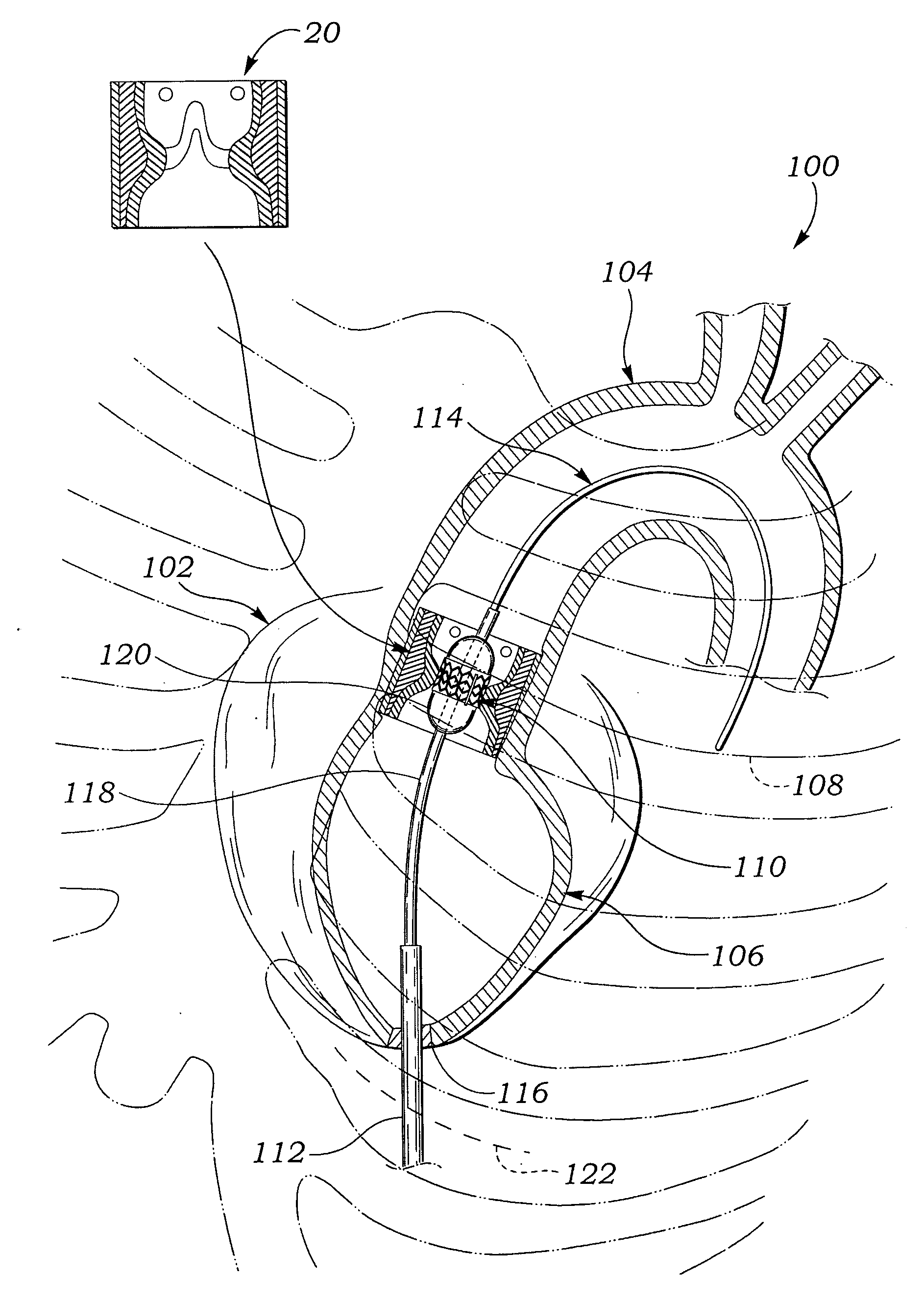
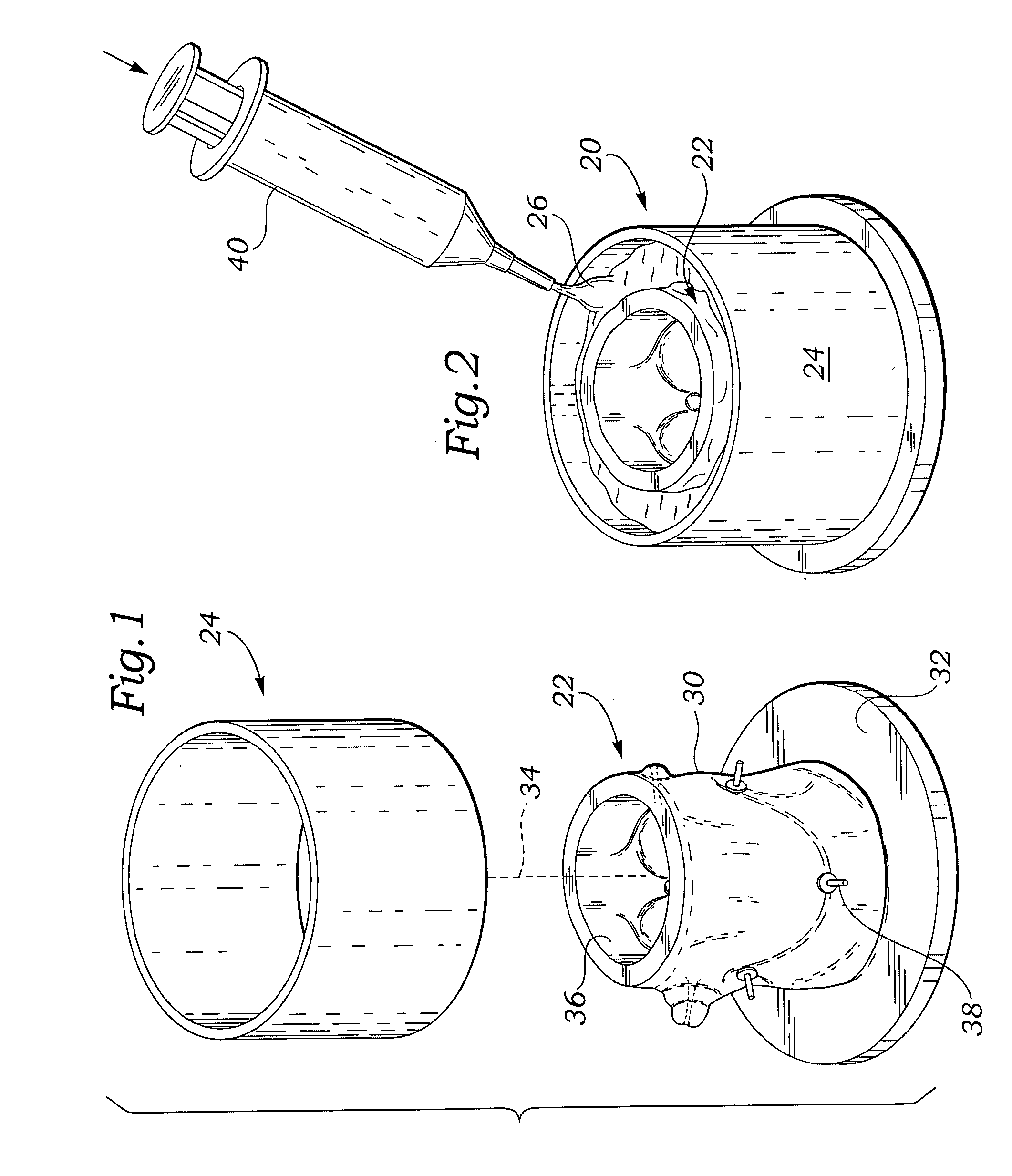
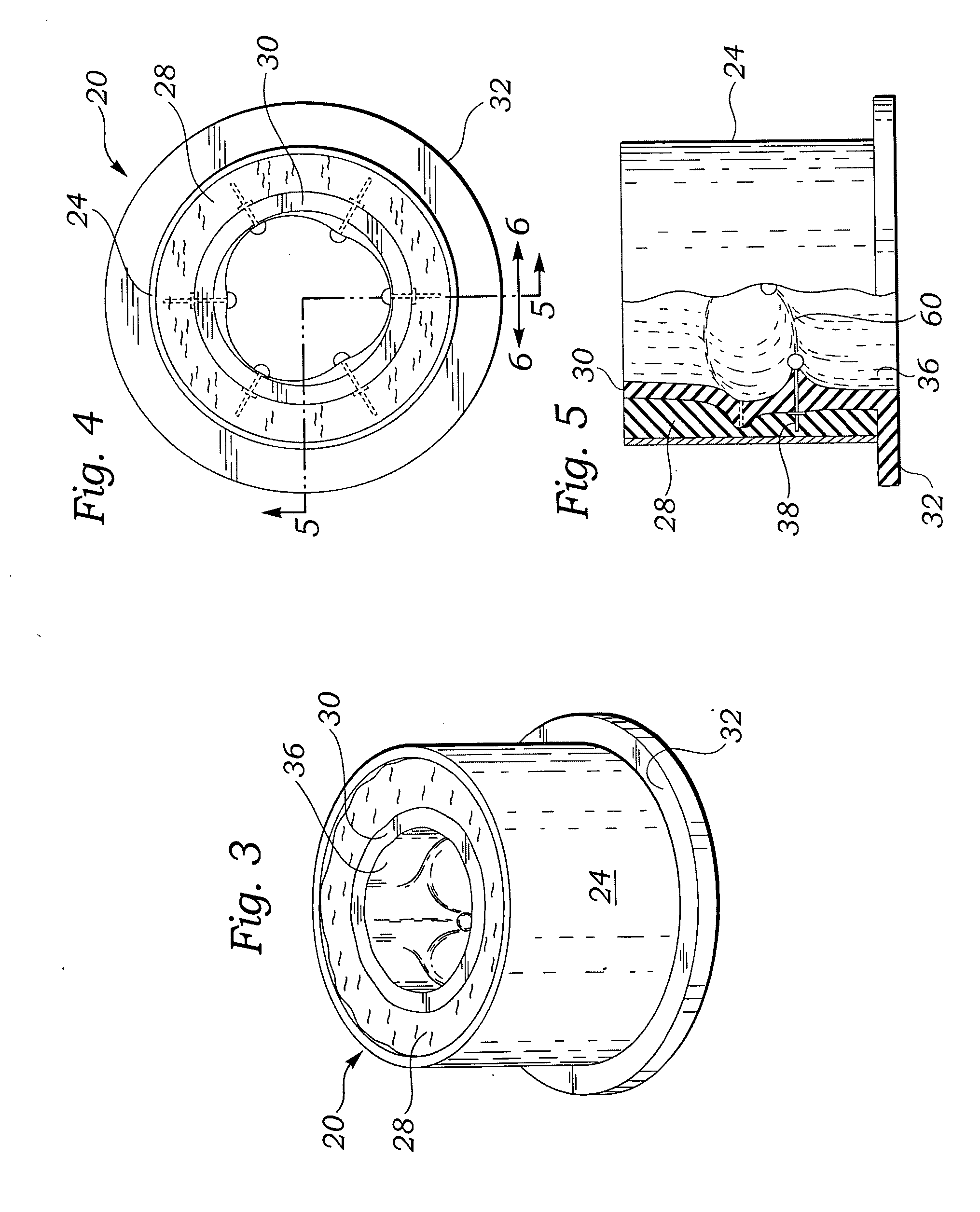
[0028] The present invention provides a simulated aortic root that is constructed to more realistically mimic the diseased annulus of the typical patient. No specifically, the simulated aortic root of the present invention provides calcification and other anatomical abnormalities that more faithfully re-creates the diseased aortic root. These simulated features are critical in teaching positioning and deploying both MIS and percutaneous aortic valves. Moreover, the more realistic aortic root provides an invaluable tool for in vitro testing to assess the paravalvular and migration performance of MIS and percutaneous prosthetic valves.

[0029] In the context of the present invention, the term “aortic root” refers to at least the tubular section of the aorta (the large artery leaving the heart) that is attached to the heart. The natural aortic root includes the annulus (tough, fibrous ring) and leaflets of the aortic valve, and the openings where the coronary arteries attach (coronary o...


PUM


Abstract

A simulated heart valve root used for training physicians in techniques of implantation of prosthetic heart valves as well as for more realistically testing the efficacy of prosthetic heart valves. The simulated heart valve root is made of the flexible, tubular body having an inner wall defining an annular ledge within which the prosthetic heart valve is implanted. Discrete nodes or areas of simulated calcification may be provided on the annular ledge. A simulated aortic root includes alternating cusps and commissures with calcification simulated at least at one of the commissures. A tear in the annular ledge may also be provided which simulates a tear that might occur from a valvuloplasty procedure. A reinforcing sleeve may surround the flexible tubular body to provide rigidity or hoop strength thereto. A method of testing includes mounting the simulated heart valve root in a flow conduit, implanting a prosthetic heart valve in the root, applying pulsatile flow to the assembly, and monitoring for leaks. The simulated heart valve root may also be incorporated within a larger simulated heart for use in training physicians to remotely implant prosthetic heart valves.

Description

FIELD OF THE INVENTION [0001] The present invention relates to fixtures and methods for testing the performance of prosthetic heart valves and, in particular, a simulated diseased heart valve root for a more realistic test. BACKGROUND OF THE INVENTION [0002] Heart valve replacement may be indicated when there is a narrowing of the native heart valve, commonly referred to as stenosis, or when the native valve leaks or regurgitates, such as when the leaflets are calcified. In one therapeutic solution, the native valve may be excised and replaced with either a biologic or a mechanical valve. Prosthetic valves attach to the patient's fibrous heart valve annulus, with or without the leaflets being present. [0003] Conventional heart valve surgery is an open-heart procedure that is highly invasive, resulting in significant risks include bleeding, infection, stroke, heart attack, arrhythmia, renal failure, adverse reactions to the anesthesia medications, as well as sudden death. Fully 2-5% ...

Claims


Application Information

Patent Timeline
IPC IPC(8): G09B23/28
CPCG09B23/32A61F2/2472
Inventor LAFRANCE, HUGUESSTOBIE, ROBERT
Owner EDWARDS LIFESCIENCES CORP
Why Eureka
Try Eureka