
Curved needle assembly for subcutaneous light delivery

Inactive Publication Date: 2008-01-10
DELIANIDES THEODORE PHILIP +1
View PDF11 Cites 16 Cited by
  • Summary
  • Abstract
  • Description
  • Claims
  • Application Information

AI Technical Summary

Benefits of technology

[0016]Second, the integral fibers function to deliver light below the surface of the skin to the tissue at the tip of the needle. This is a particularly advantageous configuration for photoplethysmographic measurement techniques such as pulse oximetry. In a photoplethysmographic sensor of this configuration, the emitted light must travel from the emitter, in this case the distal end of the needle, through the intervening tissue before it can be incident on the detector situated in the base of the sensor body. This eliminates the possibility of light shunting directly from the emitter to the detector as sometimes occurs in “reflectance mode” pulse oximetry sensors. With reflectance sensors, whether for fetal or adult use, light can travel directly from the emitters to the detector, without ever entering the tissue-under-test, thereby causing erroneous measurements.
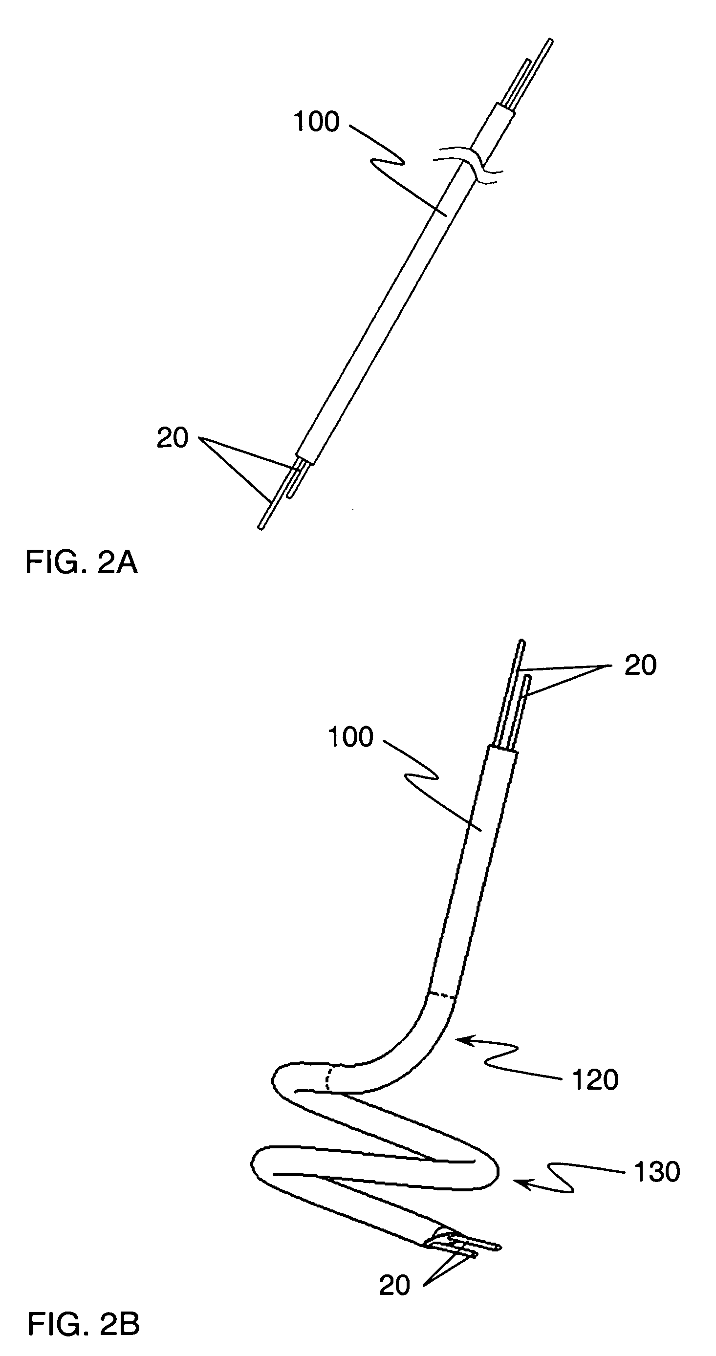
[0019]Two main elements of this invention make this a manufacturable assembly. The optical fibers are inserted into the hollow metal tube, or needle, prior to bending the tube into its curved shape. This eliminates any source of resistance while inserting the fibers into the needle. Once the fibers are in place inside the needle, the assembly is bent around a mandrel into its final form. In the current embodiment of this needle assembly there are two separate bends required. The first sweeps the needle from the straight portion that will extend through the sensor body towards the helix, and the second bend is the helical portion of the needle assembly, the portion that is rotated into the tissue-under-test.
[0020]The second element of this invention involves creating the pointed or beveled end of the needle assembly. The distal end of the needle, the end which is first inserted into the tissue-under-test, must be pointed to allow easy insertion into the tissue. To create this pointed end of the needle, the tip of the distal end is polished to create a bevel. If the polishing operation is performed anywhere on the helical portion of the assembly, however, the fibers will fracture or shatter along the beveled portion. This diminishes the optical transmissivity and could allow small pieces of the fibers to be left behind when the sensor is removed from the tissue-under-test. This fracturing occurs because the bent fibers are under stress and the polishing or cutting process used to create the bevel causes scoring of the fibers. The fibers then fracture along the score lines to relieve the bending stress in the glass.

Problems solved by technology

With reflectance sensors, whether for fetal or adult use, light can travel directly from the emitters to the detector, without ever entering the tissue-under-test, thereby causing erroneous measurements.
Such a configuration would have some disadvantages in photoplethysmography in that the detecting fiber would only pick up a small amount of the light compared to the amount that would be received by a typical photodetector conventionally used in pulse oximetry.

Method used


Image

  • Curved needle assembly for subcutaneous light delivery
  • Curved needle assembly for subcutaneous light delivery
  • Curved needle assembly for subcutaneous light delivery

Examples


Embodiment Construction

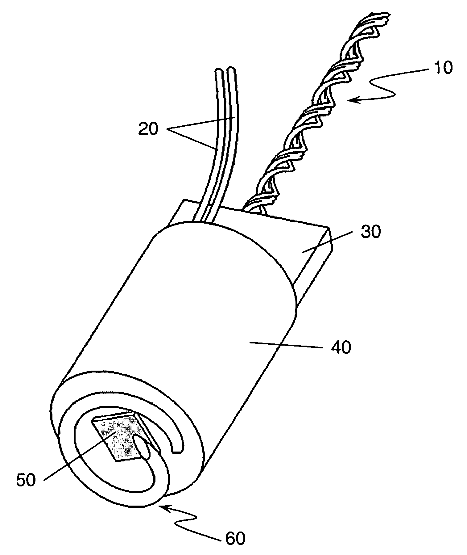
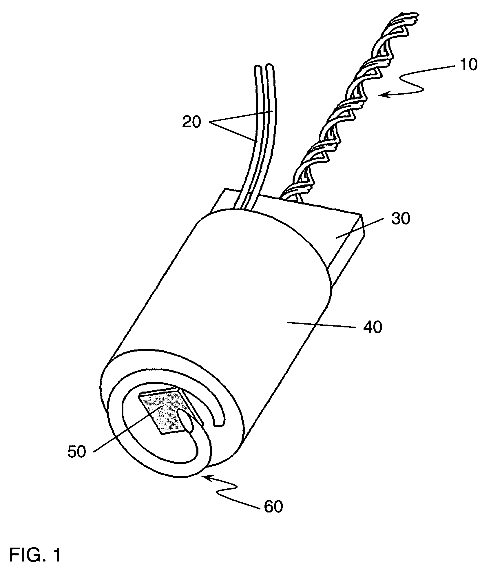
[0028]Helical needles have been in use for many years for the purpose of monitoring the fetal electrocardiogram (ECG). These devices were referred to as spiral needle electrodes or fetal spiral electrodes. This spiral electrode is a solid needle that is rotated into the fetal scalp. The spiral needle provided one of the electrodes necessary for monitoring the fetal ECG. The “spiral” needle electrode is actually helical in shape. This helically shaped electrode is similar to the one shown as item 60 on the sensor pictured in FIG. 1. In the conventional spiral needle electrode the needle was solid, not hollow, and the distal end was beveled to create a point.

[0029]A complete sensor incorporating the needle assembly of this invention is depicted in FIG. 1. This sensor would allow the monitoring of the fetal ECG in conjunction with the monitoring of fetal arterial oxygen saturation. Such a sensor would consist of both optical and electrical components. The needle assembly 60, of this in...


PUM


Abstract

In this invention, a substantially straight needle containing one or more optical fibers is bent into a curved shape, typically including a helical portion, after the insertion of optical fibers into the needle. This curved needle, with its integral optical fibers, allows the subcutaneous delivery of light to a tissue-under-test. The preferred embodiment of this apparatus performs three functions: subcutaneous delivery of light to a tissue-under-test, attachment to the tissue-under-test, and subcutaneous electrical contact. In this invention the optical fibers are inserted into the needle prior to needle bending which prevents fiber breakage because the needle is not curved or in a helical form and therefore provides no resistance to the insertion of the fibers.

Description

STATEMENT REGARDING FEDERALLY SPONSORED RESEARCH OR DEVELOPMENT[0001]This invention was made with government support under R44 HL081866 awarded by the National Institutes of Health. The government has certain rights in the invention.BACKGROUND OF THE INVENTION[0002]The present invention relates generally to the field of fetal monitoring and more specifically to the photoplethysmographic measurement of oxygen saturation and heart rate from a fetus during labor and delivery.[0003]Pulse oximeters are commonly used in adult and neonatal care to provide a measurement of arterial oxygen saturation. A pulse oximetry system typically consists of a sensor which is applied to the patient, a monitor on which the measurements of arterial oxygen saturation are displayed, and a cable which connects the sensor to the monitor. The sensor typically contains light emitting diodes whose output light is incident on the surface of the tissue-under-test and a photodiode detector that measures the intensi...

Claims


Application Information

Patent Timeline
IPC IPC(8): A61B6/00
CPCA61B5/0448A61B5/14542A61B5/6814A61B5/4362A61B5/1464A61B5/288
Inventor DELIANIDES, THEODORE PHILIPVERHEIJ, ADRIAAN GERARD
Owner DELIANIDES THEODORE PHILIP
Learn More