Motion-guided segmentation for cine dense images

a motion-guided segmentation and image technology, applied in the field of myocardial tissue tracking, can solve the problems of lack of automation in clinical use of automated myocardial contour detection techniques based on image intensity, no segmentation algorithm currently exists for cine dense,

Active Publication Date: 2008-01-17
UNIV OF VIRGINIA ALUMNI PATENTS FOUND +2
View PDF11 Cites 36 Cited by
  • Summary
  • Abstract
  • Description
  • Claims
  • Application Information

AI Technical Summary

Problems solved by technology

These techniques are potentially useful for the diagnosis, prognosis and management of heart disease, but their clinical use is currently limited by lack of automation.
For cine DENSE these are typically manually delineated for all cardiac phases, which is a laborious process and is currently the most time-consuming component of the cine-DENSE image analysis.
Automated myocardial contour detection techniques based on image intensity are ill-suited to cine DENSE because: 1. boundaries between the myocardium and adjacent tissue (e.g. the liver) are often indiscernible; 2. a T1-related decay in signal-to-noise ratio (SNR) with time is often present; and 3. high signal is present in the encoded blood of the first few frames, before it is washed out of the image plane.
A number of advances have been made towards automating image segmentation for myocardial tagging and velocity encoding, but no segmentation algorithm currently exists for cine DENSE.
Their method performed well for detection of the endocardial boundary in both short- and long-axis images, but limited manual correction was sometimes required for the epicardium.
Although PC velocity encoded magnitude images are not interrupted by tag lines, they are “white blood” sequences and thus suffer from a lack of contrast between blood and myocardium.

Method used


Image

  • Motion-guided segmentation for cine dense images
  • Motion-guided segmentation for cine dense images
  • Motion-guided segmentation for cine dense images

Examples


Embodiment Construction

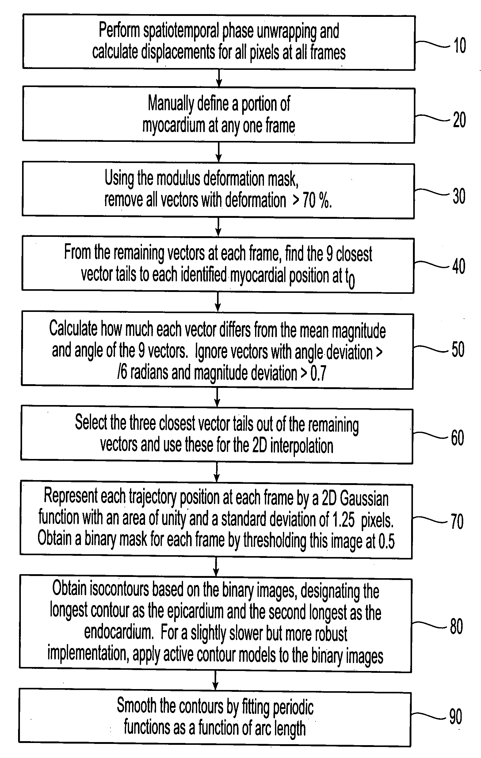
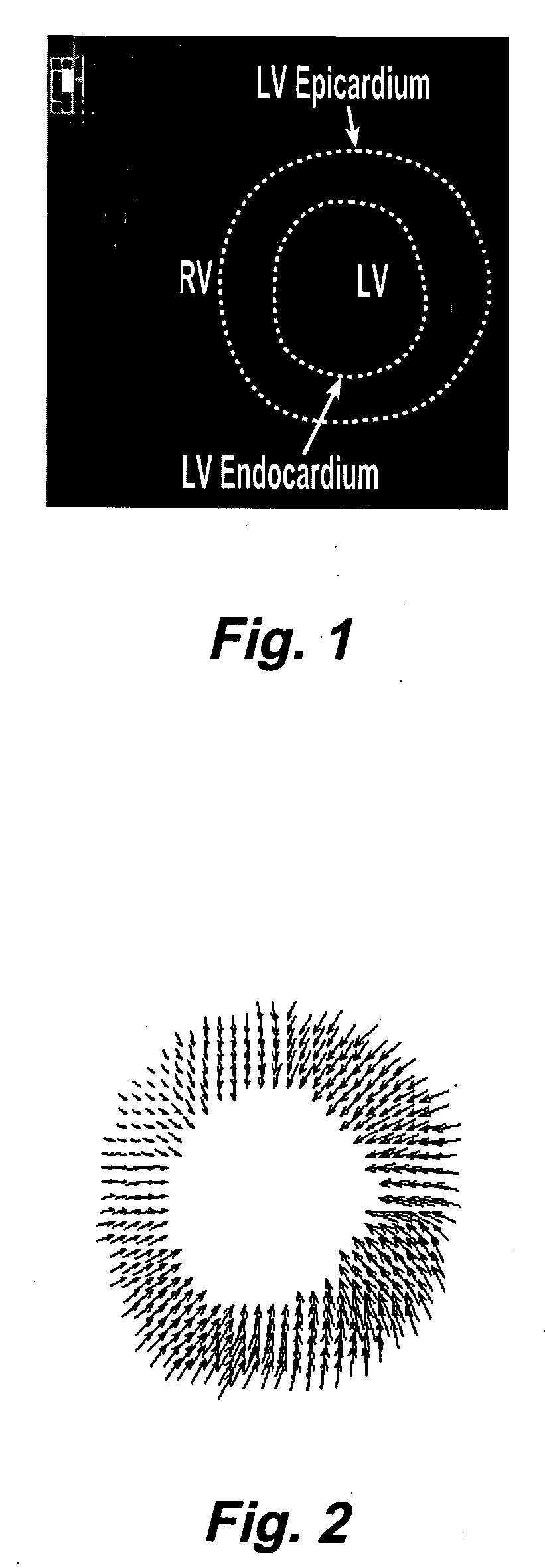
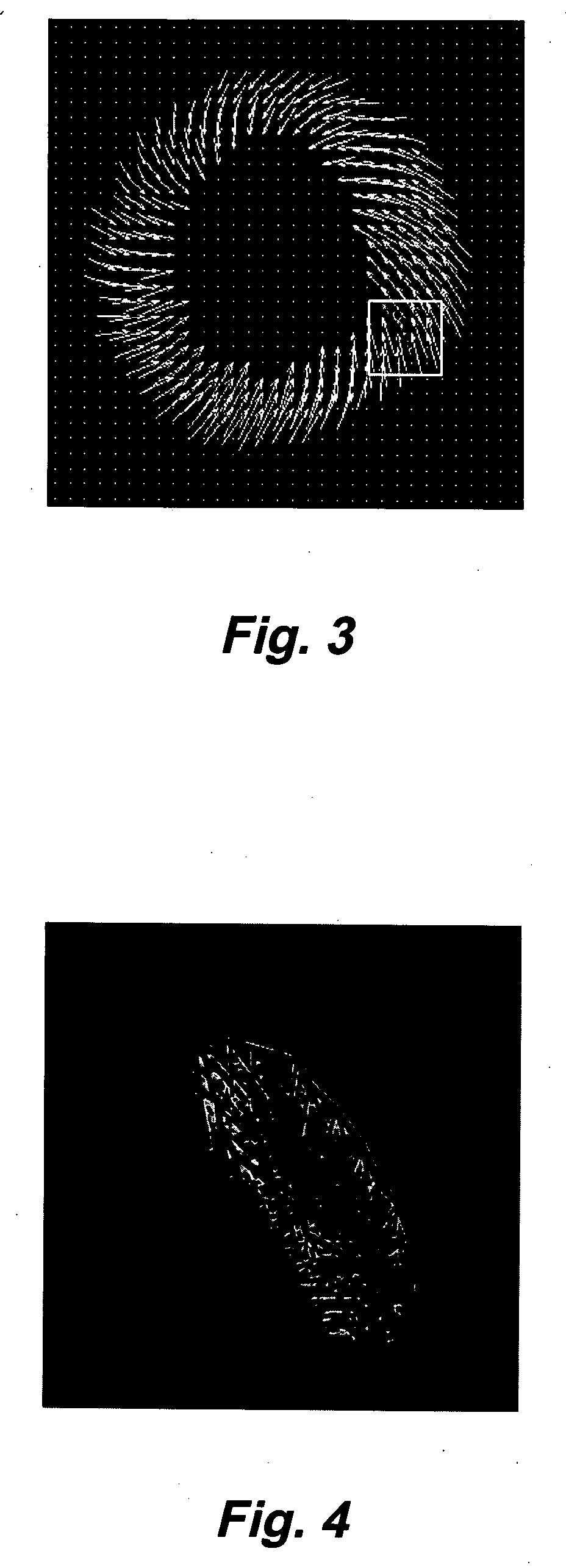
[0033] DENSE uses the phase of the stimulated echo to monitor myocardial motion and deformation at a pixel resolution. The magnetization is initially position encoded using a 1-1 spatial modulation of magnetization (SPAMM) tagging sequence, which is typically applied at end-diastole. Displacement is then accrued as a phase shift during the time between the SPAMM sequence and readout, resulting in images with phase proportional to displacement. Cine DENSE (see, e.g., Kim, D., Gilson, W., Kramer, C., Epstein, F., “Myocardial tissue tracking with two-dimensional cine displacement-encoded MR imaging: Development and initial evaluation,”Radiology 230, 862-871, 2004) provides a time series of such displacement measurements.

[0034] Cine DENSE allows for displacement encoding in any direction, but 2 or 3 orthogonally encoded directions are typically applied. Since only the final phase accrual of the spins is measured, MRI phase is inherently confined to the range−πIEEE Transactions on Medic...


PUM


Abstract

Myocardial tissue tracking techniques are used to project or guide a single manually-defined set of myocardial contours through time. Displacement encoding with stimulated echoes (DENSE), harmonic phase (HARP) and speckle tracking is used to encode tissue displacement into the phase of complex MRI images, providing a time series of these images, and facilitating the non-invasive study of myocardial kinematics. Epicardial and endocardial contours need to be defined at each frame on cine DENSE images for the quantification of regional displacement and strain as a function of time. The disclosed method presents a novel and effective two dimensional semi-automated segmentation technique that uses the encoded motion to project a manually defined region of interest through time. Contours can then easily be extracted for each cardiac phase.

Description

CROSS-REFERENCE TO RELATED APPLICATIONS [0001] This is a non-provisional of co-pending provisional patent application Ser. No. 60 / 800,353, filed May 15, 2006 by Spottiswoode et al., the entire contents of which application is incorporated herein by reference.FIELD OF THE INVENTION [0002] This invention generally relates to methods for myocardial tissue tracking, and more particularly to the use of tissue tracking for projecting a single manually defined set of myocdardial contours through time to give the position of the myocardium at all cardiac phases. BACKGROUND [0003] A number of MRI techniques have been developed to quantify myocardial motion, including myocardial tagging (see, e.g., Zerhouni, E. A., Parish, D. M., Rogers, W. J., Yang, A. Shapiro, E. P., “Human heart: tagging with MR imaging—a method for noninvasive assessment of myocardial motion,”Radiology 163(1), 59-63, 1998; Axel, L., Dougherty, L., “MR imaging of motion with spatial modulation of magnetization,”Radiology 1...

Claims


Application Information

Patent Timeline
Patent Type & Authority Applications(United States)
IPC IPC(8): A61B5/055G06K9/00
CPCG06T2207/30048G06T7/2006G06T7/215
Inventor EPSTEIN, FREDERICK H.MEINTJES, ERNESTA M.SPOTTISWOODE, BRUCE S.
Owner UNIV OF VIRGINIA ALUMNI PATENTS FOUND
Why Eureka
Try Eureka