Methods and systems for optical imaging or epithelial luminal organs by beam scanning thereof

Active Publication Date: 2008-01-24
THE GENERAL HOSPITAL CORP
View PDF98 Cites 208 Cited by
  • Summary
  • Abstract
  • Description
  • Claims
  • Application Information

AI Technical Summary

Benefits of technology

[0009] To address and/or overcome the above-described problems and/or deficiencies, exemplary embodiments of arrangements and processes can be provided that for optical imaging of epithelial luminal organs by beam scanning thereof. These exemplary embodiments of the arrangements and process can utilize a probe and/or disposable portion thereof or of another device which can utilize the following elements and/components for optical imaging...

Problems solved by technology

Certain challenges exist when using scanned, f...

Method used


Image

  • Methods and systems for optical imaging or epithelial luminal organs by beam scanning thereof
  • Methods and systems for optical imaging or epithelial luminal organs by beam scanning thereof
  • Methods and systems for optical imaging or epithelial luminal organs by beam scanning thereof

Examples


Embodiment Construction

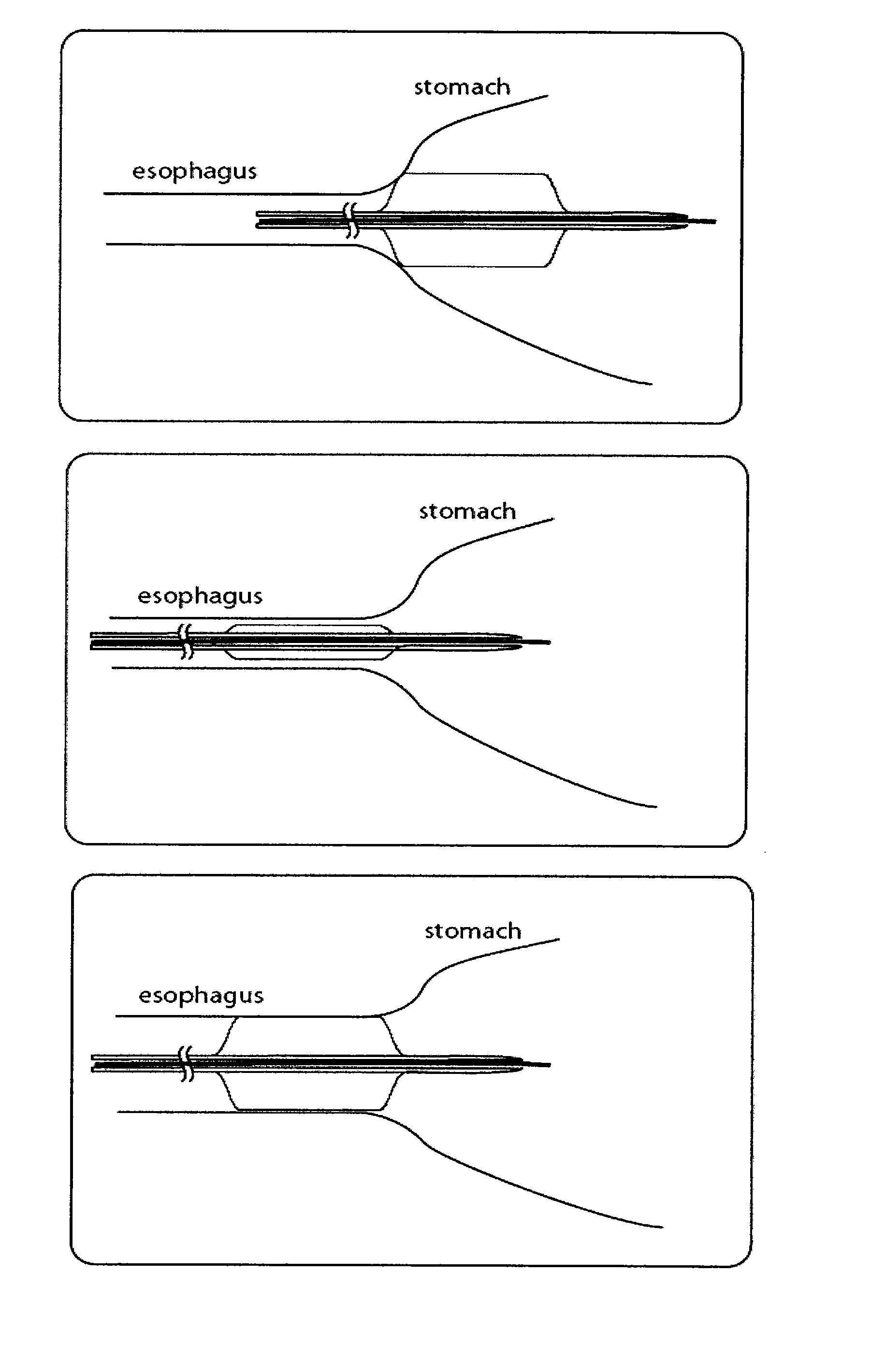
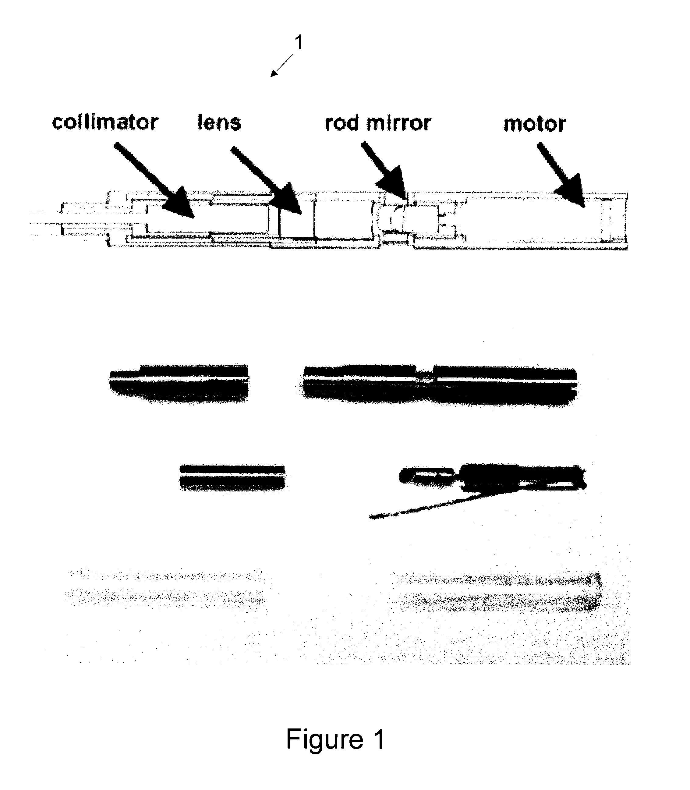
[0059] An exemplary embodiment of a prototype esophageal probe 1 in accordance with the present invention was constructed to investigate the feasibility of obtaining images of the entire distal esophagus, the schematic diagram of this exemplary probe is illustrated in FIG. 1. Such exemplary prototype esophageal screening probe 1 was designed to enable acquisition of images of the entire distal esophagus while operating independently of endoscopy, in standalone mode. Imaging of the entire distal esophagus, however, can be a challenging task as the distance between the catheter and the esophageal wall may vary significantly, even under optimal conditions. Since the Rayleigh range over which the images remain in focus is approximately 1 mm (˜35 μm spot diameter), the esophageal lumen should be made as circular as possible, and the probe should generally be centered within the esophageal lumen.

[0060] In such exemplary prototype screening probe 1, an esophageal balloon centering cathete...


PUM


Abstract

Arrangements, apparatus, systems and systems are provided for obtaining data for at least one portion within at least one luminal or hollow sample. The arrangement, system or apparatus can be (insertable via at least one of a mouth or a nose of a patient. For example, a first optical arrangement can be configured to transceive at least one electromagnetic (e.g., visible) radiation to and from the portion. A second arrangement may be provided at least partially enclosing the first arrangement. Further, a third arrangement can be configured to be actuated so as to position the first arrangement at a predetermined location within the luminal or hollow sample. The first arrangement may be configured to compensate for at least one aberration (e.g., astigmatism) caused by the second arrangement and/or the third arrangement. The second arrangement can include at least one portion which enables a guiding arrangement to be inserted there through. Another arrangement can be provided which is configured to measure a pressure within the at least one portion. The data may include a position and/or an orientation of the first arrangement with respect to the luminal or hollow sample.

Description

CROSS-REFERENCE TO RELATED APPLICATION(S) [0001] This application is based upon and claims the benefit of priority from U.S. Patent Application Ser. No. 60 / 761,004, filed Jan. 19, 2006, the entire disclosure of which is incorporated herein by reference.STATEMENT REGARDING FEDERALLY SPONSORED RESEARCH [0002] The invention was made with the U.S. Government support under Contract No. RO1CA103769 awarded by the National Institute of Health. Thus, the U.S. Government has certain rights in the invention.FIELD OF THE INVENTION [0003] The present invention relates to methods and systems for optical imaging, and more particularly to optically imaging epithelial luminal organs by beam scanning thereof. BACKGROUND OF THE INVENTION [0004] Screening for diseases is a process whereby a person who is not known to have one or more possible diseases undergoes a test to determine whether or not the person has any such diseases. Screening is often conducted on a large population, and therefore is like...

Claims


Application Information

Patent Timeline
IPC IPC(8): A61B1/00
CPCA61B5/0062A61B5/0066G06T7/0012A61B5/6852A61B5/0084A61B2562/0247A61B5/0068A61B5/7271G06T2207/10056G06T2207/10101
Inventor TEARNEY, GUILLERMO J.SHISHKOV, MILENBOUMA, BRETT E.VAKOC, BENJAMIN J.NISHIOKA, NORMAN S.
Owner THE GENERAL HOSPITAL CORP
Why Eureka
Try Eureka