System and Method for Detection of Breast Masses and Calcifications Using the Tomosynthesis Projection and Reconstructed Images

a tomosynthesis and projection technology, applied in the field of computer aided detection of masses and calcifications in tomosynthesis images, can solve the problems of difficult mammogram interpretation in younger women, unsatisfactory, and radiation exposure associated with mammography as a potential screening risk

Active Publication Date: 2008-01-31
SIEMENS MEDICAL SOLUTIONS USA INC
View PDF3 Cites 61 Cited by
  • Summary
  • Abstract
  • Description
  • Claims
  • Application Information

AI Technical Summary

Benefits of technology

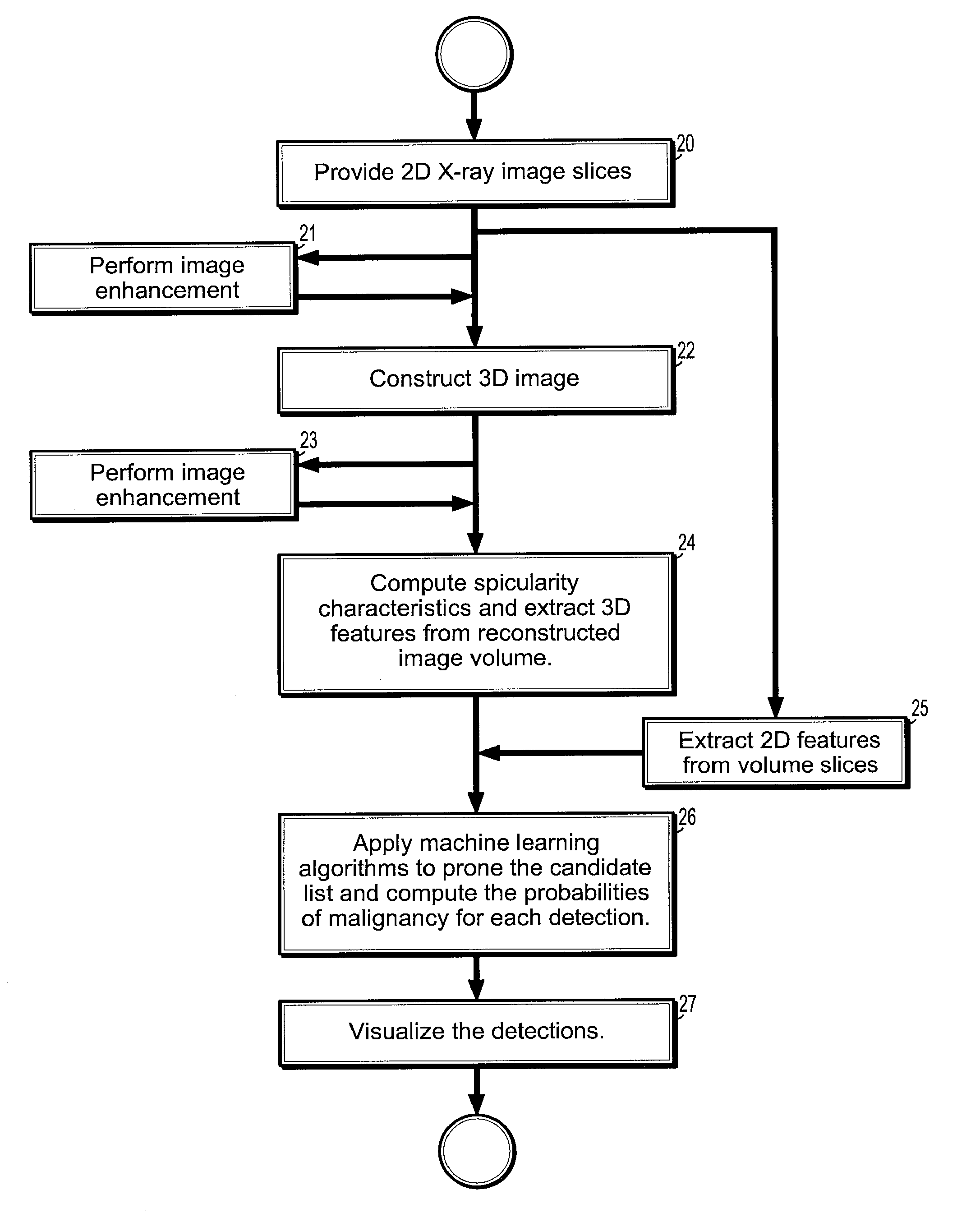
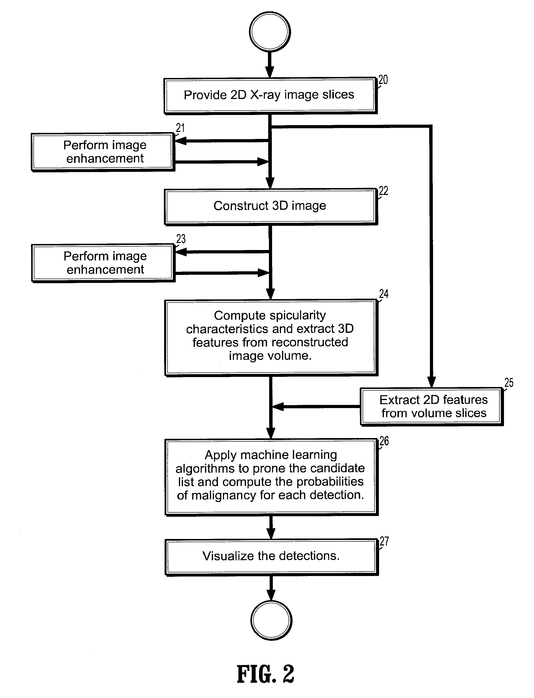
[0021] According to a further aspect of the invention, the method comprises enhancing said X-ray breast images prior to constructing said 3D image volume.
[002

Problems solved by technology

The radiation exposure associated with mammography is a potential risk of screening.
While mammography is the only breast cancer screening method that has been shown to save lives, it is not perfect.
Reasons for not seeing the cancer include observer error, but more frequently it is due to the fact that the cancer is hidden by other dense tissue in the

Method used


Image

  • System and Method for Detection of Breast Masses and Calcifications Using the Tomosynthesis Projection and Reconstructed Images
  • System and Method for Detection of Breast Masses and Calcifications Using the Tomosynthesis Projection and Reconstructed Images
  • System and Method for Detection of Breast Masses and Calcifications Using the Tomosynthesis Projection and Reconstructed Images

Examples


Embodiment Construction

[0026] Exemplary embodiments of the invention as described herein generally include systems and methods for computer aided detection of masses and calcifications in tomosynthesis images. Accordingly, while the invention is susceptible to various modifications and alternative forms, specific embodiments thereof are shown by way of example in the drawings and will herein be described in detail. It should be understood, however, that there is no intent to limit the invention to the particular forms disclosed, but on the contrary, the invention is to cover all modifications, equivalents, and alternatives falling within the spirit and scope of the invention.

[0027] As used herein, the term “image” refers to multi-dimensional data composed of discrete image elements (e.g., pixels for 2-D images and voxels for 3-D images). The image may be, for example, a medical image of a subject collected by computer tomography, magnetic resonance imaging, ultrasound, or any other medical imaging system...


PUM


Abstract

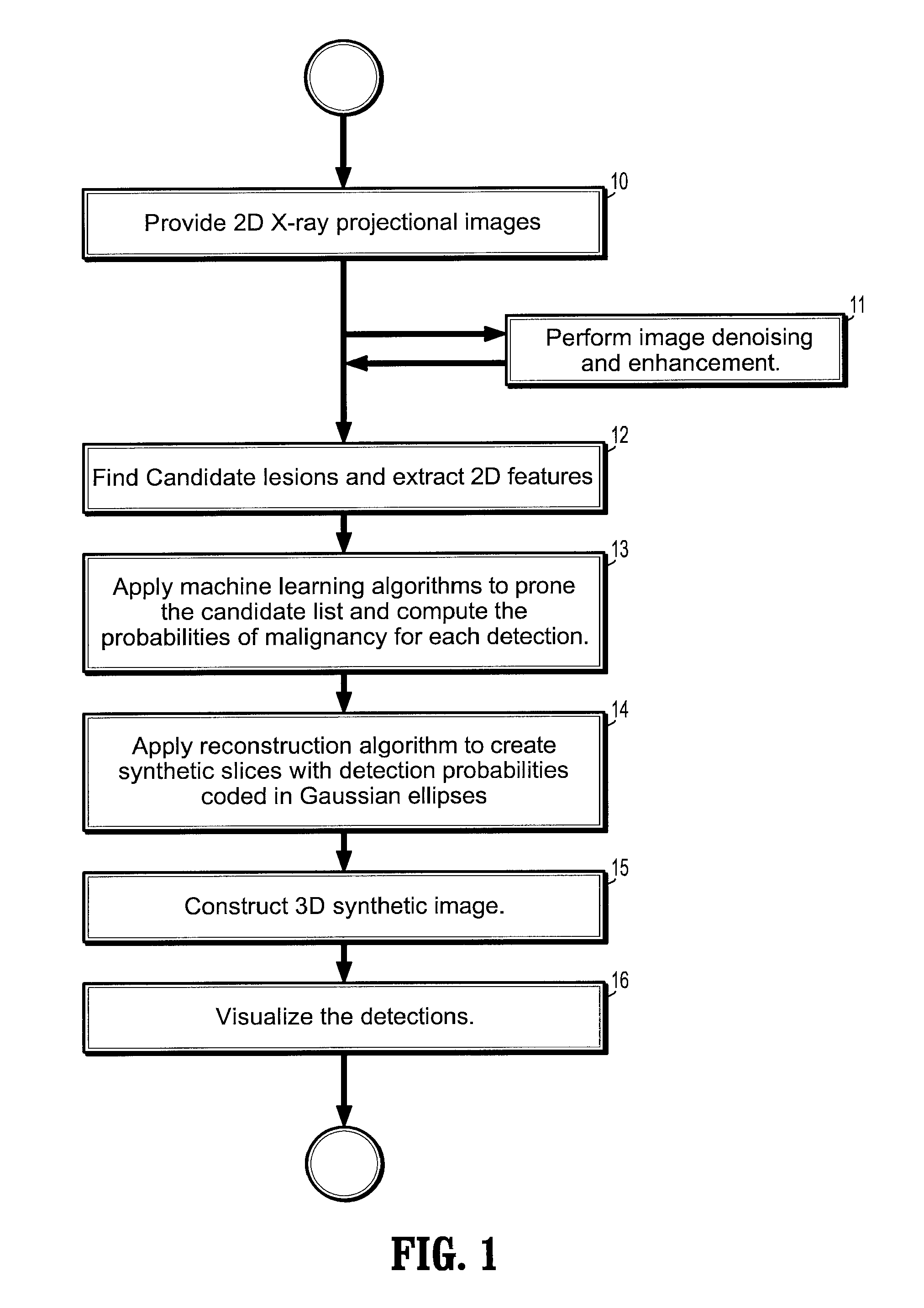
A method of detecting breast masses and calcifications in digitized images, includes providing a plurality of 2-dimensional (2D) digital X-ray projectional breast images acquired from different viewing angles, extracting candidate lesions and 2D features from said 2D projectional images, computing spicularity characteristics of said candidate lesions, including location, periodicity, and amplitude, applying learning algorithms to said candidate lesions to predict a probability of malignancy of said lesion, receiving from said learning algorithm a probability map of detections for each breast image, said detections comprising associating pixels with a probability of being associated with a malignancy, creating a synthetic 2D slice for each X-ray image wherein malignant regions are indicated by ellipses on a non-malignant background, and constructing a synthetic 3-dimensional (3D) image volume from said 2D synthetic slices.

Description

CROSS REFERENCE TO RELATED UNITED STATES APPLICATIONS [0001] This application claims priority from “A method for detection of breast masses and calcifications using the Tomosynthesis projection and reconstructed images”, U.S. Provisional Application No. 60 / 816710 of Jerebko, et al., filed Jun. 27, 2006, the contents of which are herein incorporated by reference.TECHNICAL FIELD [0002] This disclosure is directed to computer aided detection of masses and calcifications in tomosynthesis images. DISCUSSION OF THE RELATED ART [0003] Mammography is the process of using low-dose X-rays (usually around 0.7 mSv) to examine the human breast. It is used to look for different types of tumors and cysts. Mammography has been proven to reduce mortality from breast cancer. Like all x-rays, mammograms use doses of ionizing radiation to create this image. Radiologists then analyze the image for any abnormal growths. It is normal to use longer wavelength X-rays than those used for radiography of bones...

Claims


Application Information

Patent Timeline
IPC IPC(8): G06K9/00
CPCA61B6/03A61B6/466A61B6/502G06T7/0012G06T2211/436G06T11/005G06T2207/10112G06T2207/10116G06T2207/30068G06T7/0022G06T7/97
Inventor JEREBKO, ANNAKRISHNAN, ARUNZHOU, XIANG
Owner SIEMENS MEDICAL SOLUTIONS USA INC
Why Eureka
Try Eureka