

Application-driven optimization of acquisition and reconstruction of SPECT/PET projection data
a projection data and spectroscopic technology, applied in the field of nuclear medicine, can solve the problems of increasing clinically significant image resolution, image quality may suffer, and insufficient acquisition of projection data, and achieve the effect of reducing the noise power of the reconstructed imag
- Summary
- Abstract
- Description
- Claims
- Application Information
AI Technical Summary
Benefits of technology
Problems solved by technology
Method used
Image

Examples
Embodiment Construction
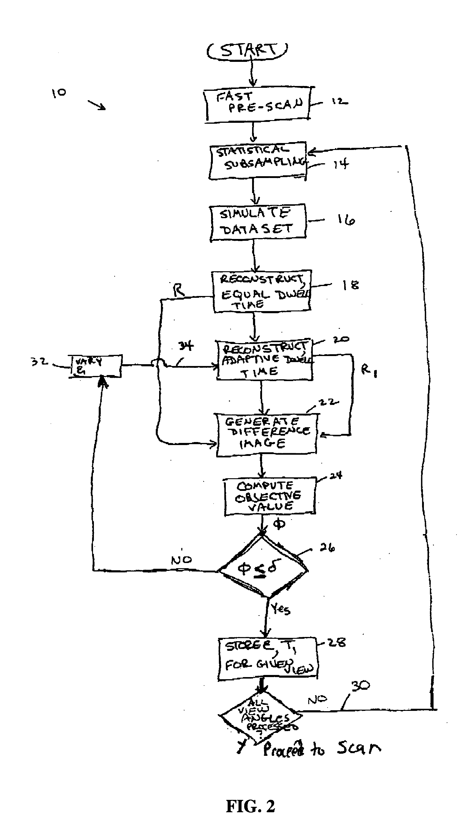
[0015]An embodiment 10 of one method in accordance with the concepts of the invention is illustrated in FIG. 2. According to this method, an initial “fast” pre-scan 12 is first conducted for total time T to accumulate statistical projection data. The fast pre-scan is conducted over a defined ROI in the projection image. The ROI may be defined automatically, such as through the use of various known methods such as automatic segmentation methods in heart studies. The ROI also may be defined semi-automatically, such as having a user define an ROI in one projection view, from which a software program can infer ROIs in all other projection views. Alternatively, the ROI may be identified manually by the user in each projection view.
[0016]During this initial pre-scan, the dwell time ti per angular view is held constant over the entire scan range (either π or 2π radians, depending on the type of scan being conducted) such that:
T=Σti, i=1, . . . N, N=number of views, t=ti, ∀i.
[0017]For each ...
PUM
 Abstract
Description
Claims
Application Information